Can I load this implant?
I extracted an impacted canine and immediately installed an implant with TCP, PRF and DBM graft. Radiograph is 4-months post-op, looks okay. However, CBCT does not show complete osseointegration. Do you recommend that I go forward and now load this implant?

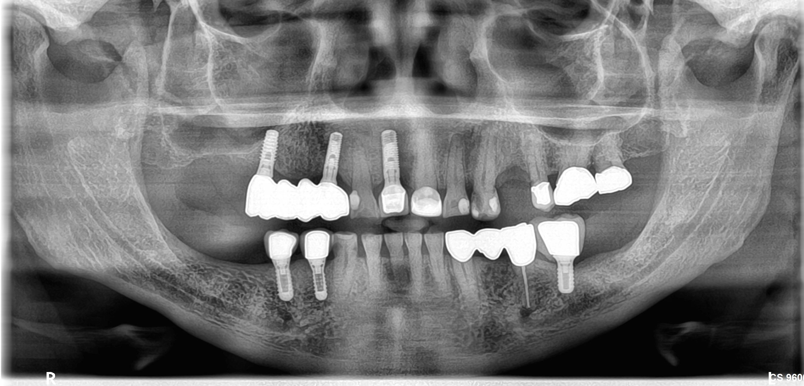
 Follow Up Case Photos


21 Comments on Can I load this implant?
New comments are currently closed for this post.
Julius
11/14/2015
Dear colleague, my suggestion to you:
use a fancy word to educate your patients - and tell them that it is now a time for BONE TRAINING (simulate bone by loading, to facilitate implant osseointegration) - and make a provisional
At prosthetic point of view, never forget to go with provisional at upper anterior region.
what i am doing in similar cases:
-provisional - soft tissue contouring to you, bone training for patient
->if you got the screw hall in esthetic part, just use composite, go to form exelence, rather than color
->show the patient what is going to look like
->you may have to thicken something or reduse (sorry, photos are made with phone, as i see)
-if patient happy, you are happy with soft tissue esthetics, forms and functions - make a soft tissue contouring for min 3 weeks, max 1 month and 2 weeks
->individualize a transfer - take implant analog, screw the provisional on it -> sink it in impression puty -> unscrew provisional and place it in the mouth-> take a transfer place it on analog and puty complex-> individualize transfer with flowable composite, cure it fully, by curing oxide inhibition layer or polish it after that. if you are not happy with something, you are able to fix, just apply some composite
-> place individualised transfer in the mouth on implant, take your loops (recommend), and take of composite at the level of soft tissue, to make your dental technician where the gingiva is. If you are using chair side cad cam, you are on your own, besause i do not know how it is working, for now :)
choose abutmet materials wisely and you will be okay
Satish
11/14/2015
Thank you for your comments.
And for taking time out to write a lengthy answer.
Much appreciated
CRS
11/14/2015
Is the CBCT current? if so there is no osteointegration. One thing about peri apical films, one can't assess the buccal plate. If the Cone beam is accurate then when you open the flap and place the impression coping the implant will come out. Looking at the angle of the implant it is not restorable. Most likely there will be a significant defect when the implant is removed patient will need grafting in an esthetic area. One Pearl when a canine is impacted the alveolar process does not develop completely and this lack of fully developed crest need to be taken into account. I advise referring this case it is compromised.
Satish
11/14/2015
Thank you for your comments
Satish
11/14/2015
Also the Implant position was the one designed in the pre op cbct through software
Is the positioning wrong?
Thank you
CRS
11/15/2015
I'm confused you placed an implant for a central incisor in an impacted canine extraction site? Do you have a preop film with the impacted canine? The implant appears to be angled to the buccal ideally I like to align it with the cingulum of the adjacent teeth. If that post op cone beam is accurate there is no bone around the implant with a defect in the buccal plate or it is quite an artifact. You know what,open up the area and take your impression then post what was the outcome then we will all know for sure. Good Luck.
Satish
11/15/2015
Thank you
Wil do it tmw and post updates
Alex Zavyalov
11/14/2015
Nothing is said about the implant clinical mobility/stability which is crucial as the implant is surrounded with grafting material mostly. If it's even a minimally mobile, I would refrain from proceeding.
Satish
11/14/2015
I had got excellent primary stability with threads engaging the bottom 3 threads and top 3 threads and 50 N
Thank you for ur comments
PeterFairbairn
11/17/2015
Hi Satish always difficult removing an impacted Canine , place and graft at one go . I have just placed a500 case study on this protocol and understanding materials and protocols is important ...
I think you have too many materials here and maybe missed getting graft stability which is the key ...... motion inthe particles could led to Mesechymal cell differentiating to fibroblast .....hence what you have here is a lot of granulous tissue with some particles in the mass as see in your PA ....
This needs a re-graft or to start again ..
Just some ideas
Peter
pierre
11/18/2015
in cases like these I rememder Ed Rosenbergs advice......do one miracle at a time ......and to remove the impacted canine first ,augment,for enough bone and the implant later
Satish
11/18/2015
Thank you all for your valuable comments.
Today I exposed the implant and the implant was stable and covered by bone all around the cover screw.
Abutment was placed and finger torqued to maximum,still stable.
Is there anyway to upload photos for further comments.
Thank you again everyone!
Satish
11/18/2015
Keeping fingers crossed now.
Satish
11/18/2015
Exposed implant
Implant stability very good
Good bone cover clinically around implant
Placed abutment,maximum finger tightening done
Implant still stable
Anyway to post photos?
Thank you everyone for your inputs and advice
Much appreciated
OsseoNews
11/19/2015
To post case photos, simply click on the "Post a Case" link in the website menu. In the case description, just let us know that you want to add these photos to the current case. Be sure to use the same contact information you used to post the original case.
OsseoNews
11/19/2015
Follow up photos have been added for this case. Please just refresh your page and scroll up to the original question to see the follow up photos.
Richard Hughes, DDS, FAAI
11/26/2015
You may it tried to many procedures into one visit.
You mostlikely will have to vemove the implant and regraft. Mp lace the implant later. The maxillary cuspid is a difficult site.
Richard Hughes, DDS, FAAI
11/28/2015
I suggest to always line up the platform with the adjacent teeth when viewing from the occlusal. This will usually result in the implant being in the correct F-L orientation.
According to your CBCT there appears to be poor integration. One should probe the osteotomy sites after each bur or prior to placing the implant. If you feel a perforation then act accordingly.
I'm a bit confused. I do not see this implant in a cuspid site. Please elaborate!
Richard Hughes, DDS, FAAI
11/28/2015
I suggest to always line up the platform with the adjacent teeth when viewing from the occlusal. This will usually result in the implant being in the correct F-L orientation.
According to your CBCT there appears to be poor integration. One should probe the osteotomy sites after each bur or prior to placing the implant. If you feel a perforation then act accordingly.
Dr Richa Raj
4/27/2016
Hi,
I was following your case. want to know that have u loaded the implant. how is it after loading. can u share some pics. Thx
Satish
5/2/2016
Thank you for your query
Yes we loaded the implant
Acrylic crown for 6 months
Off occlusion
Pt very satisfied with prosthesis
Implant reviewed every month
Doing well
Keeping fingers crossed
Wil try to send photos and xrays