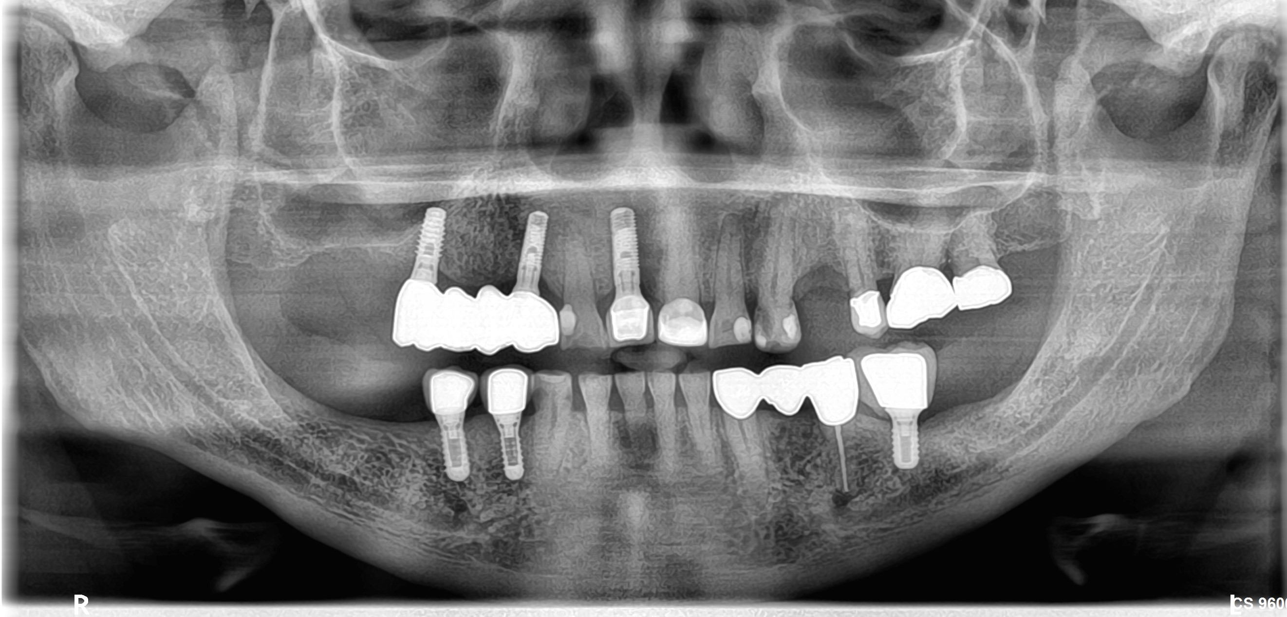
CBVT Scan for Small Implant Cases?
Dr. S. asks:
I am new to the surgical placement of implants. I currently do only small and easy cases where I do not have to worry about complications from the inferior alveolar nerve, maxillary sinus, mental foramen, etc. This really limits my range of operations. I still understand the potential for complications and I certainly want to avoid them. My oral surgeon has been encouraging me to do more. He recommends that I learn how to use a cone beam volumetric scan to plan my cases to avoid complications. Is this really necessary for easy implant cases? Can I get enough information from the panoramic radiograph?
33 Comments on CBVT Scan for Small Implant Cases?
New comments are currently closed for this post.
A.Romano dr.med.dr.dent I
5/19/2009
i wuold say that you have to learn how to read in the best way a cbvt scan imago. this is very important and you can study and learn this in an important rx management.
for simply or mid-simply implants you can perfectly use a panoramic rx but you must have anyway a good frequentation with a surgeon with good experience.
for instance if you are not a medician you cannot be face to face with the many complications of surgeon practice. and you have to be a good chemist so to treat in the best way yor patient.
sincerely good luck. alex
Thomas Gibbs
5/19/2009
Pan's still have significant distoration. I went with the Instrumantarium OP200 with VT, or volmetric tomography. With a VT you can get good images of a single quadrant without the distoration of a pan. The unit cost about 65,000. Still a good price, but not nearly as expensive as a ICAT (170,00 at that time). At the time I bought (year ago), upgrading the other Pans was too expensive. Most cases are just 1 to 3 implants and this works fine.
For larger cases or when guided surgery is needed, I still get a full scan done and use Simplant.
Joseph Kim, DDS
5/19/2009
Pans are good when you have a reference object or when you know the amount of magnification/distortion in your particular machine. In all honesty, a PA will provide you with an accurate distance to most vital structures. You can also take a working PA leaving a depth gauge in the 2 mm osteotomy. Make sure the depth gauge has grooved markings that will show up on the PA. This is the most accurate technique.
A final option is to use implants that are 6.5, 8, and 10 mm long. This was presented at the last AO meeting in San Diego rather convincingly. The modern literature on acid etched fixtures supports the use of shorter fixtures. Don't get a CT scan for a single implant. If you feel you really need it due to a large bony defect, it may be a case you would be more comfortable referring anyways at this stage in your practice.
CT has distortion issues as well, so don't think that it is a fool proof way of avoiding vital structures. Also, how do you plan to transfer the information you see on the CT to the live clinical situation? By the time you order the CT, make a surgical stent from the CT, you have added a significant sum of money and time to the otherwise simple procedure. Just food for thought.
Rik Vanooteghem
5/19/2009
First, let me state that according to a legal opinion, from a "standard of care" standpoint, any neurovascular complication in the mandibular posterior will be indefensible in court.
I have had an I-Cat in my office for the last year and a half and it has changed the way I place dental implants and the way my patients view dental implants.
I take a scan for every implant case and feel more confident in my procedures and I am more accurate. Also my patients appreciate the added safety and my concern about their well being. Even though this is an "out-of-pocket" expense rarely does a patient have an objection to doing a scan. From a technical standpoint, particularly in immediate implant cases anywhere in the mouth, in the mandibular symphysis, above a nerve, below a sinus, the pre-op CBCT is very important in the planning of the case.I use Simplant routinely as an implant plannig platform.
My 2 cents.
Richard Hughes DDS, FAAID
5/20/2009
Good points Dr. Kim, I use 5mm. stainless steel ball bearings and some basic algebra for this. You are correct PA's are excellent. I wonder how many CBCT's are in use in the USA. Some people imply that this is the standard of care, but for something to be the standard of care it has to be very commonly used.
steve c
5/20/2009
If you begin to use CBVT for some of your cases, you'll soon want the info. it provides for all cases in any part of the mouth, including single implant cases. I agree that almost all patients readily accept tomography when they realize the benefits it offers. Before I enter any implant case I have full information on what I will encounter. I'm never surprised by a narrow bony ridge under thick gingiva. I always know when there is an undercut or narrowing in the ridge further apically. I am much more secure with implant selection for each case. My patient is fully aware of the fact that I will need to augment a slightly narrow ridge at the time the implants are placed. I almost never enter a treatment expecting to place implants only to find it will be necessary to perform a lateral ridge augmentation instead. I rarely have to change direction of treatment due to unforseen anatomy and thereford don't face telling the patient that their treatment will require extra steps, time and expense. I know this beforehand through CBVT. To me this small diagnostic step takes away most of the unknown and makes implant surgery much more enjoyable and definitely less stressful.
Russell
5/21/2009
Rik;
Ditto!
Empirical Medicine
5/26/2009
I have a CBVT and I use it when indicated, However....
In my opinion, it is not the standard of care!
To suggest that placing an implant without a CT is indefensible.... well, that sounds like someone selling equipment.
As always, the standard of care to ensure you have measured and determined the available anatomy using an calibrated, diagnostic image.
This is a small part of a larger treatment planning session which culminates in a true informed consent process with the patient which helps them actually understand the risks, benefits and alternatives of their care.
Dr. Powers
5/26/2009
Question #1=No
Question #2=Yes
I'm going to guess that WAY over 95% of all implants placed are done with a PA and or Pano. Is cone beam better? Yes. Do you have to have one? No. Save yourself $100,000+ and refer the tricky ones over to your OS buddy.
Brian James
5/26/2009
I think there is real merit in learning how to interpret CBCT scans, whether they be in printed/PDF format from a radiology practice, or via one of the interpretation programs such as iCAT Vision. As has been said, it can and will change how you think in three dimensions, and the more you know the less surprises you will encounter. It will also have benefits in assessment of pathology, third molar removal etc. You do not have to buy one to use the technology. The comment about distortion and CBCT is erroneous. There is zero distortion from the iCAT and this has been verified. Just because it's new doesn't mean it should be avoided. I have a machine in house, but I think a general dentist doing implants would find it difficult to justify, and it is NO replacement for a screening OPG, as the radiation is considerably greater. Great tool, in its right place.
Jay Reznick
5/27/2009
Just as panoramic radiography changed dental diagnostics in the 1960's, CBCT is changing the way dentists look at anatomy. Our technology and materials are constantly advancing and improving. When a panoramic was the best we had, then it was a great way to evaluate patients for dental implants. CT scans and DentaScan were introduced to dentistry in the 1980s, and gave us information that was not previously attainable. But, it was a pain to get the patient to the CT scan, and not every radiology center had the software, and the huge radiation dose did not make sense for "simple" cases. The late 90's brought us SimPlant software, and the turn of the millenium brought us CT-guided implant surgery.
I embraced this technology about 4 years ago, and cannot go back to doing things the "old-fashioned" way. The incredible increase in diagnostic information is invaluable. I do not have "surprises" any more in surgery. I can plan an implant case with great precision, knowing the 3D anatomy of the ridge and anticipating the need for grafting and any other challenges before I have touched the patient. Obviously, having ready access to a CBCT machine is essential for this.
I have had a cone beam in my office for about 1 1/2 years, and it has changed the way I practice. About 95% of my implant cases are done using CT-guided surgery. Sure, the machine was expensive, but it more than pays for itself in increased case acceptance, shortened surgical times, fewer complications, and fewer cases of having to "eat" the cost of a bone graft because I did not anticipate it before surgery.
My charge for a CBCT scan is not much more than for a pano, and I provide the surgical stent at cost, so the patient gets the benefit of this technology at very little increased cost, compared to the "traditional" method.
Once you start using this technology for your implant cases, you will not want to do them any other way. I think a lot of the obstacles to this technology are psychological. We have a hard time changing our ways when we feel that we have been getting good results. But, once you start using this technology for your implant cases, you will not want to do them any other way.
Richard Hughes DDS, FAAID
5/27/2009
I think CBCT is a great tool. However I use PAs and PANOS solely for 99% of my cases and refer to an orthodontist for the times I need one. Is it the standard of care???????????? Perhaps in certain cases, but not all the time.
Dr. SDT
5/27/2009
Dear Dr.S
My point of view is that CBCT it is just another tool that we as Doctors can use for a bettter diagnosis and consecuently a better Treatment plan for our patients, I have been using CBCT for Tx planning for almost 5 years now. You do not have to own a CBCT to take a spet forward in the fiel of imaging. This is not only for Implants, there is a lot more out of this technology. This will give you the confidence you need for more complicated cases. Best of luck
Dr. SDT
5/27/2009
Correction: ..to take a step forwad in the field*
Dr Z
6/6/2009
I placed two implants in the anterior maxilla ,11 and 21 region the 21 area had been augmented with a bock bone graft 6 months prior to implant placement. when i placed the implant in the thickend ridge the bonegraft cracked but didnt detach. i left the implant inplace aumented the crack with bio-oss and placed a membrane. any advice on how to handle these cases and does this commonly happen in block bonegrafted regions ?
John Stedmen DMD MD
6/6/2009
Most cases can be done with a pano and pa. However, as cone beam gets cheaper in price, and they will get cheaper, just think about plasma tv's when they first came out, they will probably change the face of dentistry.
tony collins
6/21/2009
In Australia we call it "Cone Beam Volumetric Tomography" to distinguish it from "Computerised Tomography". One prime difference is the vastly reduced amount of radiation for a CBVT compared with CT.
I have a local radiographer with an ICAT who accepts the Government fee so no charge to the patient. The anatomical information on a scan is impressive and I won't place an implant, or do ANY surgical procedure without one.
PAs and OPGs will never disclose the maxillary incisive fossae (labial inrolls), nor will they alert you to the presence of a pronounced mylo-hyoid ridge sitting above soft tissue in the posterior mandible (just waiting to be penetrated with your drills - think Ludwig's Angina ! That will really spoil your day and perhaps your patient's life. The Mental foramen is a breeze to locate and the IAN is more easily traced (still some way to go in refining the definition though).
Just last week I had two patients with apical pathology of upper anteriors that was only detected on the 3-D reconstruct by turning the image of the skull and looking into the nasal cavity at the inner aspect of the piriform rim. Try that with PAs and OPGs.
My radiographer still sends me back an OPG and I still appreciate the diagnostic qualities of a PA, but the way forward is with a CBVT and I consider it the standard of care in my practice.
Dentist North York
7/15/2009
Using a PAN or PA is sufficient for standard cases. For more difficult cases, I suggest referring to your local oral surgeon if you don't use CT
Robert
7/17/2009
There is now a mobile CT Scanner operating in the New York City area called Facial Imaging Mobile. They are a good source for Dental CT Scans, Simplant conversions and NobelGuide cases. They provide a radiology report as well. Most importantly you do not have to send the patient to another doctors office for the scan. Very convenient and fast turnaround time
Dr.T
8/12/2009
In my opinion, based on radiological and clinical experience, one can called an implant case "easy" ONLY when you know the 3 dimensional anatomy of the area of surgery and that is only achievable by any type of 3D imaging, before that just make sure you have a great liability coverage. You will be amazed of how many interesting cases that were called easy cases, ended up in failures just because there was a lack of diagnostic aids. Think oyur self as a patient, what would you like to be done if you are the onne that needs an immplant???
Making your patients smil
8/12/2009
There is not need to buy such an expensive equipment, why to delay your retirement? specially when times are tuff. I received a mailer from an Mobile Imaging center in Florida called 3D Mobile Scan Corp, they come to my office and scan my patients, in just 20 minutes I have all the information I need. This allow me to do a more comprehensive treatment planing for my patients, they feel that I really care about them. I also consider that is better to pay a fee for that service and this way reducing my liability.
Ilya Benjamin
9/5/2009
I use Cone Beam for every case, I dont know how people choose implants sizes without knowing the volume (depth, height, width)
And I have allowed my Cone Beam to be used amongst Local Dentist via mobile delivery.
Great service!
Richard Hughes DDS, FAAID
9/5/2009
CBVT is a nice tool to have when it is needed. I try to avoid using it routinely due to the cost to the patient. I use 5mm. ball bearings, bone calipers, PA radiography, study models etc. However there are times when I have used CBVT an happy that I did so.
dr.ws
9/9/2009
dear friends,
i just started implants[n.bio]did one last month,joined this practice,have few implants pt's ready.started rx planning and trying to get cbct for every case,just to be on safer side,do we really need n.guide/simplant software,any suggestions about an easy readable software for the scan reports.do you still need a s.stent[lab made] after a scan for simple cases.thnx in advance''
DrD
9/10/2009
Dear Dr WS, it is great that you are thinking safe.
There is different ways of taking advantage and working with CBCT. It is necessary to understand the concept of: Planning software, scan appliance, surgical guides.
Having mentioned that and after using CBCT for treatment planning for years on a regular basis, I think:
1. Using a Planning software is a great tool, now days there is a lot of them, some more user and cost friendly than others. However you can evaluate the images from a CBCT just using a "Free viewer" which will allow you to take distances, see 3D images, etc (I-Cat Vision, NNT Viewer etc.)
2. Scan appliance will make your Dx easier, and it is a non expensive aid (a vacuum formed stent with Rx opaque teeth will work, using the pt's own partial or denture and placing gutapercha)
3. Surgical guides are useful for cases with multiple implants, difficult angulations. But I don't think it needs yo be used for all cases.
Finally, make sure that you have a Radiology report for all your CBCT cases.
dr.ws
9/10/2009
DEAR DR D,
thanks a lot for your suggestions and time,i believe its great to be helping/encouraging others,especially when they are out of school and have nowhere to go/ask.i really appreciate that.keep it up''
thanks again,
cheers''' dr ws.
William Reeves DDS PC
9/19/2009
I cannot imagine doing implants without a cone beam. Every case I execute is planned with my cone beam CT. I do not charge my patients for my scans or radiology reports. Having come from using panoramics, it is a night and day comparison. I use Keystone Easy Guide with their x-marker for my surgical guide with great results. It makes me a better dentist, giving me the ability to screen for obstructive sleep apnea/hypopneas with the click of a button, TMJ is 3D, Hypertrophic tonsils and turbinates, Calcified carotid atheromas, cysts & mucocoeles, tonsilleths so big you would swear they swallowed some rocks, etc, etc......day in and day out it happens and I shake my head, wondering how I practiced before without it. Referrals to the correct Medical/Dental specialists have my patients thanking me for "not just looking at their teeth." It makes a difference knowing that you caught something that will allow your patient to live a longer, healthier life.
Sorry, I started rambling...to answer the original question---yes, I believe that as experience/confidence builds and you want to execute cases that are increasing in difficulty, CBCT is necessary. Freehanding multiple implants can make for a restorative nightmare. Also, I hate to see an implant case go wrong that makes the dentist never want to touch another one out of fear. For what it's worth, CBCT is a vital tool, helping keep you and your patient safe. Sincerely, Dr William Reeves
Dutchy
10/25/2009
What about the legal site of using CBCT-scans? Standard of care is also that you are able to read all the other information that's on a scan So with the large FOV you must be willing to learn more about for example stenosis in the brains or tonsilla and do differential diagnosis for these kind of things. So If you want to buy a device like the CBCT scan you also have to ask which field of view you are comfortable with. I use in my practice a small FOV and for lager guided cases I refer tot someone who also can interpret the structures I don't have any knowlegde of, but in some way you are still responsible for when taking a CBCT scan with a large FOV. Last but not least, I don't know if it is the best for your patient if you take the benefits-costs ratio into account and what you can do with normal rx-photo's, but it gives you as a surgeon more confidence because you get less surprices during operation time. In this way you know the complications more before starting, but you still have to manage them. So Maybe it is more a device for the surgeon then for the patient.
Dr. A- OMFS
2/25/2010
Very nice comments all around.
Notice that it seems most of the nay-sayers for using CBCT routinely are the General practitioners that might not have as much access. If you are adding implant placement to your armamentarium, or do them routinely now, I would implore you to treatment plan a few cases with CBCT done by a colleague specialist, or a mobile unit (alot of them in larger areas now). I have had an iCAT for 5 years, and agree with those that state it will totally change the way you treatment plan. Just to prove it to yourself, take 5 completed cases and do post-op CBCT on them. Can almost gaurantee that you will see things you never appreciated on plain film views, i.e. implant width excessive, displaced too far facial/lingual, angulation off more than you thought. And this will be on successful, completed cases that you thought were "text book" perfect on a regular pano.
Also agree not standard of care YET, but think that will change as lawyers grasp this issue and, unfortunately, make it one via lawsuit outcomes.
Dr. A- OMFS
2/25/2010
Dutchy-
There are multiple sites available online that for a relatively small fee you can email the scan, radiologist reads, and you get report. Have a radiologist in my area that does same service and it's cheap. This is also somewhat overblown, in my opinion, as the limitations of the CBCT are such that it really only shows bony structures in a diagnostic fashion. While I have heard people state the same worry, have yet to hear of any actual case involving this.....
Has anyone else?
Richard Hughes, DDS, FAAI
2/26/2010
Dr. A OMFS, Good points made. I myself do not use CBVT for all cases but it's nice to have when one may have questions as per anatomic structures/ surgical limitations and even to evaluate prior placed implants without laying a flap etc. Like any thing new, it will take time for it to be accepted and appreciated. The pano and periapical films are also valid tools. Manual palpation and bone calipers are also great methods and tools for diagnostic needs.
Robert
3/26/2010
As far as large FOV machines are concerned, it is no different than the FOV in a panorex. CBCT only images boney structures, no soft tissue. Anyone who has done a scan can testify that you can't see the eyeballs, nerve structures of the ear or the brain. The liability concerns are 100% marketing, that has been propagated by the "focused" field of view manufacturers like Kodak, and Prexion. What these companies fail to point out is that if you want to do a dual arch full field of view scan, you have to scan the patient multiple times, exposing the patient to higher radiation levels than needed. Against ALARA (As Low As Reasonable Achievable) concept. Additionally, from a patient perspective, if there was a differential diagnosis seen on a CBCT scan, and picked up by either the dentist or radiologist, it is absolutely beneficial to the patient to learn about and seek treatment for a condition discovered by a routine scan. The attorney Art Curley, the countries leading expert on dental liability stated at the Idontics Symposium in NYC in 2007 that a dentist is only liable for diagnosing diseases and conditions that he has been trained to diagnose. A dentist is liable for what he is an expert in-the diseases of the oral cavity. He is not liable for structures outside his area of expertise. Art Curley writes the guidelines for every major OS and Dental malpractice policy in the United States. Conversely, if a dentist places an implant incorrectly, causes parathesia, or any other conditions within the oral cavity by NOT using a CBCT scan he is also liable.
Benjamin D. Oppenheimer,
7/20/2010
I use cbct in very case now that I have access to one. Don't know how I would practice without it. I feel it is important to get the best information possible and the cb gets me a 3d image of my 3d patient...just like the md's. Imagine them doing surgery without it. I also get to do more flappless that way...and with no guide! I read the scan and orient myself with a bone mapping caliper. Works great and very cost effective. Please, use cb images with or without guides....it's going in that direction anyway.