Large cyst: proper treatment plan?

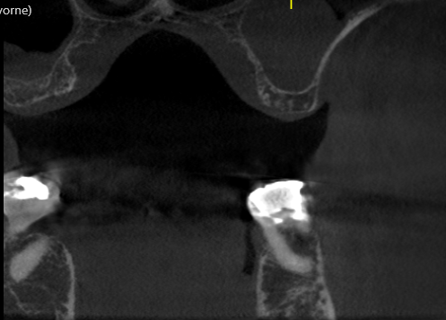
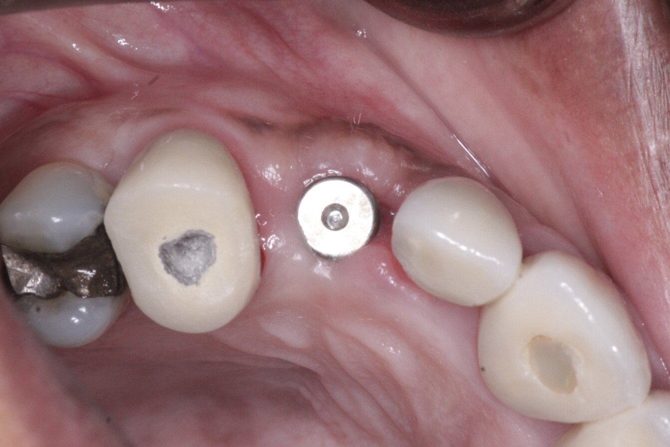
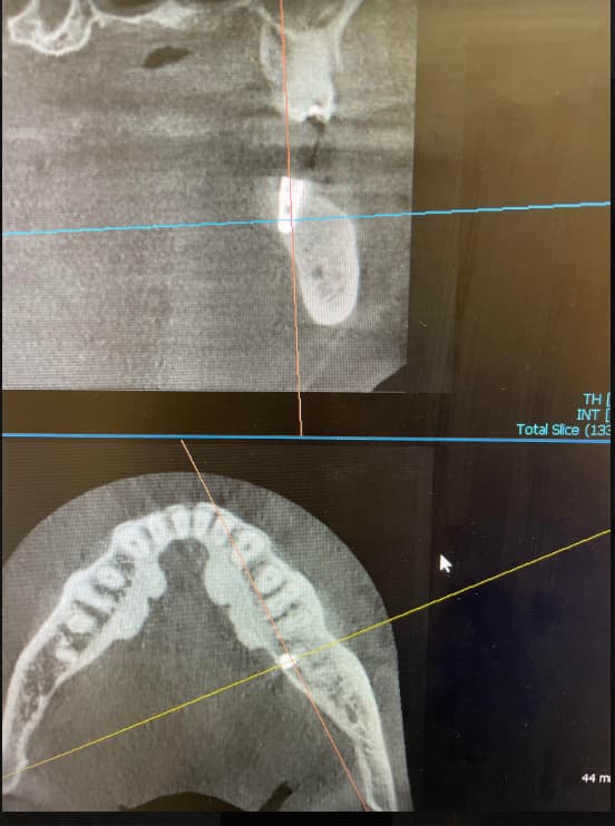
This is a 50 year old patient. He presented with the upper left central incisor missing. The tooth was avulsed the day before. The same tooth was knocked out 20 years ago and was put back in. The day he came in there was a draining sinus between the upper left central incisor (21) and the upper left lateral incisor (22), but it had disappeared after one week. The tooth 22 did not respond to cold but responded well to electric pulp test. A resin bonded bridge was made as a temporary. The patient wanted to have an implant supported crown. The Ct scan showed a large cyst under the teeth 21 and 22. I am planning to root canal treat the tooth 22 and lean out the cyst. What is the best bone graft material to graft the defect? Is it possible to place an implant at the same visit when the cyst is curreted if there is enough bone for primary stability?


![]Head^03_Sinuses-Adult-0001](https://osseonews.nyc3.cdn.digitaloceanspaces.com/wp-content/uploads/2014/02/Head%5E03_Sinuses-Adult-0001-e1392050454110.jpg)


![]Head^03_Sinuses-Adult-0003](https://osseonews.nyc3.cdn.digitaloceanspaces.com/wp-content/uploads/2014/02/Head%5E03_Sinuses-Adult-0003-e1392050431540.jpg)


![]Head^03_Sinuses-Adult-0004](https://osseonews.nyc3.cdn.digitaloceanspaces.com/wp-content/uploads/2014/02/Head%5E03_Sinuses-Adult-0004-e1392050411460.jpg)


![]Head^03_Sinuses-Adult-0006](https://osseonews.nyc3.cdn.digitaloceanspaces.com/wp-content/uploads/2014/02/Head%5E03_Sinuses-Adult-0006-e1392050491920.jpg)


![]Head^03_Sinuses-Adult-0008](https://osseonews.nyc3.cdn.digitaloceanspaces.com/wp-content/uploads/2014/02/Head%5E03_Sinuses-Adult-0008-e1392050471126.jpg)

5 Comments on Large cyst: proper treatment plan?

New comments are currently closed for this post.
K
2/10/2014
Normally, granulamatous tissue in bone can be thoroughly clean out and you can proceed with bone grafting procedure as long as the surrounding bone is in good condition. However, in your case, there is high chance that the radiolucency is caused by the necrotic upper left lateral incisor (no response to cold test). In this case, I'd perform a perfect RCT on the lateral incisor first. Once you are convinced that the lateral incisor will not cause any infection, you can go ahead and perform the surgery. If the labial plate is missing or damaged, i'd wait on placing an implant but rather just perform socket preservation. I usually use all cortical allograft with particle size .25 - 1 mm. Do not use anything smaller than .25 mm. Studies show that allografts smaller than .25 mm will cause inflammation. In some cases where all the bony housing is intact and if i can achieve excellent primary stability, I place the implant and immediately connect a screw retained provisional crown and get it out of occlusal contacts as done by numerous other dentists around the world.
CRS
2/10/2014
Remove the lateral there is no bone support and it is displaced by the pathology. Remove and biopsy the cyst, graft and see what you get back, then place the implants. You may want to consider treating the other posterior cysts which would be more amenable to root canal therapy. Not wise in this situation to place implants without knowing the diagnosis, lack of bone and active infection. Not likely for the cyst to resolve with the lateral present with his much pathology. Thanks for reading.
Bill Woods
2/25/2014
I would be concerned about any cyst displacing a tooth. I agree with delaying any implant placement until something is known about the pathology. JM2C. Bill
Birgitta W.
3/11/2014
There is significant thinning and expansion on the buccal aspect of your lesion, also in one view it looks like there are radiopaque densities occurring within your lesion I would not do anything until you have a full path report. This may not be a radicular cyst. The thinning and the expansion of the buccal aspect of the alveolar crest is concerning. Normally periapical cysts do no cause that much expansion of the buccal plate. What are you doing about the sinus pathology in the frontal sinuses? From the images I can see your sinuses are not well aerated.
LN
3/12/2014
Hi, thank you for all your advice. i did the RCT the tooth 22. Surprisingly, the pulp was still vital. I also used a 25 gauge needle to penetrate the cystic area to see if i could draw any fluid out of the cyst. There was no fluid drawn out. I have also advised the patient of the other radiolucency. He is aware of them. Since he is in no pain or problem with sinus, he is not in a rush to get treatment. The reason he came in to get the tooth 21 implant is because it's an insurance case. My next step would be to clean out the cyst, graft the defect and to get a path report. many thanks for advice

Featured Products

OsteoGen Bone Grafting Plug
Combines bone graft with a collagen plug to yield the easiest and most affordable way to clinically deliver bone graft for socket preservation.
CevOss Bovine Bone Graft
Make the switch to a better xenograft! High volume of interconnected pores promotes new bone. Substantially equivalent to BioOss and NuOss.