Lingual plate fracture and minimal buccal bone: will this case fail?
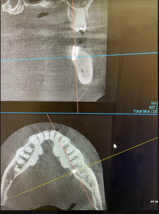
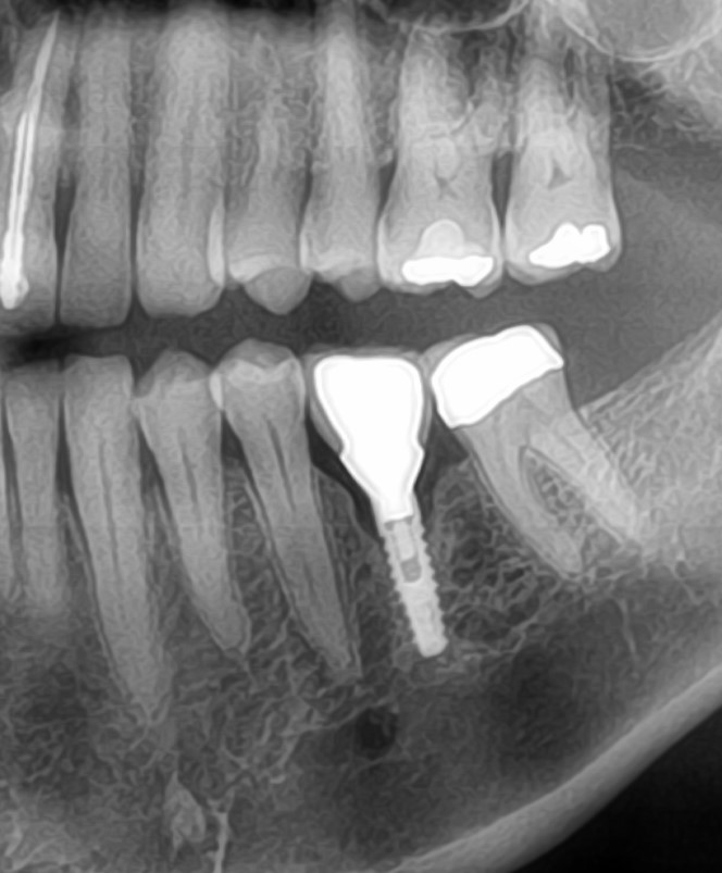
Patient has 2 implants in the mandibular right edentulous areas of #29 and 28 [mandibular right second and first premolars; 45, 44]. We noted that both are placed too far to the buccal creating a very thin layer of covering buccal bone. Will this case fail as soon as loaded if we leave things this way??


16 Comments on Lingual plate fracture and minimal buccal bone: will this case fail?
New comments are currently closed for this post.
dr. k
1/30/2014
There is a general consensus in the scientific community that there should be minimum of 2 mm bone around each implant in order to have a predictable long term success. If you suspect that there is very thin bone at both buccal and lingual, I recommend removing the implant and graft the site and try placing new implants at later time rather than waiting for the bone to go down and pt gets abscess.
Rafael Oliveira
1/30/2014
Do not remove them.
Its gonna be ok, but the placement was horrible. More preparation next time.
Dr. Mukhamadiev
1/31/2014
Be careful with your X-Ray measuring of bone thickness;). Because in reality it might be small amount of bone lingual.
Anyway I would check visually lingul surface of mandible. In case of absence of bone, it is better to reimplant or change position of implant/s (drilling out bone block with implant and placing in new corrected position and doing bone augmentation simultaneously).
But anyway it is important to talk with patient and explain honestly the situation).
Any other ideas?)
CRS
1/31/2014
Honestly did you use a surgical guide? The time for a cone beam is in treatment planning with a splint in place to determine where any grafting is needed to aid in the implant placement which is restoratively driven, not where the available bone is. These implants will integrate in poor position. Three dimensional imaging does not remove the responsibility of performing the surgery to obtain restorable implants. I would advise removing them prior to integration, treatment planning for ideal placement with grafting and a surgical guide. I think you are very lucky the lingual plate was not perforated with hemorrhage. A CT scan does not help a poor placement after the fact or since no one is watching just keep them, restore and hope for the best. My tone is harsh since this seems to be a pattern on the website and we all learned treatment planning, mounted models in dental school.
Marc
1/31/2014
A good way to get sufficient bone around an implant is to place a narrower diameter implant. Modern implants with conical connection of 3.5 to 4 mm are big enough.
The time spent in planning pays double in the end. First it is teeth the patient wants, so the dentist should plan teeth first. To secure the teeth to the patient, the dentist wishes to use implants. So second the implants are place below the planed teeth. Than comes the question: is there bone at that location where the implants to support the propose teeth are ? If not , ad bone. Now the icing on the cake, use all that planning and have one of those magnificent CBCT derived surgical guide made just for your patient.
Dentist sleep well, patient smile.
TW
1/31/2014
Thanks for your comments all. Implants not placed by me. I agree with importance of cbct, why we have one.
Want to know your opinions on 'survivability after loading'
Doesn't look least bit promising
CRS
1/31/2014
I agree and support your opinion.
Farhad Amini
2/1/2014
Based on the CBCT the angulations are not too bad but the size of both implants are too large for the housing. I recommend removing both of them, and re-grafts both sites (Guided bone regeneration with resorbable membrane). This way you have more control over this case in long term rather than dealing with potential complications. Don't forget that the thin layer of lingual plate most likely will be resorbed and consequently a large area of titanium will be exposed on the lingual. You have 2 to 3 weeks to back them up prior to integration.
If they are already integrated, I will uncover and torque and reverse torque them (hoping for them to come out :D),place healing collars for 3 weeks, and fabricate temporaries (lab made) so that they can stay in for more than 6 moths to a year. This way I can learn about his/her occlusion, I can evaluate how soft tissue looks like through series of maintenances, and I can teach the patient proper home care through out this time prior to fabricating the final product.
Thank you for sharing, and please let us know how it was handled. !!
Richard Hughes, DDS, FAAI
2/2/2014
You may consider placing a double layer of OsteoTape from Impladent Ltd.,on the lingual. This may yield bone on the lingual. Do you have any bone on the lingual?
CRS
2/2/2014
Richard have you tried Osteowrap, a thin cortical bone product used in orthopedic procedures, works like a charm! I will check out osteo tape thanks!
Richard Hughes, DDS, FAAI
2/3/2014
CRS, Thank you for the information.
peter Fairbairn
2/2/2014
HI TW as you know scans sometimes do not give as true a picture as we think , although a great estimate . Hence stents I feel are for the more experienced only .
The main issue is possibly the width of the Implants ...
Even in this area wider is not better , think of a screw in a block of wood , the weakness is the wood not the screw hence the more wood ( BOne ) the better .
Implants do not break easily ...
Peter
"TW"
2/2/2014
Peter- Thanks for comment. Apparently, everyone assumes I placed these. As stated above, the implants were not placed by me. I treatment planned a ridge split with immediate implant placement using piezo. I was upset when I saw this rummaging thru my cases (office shared)/ I will not enter this probable law suit case, but am happy to hear all your comments as to what you guys would do. Peter- do you think this case will fail once loaded? I don't think 'sprinkling' bone over an inappropriately sized implant and a prayer will help this unfortunate patient.
Osteo tape sounds interesting though.
Anyone else try this product?
Mike
Peter Fairbairn
2/3/2014
THe main issue may be the mucosal tissue on the lingual aspect not being attached and can lead to issues with the exposed threads resulting in failure .....
This sounds like a complex issue for you already , and things have been taken done that have made it difficult for you ...sorry to hear .
Maybe time to let patient know and remove the distal of both and replace with narrower Implants and grafting.
Or Can raise a flap and get a good look ... graft but fraught with hoping and luck.
Neobiotech remover at least makes taking Implants out easy and pain free .
Hoping and praying in Implant Dentistry is not great..
Good Luck Peter
osurg
2/10/2014
If these implants are not intergrated take them out. Simply put they are too wide and too short. The lack of buccal and lingual bone will limit your longterm prognosis. You had a lot of bone available that you could have utilized. Perhaps if you took the scan before placement rather than after this would have been an easier case. Just part of the learning curve.
camilo
3/4/2014
Soft tissue graft!!!!