Orienting Drills and Implants to Avoid Lingual Concavity?

Dr. H. asks:
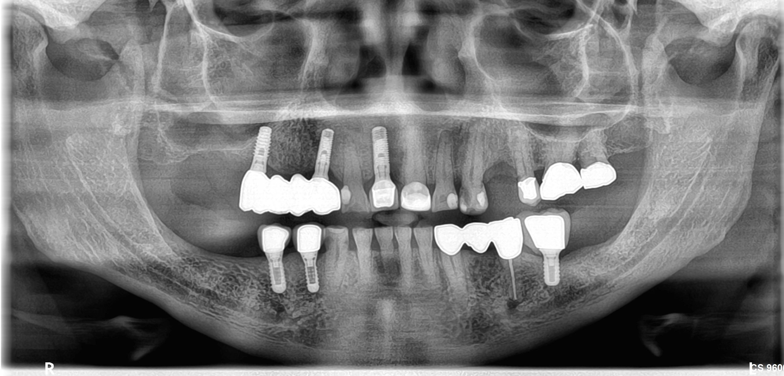
I have just started placing dental implants. I am only doing simple and straightforward cases. When I need to place an implant in the mandibular premolar-molar area I use a panoramic radiograph to orient myself. I measure the distance from the height of the alveolar ridge to the top of the inferior alveolar nerve canal. I set my stops so that I penetrate no deeper than 2mm above the inferior alveolar nerve canal. I place the implant at the center of the alveolar ridge in a buccolingual plane. So far I have not had a problem. But I know that panoramic radiographs can present a distorted image. And I am wondering about orienting the drills and implants in the proper orientation to avoid the lingual concavity. Can anyone please offer some advice? Is my protocol accurate and predictable?

17 Comments on Orienting Drills and Implants to Avoid Lingual Concavity?

New comments are currently closed for this post.
UW
12/14/2008
You should at least have a PA radiograph. Panos could give you up to 30% magnification, you could easily drill through the IA nerve. Ideally, a CBCT would be ordered to visualize the lingual undercuts and the IAN. Usually you can feel the lingual cortex or the roof of the IA canal as you get increased resistance. However, I wouldn't rely on feel. Order the appropriate radiographs.
Neda-Moslemi
12/15/2008
In addition to considering anatomic risk zones, there is one another issue: Today, implant orientation is not merely determined on the basis of "bone-driven implant" concept,but rather "restorative-driven" concept. Implant fixture should be inseted according to the future restorative matters. The posterior inferior implants should be directed to the centric fossa of the maxillary teeth (or prosthesis).
PAUL
12/16/2008
DEAR DR H A PAN IS AN ESSENTIAL RADIOGRAPH BUT USED ALONE IT IS FREQUENTLY MISLEADING. ALWAYS USE AN ACCURATE INTRA-ORAL (PARALLEL TECHNIQUE)AS WELL FOR COMPARISON OF MEASUREMENTS. AND THEN BE CONSERVATIVE. I WAS ONCE TOLD THE COMBO OF PAN/PA GIVES APPROX 65%ACCURACY IN VERTICAL MEASUREMENTS.IT WOULD BE IN THE PATIENTS AND YOUR INTEREST TO BACK THIS UP WITH CT OR CONE BEAM VIEWS AND PREFERABLY COMBINE THIS WITH A GOOD 3D PLANNING PACKAGE. ONCE YOU HAVE STARTED YOUR PILOT DRILL; DRILL TO 75 TO 80% OF YOUR PLANNED DEPTH AND INSERT A GUIDE PIN AND TAKE AN X-RAY TO VERIFY THE DIRECTION AND RE- MEASURE THE DISTANCE TO THE CANAL.ALWAYS BE PREPARED TO CHANGE OR ABORT THE PLAN IF NEEDED. IF YOU ARE INEXPERIENCED LIMIT YOUR CASES TO THOSE WITH BROAD RIDGES AND PLENTY ALTITUDE ABOVE THE CANAL. TAKE THE TROUBLE TO LOOK AT AND PALPATE A DRY MANDIBLE SPECIMEN OFTEN. IT WILL HELP YOU GET A 3D FEEL FOR THE LINGUAL ANATOMY. I AM AN EXPERIENCED SURGEON DOING TONS OF IMPLANTS AND I KEEP AND RE-LOOK A DRY SPECIMEN ALMOST DAILY.FINALLY TEAM UP WITH A PATIENT MENTOR AND READ PLENTY TEXTS AND ENJOY SOME OF THE EXCELLENT COURSES AVAILABLE GOOD LUCK
Mike Stanley, asst.
12/16/2008
Great advise from all the above. You can also add a 'radiographic ball' to your pano. Locate it intraorally near the prospective implant site. I have also seen 'pins' placed in guides that help with 3-dimensional planning. They provide a 'trajectory.' Careful measurements will help compensate for magnification effects. You're already good at that part, this just adds another layer of information.
R. Hughes
12/16/2008
You have to think 3-D, also palpatemthe bone.
Russell
12/16/2008
Having used panographic radiographs for implant placement since 1992 I understand your concerns and questions. I used cone beam scans for more difficult cases and soon came to realize that I they really are the state of the art for all implant placement planning. Save yourself a bunch of grief and make the switch and have your patients get scans. Your results will be consistent and you will sleep better and night.
Jim
12/17/2008
Although cone beam is becoming 'a' standard of care with implant placement, the radiation involved is a concern. It is no-doubt the best protection against a bad outcome. I began placing implants before the days of cone-beam. I have experienced very good success with a pre-op pan, a pre-op perapical radiograph, diagnostic casts, palpation, a guide-pin in the pilot hole to confirm axial orientation, and in the final stages an implant as a radiographic guide and as a bone-density-torq guide. This means that I may use an implant, or even two of them to 'size-up' to the 45ncm torq I want and the implant length that will fit. Therefore, I have an assortment of diameters, lengths and taper or straight implants that can be tried into place. When using Nobel, I can return the 'trial' implant and be charged $100 for a replacement. Expensive yes, but worth it to me. I sometimes use cone-beam, but this 'cowboy' method works well assuming one has experience.
vinayak
12/17/2008
if u r using opg its better to go for digital opg. in that u can get accurate length from the height of alveolar crest to the inferior alveolar canal. just ask your radiographer for the measurements he will provide it to u. if digital opg is not possible then what u can do is prepare a bite rim ( if patient is edentulous) and place a small metallic object, whose dimensions u have measured earlier, then again measure its dimension in opg. difference in both the dimensions will give an idea about the elongation that takes place in the radiograph. in the case of dentulous patient u can ask patient to just bite on the metal object and then take xray. by this method u can determine the length of implant but for determining the width ct scan is the best thing.
barry sporer
12/17/2008
if the lingual concavity is a concern, order a cbvt scan and have a surgical stent made.
dr hasan sk
12/18/2008
dear DR H dont ever forget that your are dealing with a living thing when you are trying to place a dental implant so its better for you and for your patint to make a dentascan and you will be and your patient in the safe side and the denta scan will be the true guide for you
Paul
12/20/2008
Keep in mind that 98% of bite force is distributed in the first 10mm of implant length. So, there's usually no need to place an implant any longer than this...if may need to be shorter in some cases, obviously.
dr. Alexey Shamray
12/22/2008
If you measure on PA radiograph,you can only define the height of bone and distance to mandibular canal. To avoid lingual concavity and damages of soft tissues on internal side of mandible, you mast do carefully examination of intraoral situation, angulation of mandibular crest and it dimentions together with CT-scans ( if you not shure in you expirience)dont afraid its just the beginning.Other important factor, especially at the beginning - very careful and atravmatic working whis instruments, with the observance of points, written in guidance to the system of implants. Best regards
Jay B. Reznick, DMD, MD
12/26/2008
With the easy availability of cone beam imaging and guided implant surgery technology, I do not see a reason to use the old "eyeball it" technique. Even with a good quality panoramic and study models, it can be hard to get the exact position and angulation that will keep you out of trouble. A DentaScan only gives you diagnostic information. It does not hold your hand when the implant is being placed. You can draw a nice image on the CT scan with the proper implant position and angulation, but executing that in the mouth without a guided surgical stent is leaving too much to chance. Guided implant surgery will eventually become the standard of care. This is the only reliable way to aboid the lingual concavity of the mandible.
ljungberg
12/30/2008
I think it would be a problem for the newbies, even CT scan is available; as they could not transfer the information of CT scan to the surgical effectively. They know the existence of concavities from CT, but they still don't know how to deal with them. Surgical stent would probably one of reliable guidance. If surgical stent is not available, I would suggest to use trephine bur to make a boundary about the surgical site. Then you may start to use pilot drill; you should aim at the centre of the trephine mark. The choice of trephine bur shoule be a little bit thinner than your chosen implant size.
ljungberg
12/30/2008
Also, you may lower the rpm of your handpiece; once the slower drill hits the cortical bone, the drill would not cut effectively and your hand would feel a higher resistance of drilling. Then you should stop any drilling, and rinse the socket with saline to take a look. If the lingual plate is really perforated, continuous bleeding will occur. Then you should decide whether you go ahead or not. If you are using screw-type implant, I would suffest to bone graft the location and postpone the surgery. If you are using press-fit implant, you may consider to change the angulation of drilling and continue the surgery. Before you place the press-fit implant, just remedy the perforation with bone graft material. I would suggest to finish the surgery with complete flap closure.
Dr David Harpaz
12/31/2008
in todays day af age the use of a cone beam is becoming standard of care with implant placement.God forbid something should go wrong, that is the first question that you are going to have to answer - Dr. did you use a cat scan??? But regardless of the liability, the cone beam with the use of a surgical stent with a guiding netel tube, can confirm the projectile, buccal-lingual and mesial-distal, of the drill befor you even go into the surgery. That is the only way to go anywhere in the mouth without having to worry about perferation, fenestration etc. In your qustion you did not mention the mental foramen - wouldn't you like to know its exact location !? look into "teeth in an hour" concept for the guided surgical stens for the concept. I am having my removable lab fabricationg stents like that on a weekly basis for a fraction of the cost.... good luck. Dr. David Harpaz harpazimplants.com
Shirley deLong
1/1/2009
I'm in need of (4) 3.25 STERIOSS (1997 Implants) CYLINDER HEALING CAPS. A local dentist took off the four (4) rear caps, removed posts, and then discovered he didn't have the healing caps to put into implants. Can anyone help me with this matter?

Featured Products

OsteoGen Bone Grafting Plug
Combines bone graft with a collagen plug to yield the easiest and most affordable way to clinically deliver bone graft for socket preservation.
CevOss Bovine Bone Graft
Make the switch to a better xenograft! High volume of interconnected pores promotes new bone. Substantially equivalent to BioOss and NuOss.