Small Bone Dehiscence from Collar of Implant Cover Screw: Next Steps?
« State Regulations for CBCT Machines? | Premier Implant Cement: Source of Chronic Inflammation? »
Dr. B. asks:
I placed a dental implant in the left posterior maxilla in the first molar site [#14] following augmentation of the site with a block bone graft which I harvested from the ramus. At the time of implant placement I performed an indirect sinus lift and the patient developed a sinus perforation. I placed the patient on broad spectrum antibiotics and an antihistamine. The patient responded favorably and the implant has osseointegrated.
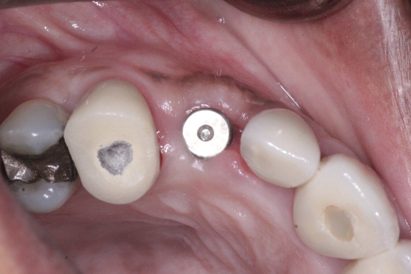
The patient is now 5 months following the implant surgery. There is a small buccal bony dehiscence of approximately 3×5mm which extends from the collar of the implant cover screw and distally to the mesial line angle of the second molar.
Today I gently removed some necrotic bone with a round burr so that the dehiscence is now concave, thinking that soft tissue may now be better able to spontaneously develop over a concave defect. The patient also just today informed me that from the time of implant placement, she wakes up approximately 1 day per week with a mild swelling under her left eye which resolves as the day progresses.
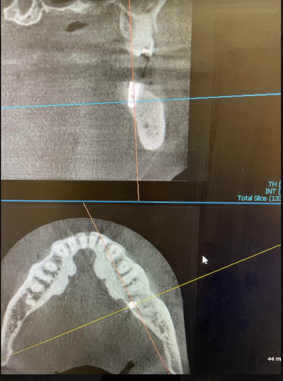
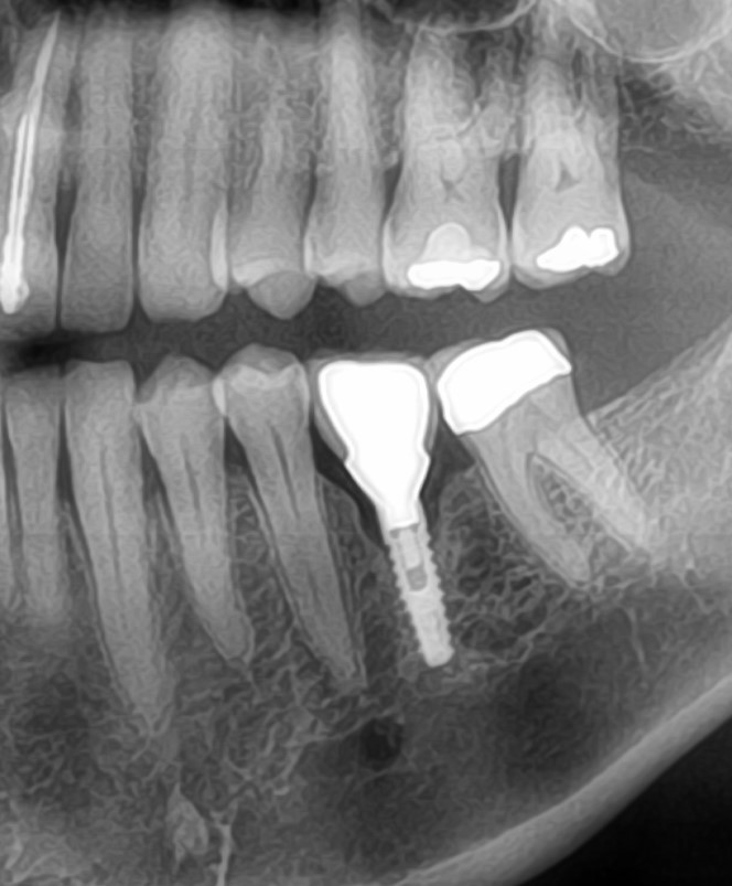
Thinking the patient has a chronic sinusitis, I took a panorex which failed to show anything significant. The sinus membrane is clearly visible and passes superiorly to the implant apex. The implant has bone surrounding it completely.
Clinically, the implant is stable, it is not sensitive to percussion and appears to have good soft tissue health excluding the dehisced area. What is your recommended next step? To remove any remaining necrotic bone and place patient on broad spectrum antibiotics and rinse or to restore the implant?
Free Daily Email Alert Click Here>>
Get OsseoNews.com Comments delivered daily! Click Here to subscribe.FREE Weekly Email
Keep current on the latest dental implant discussions! It's Free!
>>Click Here to Subscribe to OsseoNews.com Now!
-
Editor Picks
-
Popular Posts
-
Hot Topics
- Locator Attachments: Trouble Picking Them Up in Overdenture?
- Advice for Complication in Creation and Maintenance of an Interproximal Papilla?
- Implant Systems: Commercially Pure Titanium vs. Alloy?
- Type of Anesthesia for Dental Implant Surgery?
- Implants Too Close: Can I Use Orthodontics To Create Space?
- Large Defect in the Labial Cortical Plate
- Abutment Screw Gets Stuck: How to Prevent This?
- Crown 1mm Short of the Margin: Recommendations?
- Suture Lost Exposing the Implant: What to Do?
- Mini Implants in Anterior Mandible: Expectations of Longevity?
- Space Between Implant Fixture and Natural Teeth: How Much Is Enough?
- Options for Provisional Restoration?
- Loose Bio-Oss Particles: Can Implants Be Placed?
- Maxillary Overdentures: What Implant Support is Required?
- Mixing Metronidazole Solution in Grafting Material?
- Sinus Lift Complication after using Summer’s Osteotome Technique?
- Cone Beam vs. Conventional CT Scans: Radiation Levels?
- Sinus Tear: How Long to Wait for Regraft?
- Mini Implants for Long-Term Use?
- How to Avoid Hitting the Mental Foramen Nerve?
Implant Courses
>>More Implant Courses

4 Responses to “ Small Bone Dehiscence from Collar of Implant Cover Screw: Next Steps? ”
Hi
1] what length of implant is covered with bone?
2]Is the exposed area in aesthetic Zone?
3OPTIONS :
1] After polishing the threads of Implants like a polished Implant Collar Make a new crown to cover it.
2]Cover the exposed area with a graft with a membrane.
3]If clinically it is okay just polish the threads and leave it.
Hi,
I think you have your bone block not integrated. Now it is being lost, but the implant is integrated in it’s apical part.
You have to remove all necrotizing bone and then consider polishing the implamt collar and keep it or take it out and begin again, after reevaluation of what you have already lost and how can you regenerate the tissue first.
But don’t try to use membranes and ext, this will take you in deeper problems.
It would be nice to have some fotos and x-rays here!
Good luck
the exposed bone should be reduced back to bleeding bone. this should be able to granulate over now with soft tissue. a ct scan will be more useful to ro sinusitis, a plane film might be inadequate. regrafting over vasularized bone under a soft tissue flap is necesary
First of all Iwould use a 3D scan to see what it is the matter in de sinus. It can appear good on a 2 D x-ray, but a fistel of dehiscentie at the top of the implant isn’t always visible in a 2-D x-ray. If you have comlications you always have to think in a 3D dimension. Then you make a plan. To restore the dehiscentie at the collar of your implant I would use an Er- YAg laser. It is save to use on your implant as well as bone. You remove it till you get bleeding of your bone. The advantage with the laser is that there isn’t any smearlayer left to remove first by the body and it desinfect your bone and implant. Then I would regenerate the loss of bonewith a new graft taking in to account that after healing I need a collar of at least 1 mm of bone arround the implant. The other option is to remove the implant as a total failure and start again. But first I would start to get the ct or cbct scan to see what are the problems. The membrane isn’t visible on a x-ray. This is only be abble to see with an MRI or with endoscopes. So you can refer to your ENT for a check of the membrane site in the sinus.
Leave a Comment
Note: Please refrain from ad hominem attacks, and promotional comments. Outside links are not permitted in comments. Though we require an email to route questionable comments to our editors, we will NEVER publish your email or use it for any other purpose. Thank you for your understanding.
Note: At times your comment may not appear on the website immediately, because it has been sent to our editors for approval. Once approved, we will publish the comment. There is NO need to resubmit your comment, if it does not appear on the website immediately.