Zimmer 4.7 in Posterior: is this normal?

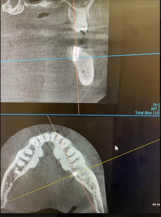
Mostly, I have placed Zimmer diameters 3.7 and 4.1 and I didn’t have any problems restoring them. In this case, when I installed the 4.7mm diameter Zimmer implant in the posterior mandible I ran into a problem. There appears to be a horizontal radiolucent area between the platform of the implant and the impression coping. I cannot tell if the impression coping is seated all the way down. What do you think? Is this normal? Also, when I view the radiograph of the final crown which is a screw retained implant crown (i.e. crown screws directly into the implant without an intervening abutment, cast to gold abutment was used) it looks like the crown is not seated all the way down on the implant platform. The proximal contacts are not too tight and the crown is seated passively. What is you opinion? Is something wrong?


4.7 impression coping4.7 impression coping
4.7 ready crown4.7 ready crown
3.7 impression coping3.7 impression coping
3.7 ready crown3.7 ready crown

14 Comments on Zimmer 4.7 in Posterior: is this normal?

New comments are currently closed for this post.
jesus x aguirre ficoi fic
10/7/2014
it would appear that the bone level has either been lost or the implant might not have been placed deep enough from the get go. i have seen this often when doctors use a punch approach versus a traditional flap. i don't think the size of the implant is a factor. the "space" between the implant and abutment I feel is not a factor and would credit it more to a slight defect in the cone angle of the xray.
Dariusz Pituch
10/7/2014
At TSV 4,7 with 4,5 platform is great implant for molars restorations. on x-ray implant abutment interface always looks like you described. Everythink is ok. good luck.
Basile Muntean
10/7/2014
I agree with Dr. Pituch. No worries. This is common and of no clinical consequence.
Esmaeel
10/7/2014
Thank you a lot dear colleagues for your comments, calmed me down. Regards
DrG
10/7/2014
I'm curious why using a punch would produce this defect. Does anyone have insight into this occurrence
Jesus x aguirre fucko fic
10/8/2014
It's not that I am opposed to the punch technique. However it is very hard to visualize the cortical bone level and assure that the implant is seated all the way down into the cortical to leave it bone level if indeed that is what you're trying to achieve. The x-ray shows that the implant could have gone down a bit further or the bone has receded away. In my experience, implants thatdid not go down all the way result more in cases where the punch technique was used as opposed to a full flap where you can visualize the bone level with the implant.in this particular case the first x-ray shows an ample amount of soft tissue making it very hard to visualize that the implant would have gone down to bone level
DrB
10/7/2014
The implant appears to be installed to the correct depth. The bone loss is a circumferential defect which should be debrided and grafted with particulate bone and closed with a membrane covering the implant. Good luck
Lance Timmerman DMD
10/7/2014
sometimes a junction can look slightly "off" due to thinner metal in the conical connection area. Since this area is superimposed over other thicker parts and connections, it doesn't look "wide open" but it IS different. Our gut reaction is to think something is wrong, but it is possible/likely that all is well. That may be the case here.
Gregori Kurtzman, DDS, MA
10/7/2014
I would agree with the prior comments it appears that there was some bone loss after placement which should be addressed as it will only worsen over time. With regards the impression head it may be the angulation of the sensor in relation to the long axis of the implant so that there is some overlap and that appear to be a radiolucent fine line, if the bone were higher i would say the bone may be hampering seating of the head but in this case thats not it unless you trapped some soft tissue between the implant and part. with regards the cast final crown it appears that the plateform of the crown was not finished with a reamer after casting to ensure that the horizontal surface was smooth and no microbleps present. if you blow up the image you can see its contacting on the outer edge which is preventing full seating. Did the lab use a gold UCLA or a Plastic one to make the pattern and cast?
Esmaeel
10/7/2014
The UCLA (cast to gold abutment was used in that cases).
Gregori Kurtzman, DDS, MA
10/7/2014
Then my question is ask the lab how did they clean the casting to remove the investment from it after casting? sandblast it off or chemically devest it?
Jesus x Aguirre ficoi fic
10/8/2014
I apologize for the initials stating my fellow status in the ICOI. M Iphone 6 has made an embarrassing self correction
SOCALOMFS
10/8/2014
I have extensive experience with the TSV implants and find them to be outstanding in most cases. This site does exhibit 3-4mm of bone loss and ideally should have been addressed before restoration. Why do we see early, pre-restoration crestal bone loss around implants? Most commonly the crestal bone was not of ideal contour or density to begin with. (Never trust grafted crestal bone that is granular in nature to remain after implant placement.) There can be inflammatory disease even when the area appears to be healthy when the implant is placed. I have seen this when healing abutments are below the tissue height and cleaning is difficult. Also in cases that were the wound was closed primarily and then dehisced over a deep cover screw where a healing abutment was not immediately placed. As for the abutment not being seated, I would agree that it could be a problem with the casting not being properly cleaned. If it is not a Zimmer part, that could be the cause. **Only use factory parts when restoring any implant no matter what the lab or the clone part maker says. Many times the clone TSV parts are subtly different to avoid patent infringement and therefore may not fully engage with the friction component of the interface. I have also seen several TSV implants where the outer tapered portion fractured and seeing this abutment not fully seated on the flat portion of the platform may predispose it to premature failure. By the way, if you see a gap on a radiograph, it is really there. If you don’t see a gap, it may still be there. What to do now is the question. I would recommend removing the crown and attempting to graft to restore the crestal bone. This is very technique sensitive and less than predictable even in the hands of experienced specialists. Check with the lab and demand they include invoices with each implant case showing that factory parts were used including the lot numbers. At least you can get the implant and prosthetic parts replaced when failure occurs. Use your team of specialists to handle cases that are anything but ideal so you have high success rates with the cases you attempt yourself. The plaintiffs bar is looking at implant failure as their next cash cow so plan for success. Regards
betsy Anonymous
8/24/2021
Phantom gap in impression xray? As long as the sides of image shows no gap between impression coping and implant , then the "long football shaped" image is probably a phantom gap.

Featured Products

OsteoGen Bone Grafting Plug
Combines bone graft with a collagen plug to yield the easiest and most affordable way to clinically deliver bone graft for socket preservation.
CevOss Bovine Bone Graft
Make the switch to a better xenograft! High volume of interconnected pores promotes new bone. Substantially equivalent to BioOss and NuOss.