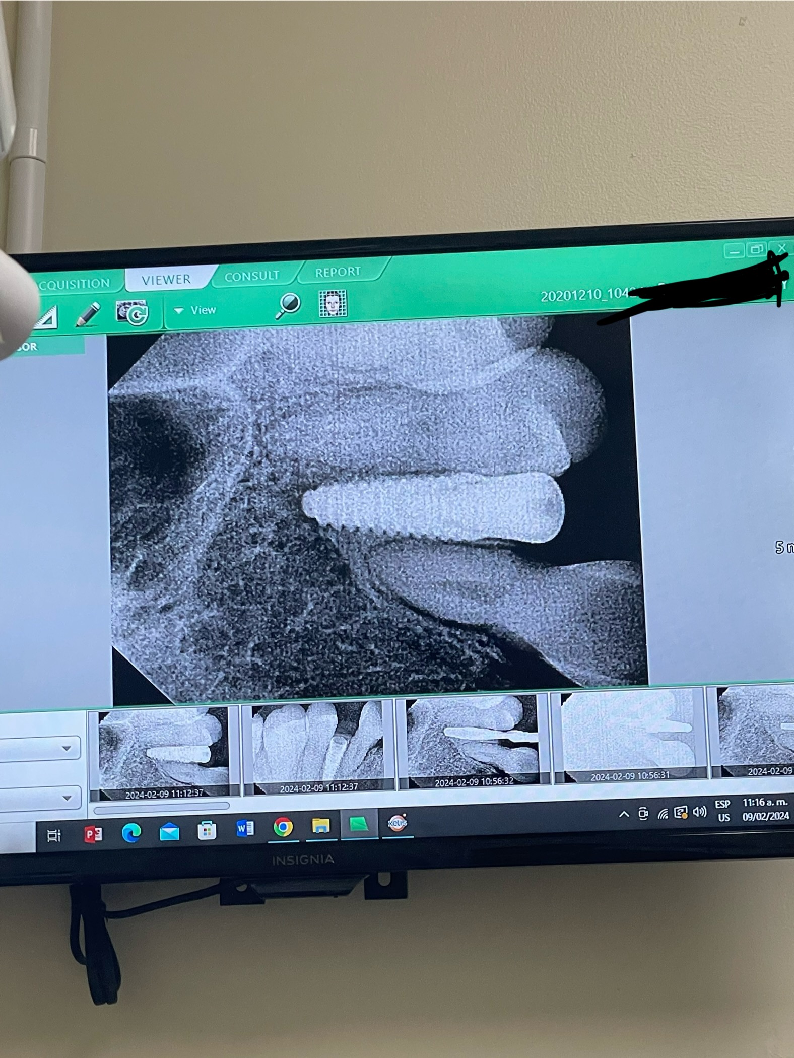
Grafting Vertical Osseous Deficiency?
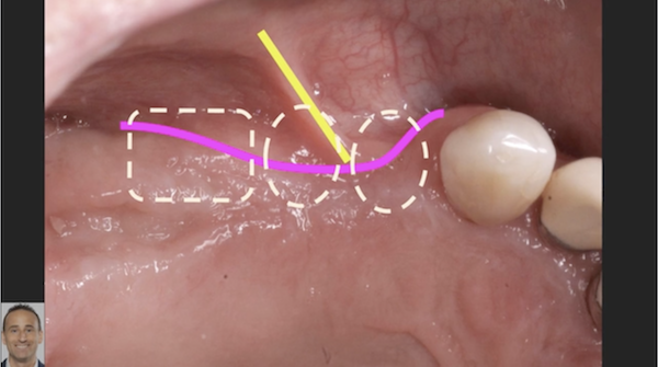
What would be the suggested way of grafting bone in a vertical osseous deficiency ( ridge dip) between a second bicuspid and second molar in a mandible? In this particular case, the tooth is missing for many years and the patient would like to replace the tooth with an implant. The vertical deficiency at the lowest point is about 5 to 6 mm. Obviously there is a simple restorative solution by adding gingival colored porcelain to the crown, but that is not the issue.
4 Comments on Grafting Vertical Osseous Deficiency?
New comments are currently closed for this post.
Alex Galo
11/8/2017
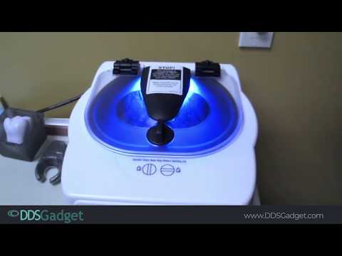
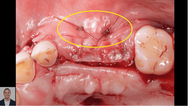
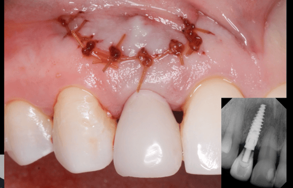
I just saw a very similar surgery done in my implant study club. The dentist reflected a large envelope flap way back to the external oblique ridge. He performed venipuncture and collected about 6 red vacuettes and two white vacuettes of blood and spun them 2700 RPM for 12 minutes. Released the buccal flap to get tension free closure. Mixed mineross and cut up PRF membrane then added liquid from white tube to make "sticky bone". Placed that on ridge overbulking quite a bit as it will shrink back as it heals. Covered with memlok membrane. Tacked membrane on buccal (2 tacks). PRF memrane over memlok. Sutured case up with primary closure with horizontal mattress for 1st suture then bunch of interrupted sutures. Super important not to put any stress on ridge therefore forget about wearing PLD as graft heals. Gaining vertical height is VERY challenging. Take new CT in 6 months to confirm new bone dimensions are adequate before placing implants. The mentor said this is much less invasive than block grafts and more predictable.
Jess
11/8/2017
Could you please repeat how he made sticky bone?what did you mean when you said white tube.could u please explain more.And is this liquid left to clot.How much time and how long to centrifuge.
Thank you
Perioperry
11/8/2017
Autogenous bone block, secured with screws, covered with a resorbable GBR membrane. Any small spaces or gaps around the block should be filled in with particulate bone, preferably autogenous. The bone can be harvested from the mandibular symphysis, or possibly the buccal aspect of the ramus. The recipient site should be decorticated, and the soft tissue flaps must be carefully managed (mobilized) to preclude excessive pressure on the graft site and to prevent opening of the wound with exposure of the underlying graft. But it would be helpful to those you are asking this question of to be able to view the area by means of CBCT images and photos. Often, in cases like this, there is an associated loss of buccal ridge dimension as well. Also, it is very likely that regaining the entire 5 - 6 mm of lost ridge height is not going to be possible, so some degree of compromise should be planned for.
Girish Bharadwaj
11/9/2017
Follow Steigmann flap. Nearly fool proof.
A tension free flap with large two layered dissection- one supraperiosteal and then sub periosteal. Lingually you will need to release mylohyoid attachment.
Always use non resorbable Ti reinforced PTFE membrane as you will need stability. Don't rely on just cancellous bone- use 30/30/30 of Cancellous allograft/Xenograft/HA or beta TCP.
If you have the skill then try repositioning IAN which is very safe and predictable as you are using host bone.
Good luck


