Sinus Membrane Perforation and Repair
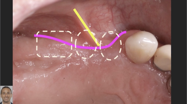
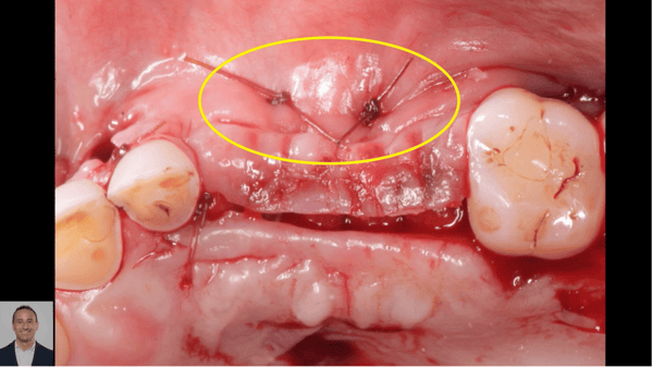
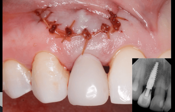
The maxillary sinus elevation is a standard and predictable procedure allowing the realization of dental implant rehabilitation in patients with severe bone atrophy in the lateral-posterior areas of the maxilla. Although the sinus lift procedure is relatively safe, some potential problems can be occur. The most prevalent intraoperative complication is perforation of sinus membrane, which can lead to graft infection and early failure. A variety of protocols and approaches have been suggested to repair the membrane. The two videos below show repairs with a resorbable membrane. The second video also shows the use of biphasic calcium phosphate synthetic bone graft for augmentation and placement of dental implants.
Our [Sinus Lift Kit](https://www.ddsgadget.com/sinus-master-kit.html) includes specially designed components to minimize risk and easily and safely lift the membrane, and reduce the risk of Schneiderian membrane perforation.
> The original technique for using a resorbable membrane in this situation is known as the “Loma Linda Pouchâ€, which consists in covering the whole sinus with a collagen membrane, and the graft material is completely covered in its centre by folding the membrane on the lateral wall. However, in this manner, an external barrier is created that totally isolates the biomaterial from the blood supply coming from the walls of the sinus . In the modified method, the cover of the sinus walls is still carried out with the support of a resorbable membrane located only on the surface of the Schneiderian membrane, leaving the bone walls free so that the blood supply from the bone can favour the vascularization and thereby the integration of the graft into this virtual space. Moreover, in such technique the resorbable membrane is fastened at the superior border of the antrostomy through titanium or surgical steel pins before being reinserted in the sinus cavity; a second membrane is positioned on the antrostomy externally, to further protect the biomaterial. It has been demonstrated that the protection of the osteotomic window increases implant survival if some prerequisites are respected: membrane stability, sterility and optimal cohesion, compactness and handiness of the graft material (1–4)
1. Fugazzotto PA, Vlassis J. A simplified classification and repair system for sinus membrane perforations. J Periodontol. 2003;74(10):1534–41. \[[PubMed](https://www.ncbi.nlm.nih.gov/pubmed/14653401)\]
2. Parenti A, Capelli M, Fumagalli L, Zuffetti F, Galli F, Taschieri S, Del Fabbro M, Castellaneta R, Testori T. Prevenzione e gestione delle complicanze. Perforazioni della membrana sinusale. Italian Oral Surgery. 2007;1:29–32.
3. Proussaefs P, Lozada J. The “Loma Linde Pouchâ€. A technique for repairing the perforated sinus membrane. Int J Periodontic Restorative Dent. 2003;23:593–597. \[[PubMed](https://www.ncbi.nlm.nih.gov/pubmed/14703763)\]
4. Testori T, Trisi P, Del Fabbro M, Francetti L, Taschieri S, Parenti A, Bianchi F, Zuffetti F. Gestione intraoperatoria di ampie perforazioni della membrana del seno mascellare. Italian Oral Surgery. 2007;1(6):21–28.
1 Comments on Sinus Membrane Perforation and Repair
New comments are currently closed for this post.
Robert
3/10/2017
With the availability of in situ hardening bone graft materials there is reduced risk of graft movement and escape when a tear occurs. Why would anyone continue to use a gritty movable and unstable particulate when such materials are widely supported with peer review and numerous successful cases?


