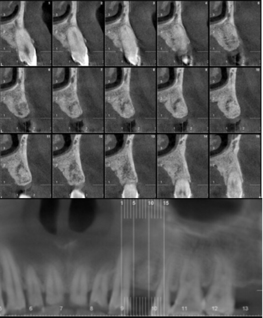
Pt's upper premolar removed 2~3years ago in another country. pt said he remembers some stuff was put into the socket after exo. cbct taken today. a radiolucent area apical to the bone graft noted. pt has never had pain from this area before. The tx plan is to place an implant here, but do I need to worry about this radiolucent area?
Dennis Flanagan DDS MS c comments:
When you do the osteotomy measure the depth and take some of this apical tissue and send it for biopsyotis comments:
whatever was grafted doesn't look like it integrated. i would flap this and remove it. probably come out in once big chunk due to the lucency following it all around, or better yet, send it to the OMFS. send that to pathologist. I would not implant this case for several reason. who knows where the crestal bone is going to end up a year from now on this esthetic zoneGregKammeyer DDS MS DABOI comments:
It looks like you didn't get all the granulation tissue out when you socket grafted. you should be able to prep the osteotomy, currette the lesion, put some graft in before the implant goes in and it'll heal fine.Rmosti13 comments:
It just means the bone graft wasn’t condensed fully into the socket.