3mm of Crestal Bone Loss: What Should I Do?
Dr. R asks:
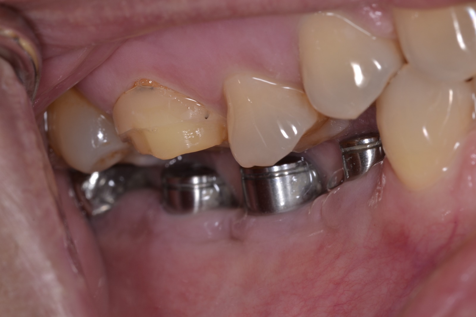
I installed this implant in a healthy 70 year old male in #30 site [mandibular right first molar; 46] adjacent to natural premolars. The surgical procedure and healing were uneventful and the implant underwent normal osseointegration. I restored the implant with a single crown. About eighteen months later, the patient presented with an acute periodontal abscess on the buccal aspect of the implant. I laid a full thickness flap and found 3mm of crestal bone loss in a crescent shaped defect. I installed two implants on the left side which have not had any problems. What is going on here and what should I do?


46 Comments on 3mm of Crestal Bone Loss: What Should I Do?
New comments are currently closed for this post.
Dr. Alex Zavyalov
2/13/2012
One of the causes is definitely occlusal overloading; the crown is massive and without an occlusal rest on the adjacent premolar creates effect of cantilever.
arnold rabin
2/13/2012
I am not there is occlusal overloading. The opposing dentition is a removable partial denture
Dr winnie
2/13/2012
May be from dental plaque, Patient cannot clean this area. You should motivate patient to clean this area and use other device such as waterpik. When you see the abscess , you must debride this area with other periodontal surgery, free gingival graft or bone graft. and prescribe antibiotics.
Mario Marcone
2/13/2012
There is not enough information given here to be able to arrive at a proper diagnosis as to the the etiology of the presented problem.
At this point, all that can be said with certainty is that a number of reasons could have contributed to the problem. But, the prime suspect has to be occlusal overload.
1- An opposing partial denture opposing a cantilevered crown on the mesial, can definitely lead to occlusal overload during function because of the nature of the food bolus, muscle dynamics during function, chewing dynamics as a result of the nature of removable prosthetic tooth relative chewing inefficiency.
2- A contributing unfavorable crown-to-root ratio especially in a first molar site as a single implant. Our colleague mentions that the left side received two implants with no problem ... this can be explained by the fact that those 2 implants are not overloaded because they are possibly splinted. But, it would have been nice to see a radiograph of the left side implant-supported prosthesis.
3- Habits ... lateral tongue thrust.
4- Choice of unfavorable implant design for the area and clinical situation. It would appear that there is bone loss to the first threads of
the implant. Is this implant over-submerged subcrestally?
5- Should there have been a bone augmentation procedure done first ... what kind of ridge morphology did this case start with prior to implant placement?
6- Has this case overlooked the possible role of an unfavorable occlusion and occlusal function.
In order to provide successful implant therapy, a clinician must insist on a proper treatment planning protocol involving consideration of all possible known medical and dental relevant patient factors and proper implant design selection.
Unfortunately, in this case, although there are a number of treatment protocols that have been suggested in the troubleshooting literature, there is no guarantee, and, usually, the prognosis is poor, at best. This implant will very likely be lost.
This unfortunate considering the patient's age.
TOBooth
2/14/2012
I think its entirely a hygiene issue.
I think in this sitiation you placed the best you could you are always going to get soem cantilevering when you use a molar sized crown. Unless you use 2 implants.
The likelihood is insufficient atached tissue poor oh. So improve oh then remove crown and do a sub epithelial connective tissue graft.
Photos would be good, and no its not definately going to fail at all in my opinion. The key is to make sure its not suppurating that kills bone!!!
When remaking the crown use a screw retained type so if teh problem happens againits a little more retrievable.
For long clinical crowns and cantilevers they work provided teh occlsuion is light in centric occlusion ie 3 sheets of shim 30 microns teeth intrude by 27 microns implanst dont. Check for interferences.
Equally you usually get marked vertical defects when teh occlsuion is too heavy- slightly in this case but not that marked.
Mario Marcone
2/14/2012
With all due respect, I wish to differ ...
It would be a reasonable assessment to make that in this case the bone loss was rather rapid.
Microbial peri-implant infection usually will not lead to such a rapid progression of bone loss.
Occlusal overload is usually the culprit with rapid bone loss.
Especially, if we have insufficient bony crest peri-implant bone morphology (insufficient bone width) and inappropriate implant design and over-submerged vertical implant placement, in combination with occlusal disease and/or unfavorable crown-to-root ratio.
Peri-implant mucositis is a possible precursor to peri-implantitis.
One must also take into consideration a possible peri-implantitis due to lack of an aseptic technique during implant surgery.
And another possible contributing factor is if this patient is a smoker.
Also, we have observed that in many earlier implants placed on ridges with insufficient keratinized attached gingiva ... there is almost never a problem as long as the hygiene is reasonably good. In conditions of poor oral hygiene, any quality of tissue, whether it is attached or non-attached, keratinized or non-keratinized, the scenario for developing a possible peri-mucositis are similar, although the case with keratinized attached tissue will have a better chance to protect the underlying bony tissue, hence, the peri-implantitis is less likely, and if it were to occur, it is a slow process.
This case will eventually fail, and, at best, it will be in survival mode with constant peri-implant suppuration, simply because this case is plagued with possibly many errors in diagnosis and treatment planning.
Mario Marcone
2/14/2012
In addition, the point I wish to emphasize is that the microbial involvement is only secondary to the existence of appropriate initial condition(s) that led to a rapid bone loss and peri-implant soft tissue pocket formation, thus setting up ideal conditions for a peri-implant mucositis and peri-implantitis.
Thank you.
Mario Marcone
2/14/2012
One further comment ... my hygienist is making the remark that if this was such a uniquely a hygiene/plaque problem that caused this issue, then why are we even placing implants in such a case? And, what is more, our colleague reports that there are implants in quad 3 with no problem ... is the hygiene so significantly different from one quad to the other that it would be the only reason to explain rapid bone loss ?
Let's think about this properly.
TOBooth
2/14/2012
It don't think this case is plagued with lots of clinical errors- thats over stepping the mark sir be constructive not offensive.
Mario Marcone
2/14/2012
To the Colleagues on this thread:
I wish to apologize if any of you have felt offended by my remarks.
But, in my own defense, I wish to state that it was not my intention to offend any of my colleagues here.
But, I will offer this additional commentary, if I may ...
I base my commentary solely on the information given initially.
Let us, first, at least, agree to disagree.
Although I cannot be 100% certain, my guess based on disclosed information is that the problem was initiated very likely by an occlusal overload situation.
Look at the neighboring teeth 45 and 44. There would appear to be severely worn cusps, and, a disto-buccal restoration on 45 that was likely a non-carious cervical lesion, possibly abfraction, caused by occlusal disease, possibly, but not with absolute certainty, after all, this is a 70-year-old patient.
Look at the possibility of a lateral tongue thrust habit or just simply an enlarged tongue due to long-standing absence of the natural molars in the area. On 2 splinted crowns, this may not present a problem, but on a single implant/crown with a compromised crown/root ratio, it is potentially a problem due to compromising lateral forces on the bone.
And, in response to the commentary made to me by colleague TOBooth:
I am quite aware of the literature that has been written over the past many years. Perhaps, we can discuss the abundance of debate that has led to no conclusive evidence about whether keratinized attached gingiva is really necessary around teeth or implants. Please do not misunderstand, I personally prefer the keratinized attached gingiva and it's potential benefits, no question.
In the present case, we are not given any information about
peri-implant pocket depths.
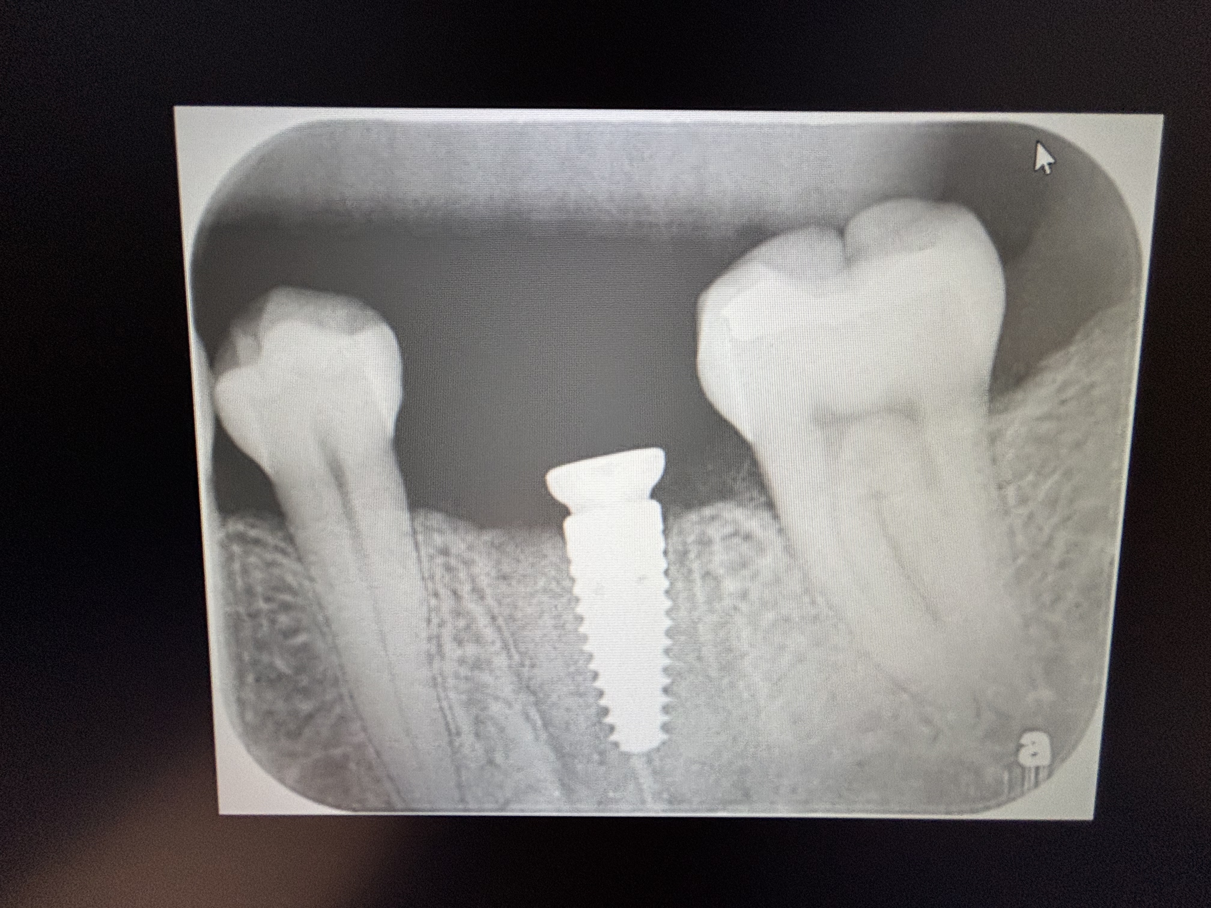
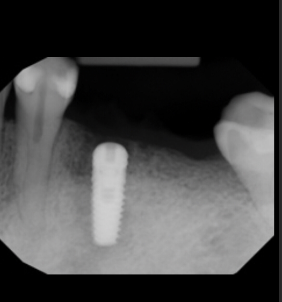
On the given radiographic image, I see bone loss with both a horizontal and vertical component.
Also, is it possible that we do not need a huge amount of keratinized attached gingiva around the molar implants ... after all, most of the natural molars I have seen in my long career usually have minimal amounts in the lower molars, naturally.
I will also agree that a peri-implantitis caused by excess cement is a possibility, and I apologize for this omission.
Also, I think I know the difference between peri-mucositis and peri-implantitis. If this "acute periodontal abcess", as it was called by mistake initially, occured, it had to start with bacterial invasion in the peri-implant sulcus ... peri-mucositis, an inflammatory condition not involving bone, will start, which may lead to peri-implantitis, bone destruction.
With my knowledge and experience, I do not intend to be arrogant with my suggestions ... I do not have time for such action; I intend to offer diagnostic possibilities. And, I stated that this case is possibly plagued with errors in diagnosis and treatment planning ... I don't think this commentary is an insult to anyone, and it should not be, it was not intended as such. It was intended to stir a debate about diagnosis, since our colleague has challenged us with such an interesting situation.
May God Bless
Richard Hughes, DDS, FAAI
2/14/2012
Four Questions: 1) how much bone was available after placing the implant? I would like to see 2mm on the buccal. 2) is the patient under any sort of occlusal trauma? 3) was the tooth that was replaced extracted due to infection? If one has a dormant infection in the bone, it can make the implant go south. 4) Is there adequate attached gingiva around the cervical area? Treat this by cleaning the area, graft with osteogen and thin Alloderm, take out of function and restore later but out of occlusion.
Jay West
2/14/2012
I have seen this with cement on the implant. May not be visible on x-ray because of it being on the buccal or lingual.
TOBooth
2/14/2012
good point.
Dr .T
2/14/2012
I think all these occlusal load ideas and plaque are a load of bull. On the placement X-ray there are clearly 2 radiopaque bone layers. One - the denser bone layer is guess where?- at the same level where the post op 18 months bone level is. The diffuse layer is at the implant connection level. This higher bone level was most likely thin lingual bone and the implant was placed in a sloped ridge. After healing the ridge almost always settles at the lowest point of bone to implant contact 360 degrees around the implant due to biological width formation. It will probably stay like this for another 10 years so I wouldn't worry. Nil bone resorption only occurs in a perfect right angle placement with flat thick bone and thick gingiva. Don't believe all the hype of micro threads and platform.
TOBooth
2/14/2012
Bull-he has just said there has been pus!!! thats not normal!
TOBooth
2/14/2012
Mario you talk about peri-implantitis and perimucositis and don't really make a clear difference. If there is puss its peri-implantitis so this case is so . I don't see much vertical defects ie occlusal associated bone loss. Up to 5mm of pocketing and bop-mucositis over 80% of implants have this!!
In addition in this situation you keep talking about increased crown to root ratio being a problem-in reality it isnt, see Bicon implants ( i don't use them personally)-provided occlsuion is correct see above post. Also you cannot grow bone or augment vertically. So in this situation the dentist couldnt avoid this situation.
Also lack of attached gingivae is a majpr cause of periimplantitis see J Lindhe article. Google it. It makes sense you don't have the intra and inter cellular junctions desmosomes and hemi-desmosomes thus allowing bacteria to the implant bone interface and bone loss. Where is the least amount of attached tissue in the human mouth? the edentulous saddle areas of the mandible.
In addition where you have lack of attached gingivae its sore when cleaning-ask your patients. So it doesnt really come into it that the patient having 2 other implants that are fine- they may well have alot of attached tissue around them.
It could well be a cement spill thats buccal in reality thats likely.
Hope that helps guys.
Renan Evangelisti
2/14/2012
I once had a patient like this, and what i found was one spicule of necrosed bone. I used metilene blue and laser therapy. One week later, the necrosis bone was gone. It came in my hand.The gengiva became fine and, of course one of gengival retraction.
Dr G J Berne
2/14/2012
The cause of the problem is crystal clear and yet so many implants are placed in total ignorance of the periodontal implications. This crown has clearly been placed significantly subgingival thereby initiating a pocket from day 1. There is no way (I hope) any dentist would place a regular crown so subgingivally on a natural tooth yet I see implant crowns frequently placed 4mm, 6mm and yes 10mm subgingivally without any appreciation of the gingival implications. They are failures about to happen. Gingiva bonds by hemi-desmosomal attachment to smooth Titanium, thereby creating a "hermetic"seal to prevent ingress of bacteria to the threads or roughened surface of the implant. You don't get this attachment of gingiva to porcelain or gold, so any prosthetics made out of these materials and placed significantly subgingival are temporary restorations,period!
Mario Marcone
2/14/2012
I agree with this comment. I have stated this in my earlier remarks as a possible contributing issue.
Dr. Vipul G Shukla DDS
2/15/2012
I think you hit the diagnosis on the head Dr. Berne!
Bonus points to you.
Not just implants, cement left in the sulcus can and will eventually eat into crestal bone around tooth-borne crowns as well. Me thinks the cemented crown margin was too sub-gingival and maybe there was a piece of cement on the buccal that caused this issue.
I think this implant has a fair prognosis with proper care.
John Kong, DDS
2/16/2012
Gingival fibers don't attach to the smooth surface of titanium either (exception being Biohorizon's implants with the very specific horizontal laser grooves solely for CT and epithelial attachment)
That said, I think you hit it right on the head - crown margin is too subgingival and when it's that subgingival, it makes it impossible to remove all the excess cement. Also this implant is not platform switch, so you're expected to lose 1.2mm of bone, the 1st year in function right off the bat.
KPM
2/14/2012
I'd like to add my two cents, if I may, regarding crown to to ratio since it was mentioned above. I am an avid user and proponent of the Bicon design. (I will pause to allow the gasping to cease!)....Well into it's third decade of positive results, the short designed Bicon implant (first to use an 8mm length and presently I believe the only implant with a 5mm length available) has demonstrated that crown to root ratio is not nearly as important, if at all, when dealing with implant restoration. There is simply no PDL for which a poor crown to root ratio to adversely affect. Surface area, solid integration and finally hygiene seem to be the main factors which contribute to an implant's long term prognosis, in my opinion. I realize I am on the side of historical implant heresy when I speak of the "short implant" so I will venture to ask, aren't we to expect at least some degree of crestal bone loss in a percentage of the implants we place? Especially "to the first thread"? At least at this point in implant placement?
Mario Marcone
2/14/2012
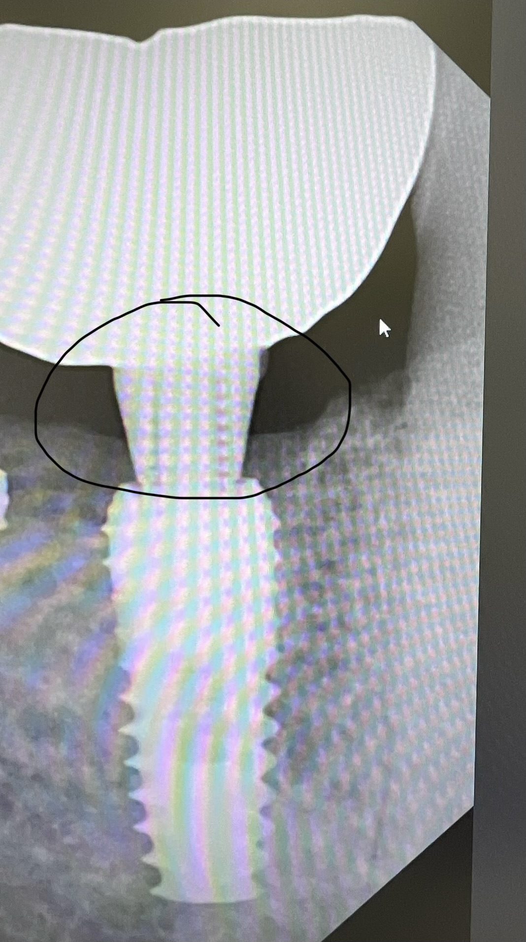
Yes, the point is that the implant is oversubmerged, and the first threads are really far from the initial crest of bone at implant placement, I suspect.
TOBooth
2/15/2012
It isn't though!! So would you leave threads exposed ???!!! Plus you can't augment vertically!! So this implant isn'rt too deep.
Mario Marcone
2/15/2012
The most coronal threads, according to the image presented, appear more subcrestal than necessary, and coronal to these said threads, there may be a machined/polished neck, which also appears to be submerged into bony tissue. This looks like a one stage tissue level implant that has been over-submerged. This may explain part of the problem.
Of course, I would not expose threads above bone crest level. However, in this case, if my guess is correct about the implant design, the most coronal threads should have been placed just apical into bony tissue; and, because we may be dealing with an irregular bony crest morphology, any exposed threads could be grafted with the clinician's preferred technique, unless it would have been preferable to flatten the crest, or maybe use an implant with the ability to have soft tissue attachment to the exposed collar area such as the LaserLok from BioHorizons.
About your insisting that one cannot augment vertically, I can only comment here that in certain situations vertical augmentation is predictable, but I will not comment on this possibility in this case simply because I do not know the case fully well to be able to assess with certainty.
God Bless.
Brandt Foster
2/14/2012
Sorry Dr Marcone. But Im siding with the bull crowd. Always difficult without seeing the case but the implant size seems more than sufficient considering that it is opposing a complete upper denture. I do think there is an increased risk of a popcorn abscess with a lack of keratinized tissue and the bone loss could have resulted from an origanal lack of with/ placement or cement. Although occ overload can result in boneloss that can then increase the risk of infection the more simple explanation would be the infection and subsequent inflammation caused the boneloss. To the best of my knowledge there were no radio-opaque implant specific cements when this was done so cement would be number one on my differential. But I also agree with Dr T. With proper treatment, I dont think this implant is going anywhere.
Mario Marcone
2/14/2012
Dr Foster, the opposing dentition is a removable partial denture, not a complete denture.
Mario Marcone
2/15/2012
Dr Foster, I did not say that implant size was the problem, my point was more about implant design and implant placement. And, with respect to the latter, we appear to have over-submerged implant-to-abutment and abutment-to-crown margins, and the ensuing possibly contributing negative sequelae, such as unfavorable crown-to-root ratio, unfavorable bone stress leading to bone loss to the first thread (and this pertains to the selected implant design), and possible retention of luting cement subgingivally at crown cementation.
May God Bless.
Dr. Don Rothenberg
2/14/2012
The question becomes ...can the implant be saved and made functional again...without future bone loss. I too believe it is NOT a crown to root ratio problem, as I too have placed many of Bicon's "short implants since 1988 (8mm) and more recently the shorter implants (6,5.7 and 5mm) the crown to root ratio of an implant is different then a tooth and we should learn this simple fact,,,,sooner then later.
What should be done?....I would remove the crown and abutment and rebury the implant ....with or without grafting (Synthograft)...leave it for 2-3 months...open and see what we have...I would not be surprise to see the bone come back. In 2-3 months place the abutmant and crown back...being careful not to have any debris collect in the area of bone loss...instruct the patient how to clean and follow ...if all goes well and the patient maintains this area...one might say we did him a nice service. One could always take the implant out and redo it after the bone heals...the down side of this is that he has no tooth for quite awhile and the expense the patient.
Theodore Grossman DMD
2/14/2012
1)Making use of the proper diameter implant will allow for platform switching .
2) Placement of the cervical aspect of the implant at the gingival margin will minimize micro gap issues.
DREAM DDS
2/14/2012
This is really not a failed case. There are many cases such as this. And what is a failure? The patient doesn't have pain and I really doubt there will ever be enough bone loss to sacrafice the implant. this doctor is trying to help the patient out. I feel the only alternative would be to place two implants, splinted or not. We need to follow the protocols of ideally one implant per tooth missing. write it down and have the patient sign it. Again, this is not bad or failed treatment. As to crestal placement: what type of implant is this, what is the collar surface type, 3 mm of bone loss is common for many well known implants. Why try to bone graft etc? Flap, debride, polish, detox, suture, give proxa brush. A problem is not a problem until someone thinks it is.
Leonard
David E. Azar
2/14/2012
Assuming all else is WNL and there was no subgingival cement then the likely reason for the 3mm "bone loss" is related to biologic width. Your crown margin is way subgingival. It is natural for the bone to remodel to 3mm from the margin. So it is not exactly bone loss but bone remodeling to its natural biologic level. Now that you have done open flap currettage I wouldn't be surprised if the problem clears up. You should expect some significant recession but tooth location should make this a non-problem. This is a basic biological reality that relates to every implant and in fact every restoration we do. Make sure to keep this in mind when planning every case, especially anterior ones as this kind of problem will definitely lead to an esthetic failure down the road.
SG
2/14/2012
With the angles of the 2 x-rays being so disparate, I don't see how any meaningful comparison of bone levels between the 2 points in time can be made.
John George
2/14/2012
CEMENT caused this. I can see it on the distal on the x-ray. Cannot possibly clean the cement out from under a margin placed this subgingival.
Paolo Rossetti Milano
2/15/2012
Mr Hughes made a good point: probably the boney crest was thin, or at least it was too thin for a 5mm implant.
Here are my considerations:
You can easily notice on the radiographs that, at time of implant placement ,the top of the boney crest was more radiolucent (the difference in radiopacity is not completely due to the presence of the oblique line), which probably means that the bone was thinner. And the implant is large (probably a 4,7-5mm body, and 5,7-6mm platform).
The implant is transmucosal, the kind of implant with a polished neck and designed so that the crown's margin fits directly on the implant platform.
The implant platform looks as if it had been placed at bone level, and consequently the polished neck is below the bone level.
If my guess is right, the bone remodeling around the polished collar, having occurred on a thin boney crest, may have induced some vertical bone loss. Moreover the crown margin, that presumably was too close to the bone at time of placement, may be responsible for the inflamation that eventually led to the acute episode (with further bone loss)
Seen from another perspective, the abscess has reestablished the biologic width, by resorbing the most coronal bone that was to close to the implant-crown interface.
If you watch the first x-ray (the one with the crown), you can see that the new bone level matches approximately with the rough surface of the implant (a bit lower on the distal).
You can't go against nature.
JIM
2/15/2012
IF, and I say IF, cement is an issue then the next logical treatment protocol would be to remove the crown, debride the area, and replace with a screw-down implant crown.
Drquintner
2/15/2012
Was there suppurations present at any of the patients maintenance visits? Annual probing around implants, while somewhat controversial can be a valuable tool if only to check for inflammation/suppuration. This was either caused by cement and/or occlusal overload. Attempt to remove the crown, if unable drill a hole in the crown so you can access your screw (which would be easy if you took a photo of your abutment prior to cementing your crown), remove the crown abutment complex, polish your margin to a mirror like finish and reinsert. Irrigate implant site with chlorhexidine when abutment is out, and place arestin after reinsertion. Check for CR interference with leaf gauge, clenching. Be sure patient is removing rpd (opposing) at night. Monitor with follow up probing and bw in 3-6 months
jim
2/15/2012
Good point.....many denture patients sleep with teir dentures.
don callan
2/15/2012
Think about the host and the ability to resist disease. Periodontal pathogens can cause rapid bone loss. I do not know of any literature that shows occlusal over load will cause rapid bone loss. Trauma causes inflamantion and that causes bone loss. It may be from bacterial, mechanical, or systemic. Look at the rest of the mouth. Bacterial is the culprit in this case from my experience.
Mario Marcone
2/15/2012
Don, occlusal overload may translate into excessive bone stress ... Bone modelling and remodelling ... Frost's Mechanostat Theory.
Once bone is lost, the secondary result may be a peri-implant tissue pocket evolving vertically apically enough to harbour pathogenic micro-organisms that cannot be easily cleansed by the patient.
In addition, it is perplexing to me that by this radiograph provided some clinicians on this thread can judge that the patient's hygiene is so terrible. I would say this is not necessarily so--based on the bone levels apical to the CEJ's of the neighboring teeth, there does not seem to be a huge periodontal issue.
God Bless
peter fairbairn
2/16/2012
That Implant will probably still be there in 20 years time , so is it a big deal? yes there are issues as mentioned above by the posts but proper cleaning and maybe poillish ing the micro threads off will be fine. As to treatment of more severe peri-implantitis there are solutions that work ( use of Easygraft on prophy cleaned implant)
As to placing the Implant higher and getting vertical bone growth , that is also very possible ( see my case , one of hundreds posted a month or so ago here ).
Materials have moved on.
Finally the idea of a flared crestal portion with a micro-thread is a fault in the design of the Implant which can cause this crestal bone loss , I have seen it on one of the systems I use , and yes they are changing it.
Peter
TOBooth
2/16/2012
Hi Peter,
where is the case of vertical ridge growth. I have to say i struggle!!
peter fairbairn
2/16/2012
Here posted a few weeks ago under vert bone regeneration case , I routinely get up to 4 to 5 mm of vertcal bone regeneration in the right cases , and have shown them around the world .
Regards
Peter
Mario Marcone
2/16/2012
The idea of attempting to grow bone vertically has been well documented in the literature especially pioneered by researchers in the late 1990's and early 2000's by Simion, Jovanovic, Tinti, Parma-Benfenati. More recently, Langer et al (2010) and Ludovichetti (2011), have published encouraging data. The lesson to be learned from all these ongoing studies is that the protocols and materials had to evolve to the point where when applied in specific situations which meet specific criteria, vertical bone growth, that is, vertical ridge augmentation, can be successfully achieved. It goes without saying that careful surgical technique, knowledge of the potential of various materials available today including bone graft materials and growth factor science, must be all on the diagnosis and treatment planning protocol.
It is important to underline the fact that specific criteria must be used to make this procedure predictable. Excellent surgical technique and knowledge of the evidence-based literature is imperative.
Jennifer Watters
2/18/2012
I think this is due both periodontal and occlusal trauma. Look at what appears to be a sharp wear facet on the neighboring bicuspid, this patient is "on their teeth"! There has been no doubt a deep soft tissue pocket interproximally, since the implant had to be placed much "deeper" in the bone than the bone level of the adjacent bicuspid; this creates a periodontal pocket from the start. Unfortunately, a pocket not being cleaned properly with heavy occlusion and parafunction, spell bone loss. A deep pocket, of course, is at risk for foreign body impaction such as a pop corn hull and an acute abscess occurs taking with it more bone. At this point, I would treat it as an "ailing-failing implant". Perform a periodontal flap procedure, debride it thoroughly, decontaminate the surface of the implant after complete degranulation, graft it with bone, cover it with a membrane and adjust the occlusion and get the patient in a hard occlusal splint, adjusted for even centric contacts. Make sure that there are no detrimental contacts on the implant crown and the same with the occlusal guard. Good luck! By the way, I have a few nearly identical cases which have gained significant regeneration, with of course, proper oral hygiene (and maintenance), perhaps a narrow or medium proxabrush, if that will fit and utilize Arestin when obvious inflammation is present at recalls. The Water Pik is useful to remove food debris and provide some soft tissue irrigation although I wouldn't recommend this for several months after grafting. Good luck!
uli.friess
8/23/2012
Take the crown off with the help of CORAFLEX and change the emergency profile of the crown.