Ailing Straumann Implant Case: Should I Cut My Losses?

Dr. Z asks:
I placed Straumann ITI bone level implant 4.1/14mm in the #9 area [maxillary left central incisor; 21] 8 weeks ago (see attached case photos). There was very limited bone volume, good primary stability, but almost 80% of the buccal bone had to be grafted with Bio-Oss [Osteohealth] mixed with autogenous bone collected from a bone trap. A Bio-Gide membrane [Osteohealth]was used.

Following the implant placement and bone graft, the patient failed to return for 8 weeks. Patient now presents with a dehiscence in the crestal region where the cover screw can be seen. Some of the Bio-Oss graft has been lost, compromising the buccal alveolar crest area. There is also a sinus tract on the buccal of the graft site which is actively draining. Patient has also complained of feeling ill. I have prescribed 500mg of amoxicillin and 400mg of metronidazole.

She will be returning in 2 weeks when I will attempt to cover the dehiscence. I will use a split-thickness flap which I will rotate from the palate over the dehiscence and attempt to submerge the implant. But, should I just be cutting my losses here, and would it be better for me to just remove the implant, graft and place another implant later? Or, should I proceed with my protocol? I really would appreciate advice.

Preop #1

Preop #2

Preop with BioOss

Postop Pic

Postop X-Ray

14 Comments on Ailing Straumann Implant Case: Should I Cut My Losses?

New comments are currently closed for this post.
Hamza
9/27/2010
Hi I have the following comments: 1. the cause of the infection should be identified, the first priority is to treat the infection 2. infected grafts resorbs and or integrate by fibrous tissue or both 3. antibiotic will mask the infection and may not eradicate it 4. the implant is positioned too high (as i can see in the provided photos), it should be 3 mm apical to the cementoenamel junction of the adjacent teeth, it is very difficult to achieve a good esthetic result with this positioning 5. maintainance of supporting bone at adjacent teeth is of paramount importance, if you get resorption it will be very difficult to have the papillae and end with black triangle overall, if I were you I will strongly consider implant removal alongwith curretage of the infection, suture, wait for 6 months, place another implant. the ratinale is implant position and infection. I have to say that my point of view is not very clear so take what i mentioned with caution. good luck Hamza
sb oral surgeon
9/27/2010
While your pictures of placement look appropriate, infected grafts need removal. Your graft is infected. I would cut my losses here. Do not do any more surgery on this site. It is infected and until the graft is removed, it will remain that way. Surgery to repair a soft tissue deficiency here is not going to help when the underlying implant - graft is infected. If I were you I would remove everything and send to a specialist. I do not question your ability, but at this point you have a compromised anterior site. Anterior sites will be your greatest successes and greatest failures. Just learn and move on!!!
Gregori M. Kurtzman, DDS
9/28/2010
I would suggest a full thickness flap and extend it making the base wider then the crestal and up past the mucogingival line. then with scoring the periosteum you can stretch the tissue to cover the site totally. After flapping you can eval if the implant has any mobility if it does remove graft and wait 3 months to reimplant. if it doesnt then treat like an ailign implant and remove any granulation tissue, place Doxy in a paste on the implant exposed threads only rinse after 1 min and repeat then treat the entire area with citric acid rinse and repeat. I would use Regenform or similar moldable graft product and cover all of the implant including the cover screw. place a peice of Ossix or other resorbable long term membrane and get primary closure and allow to heal for 3-4 months. in the mean time keep them on the antibiotics for 2 weeks post surgery.
DR. Vipul DDS
9/28/2010
Assuming that the draining fistula is not due to adjacent natural teeth, then the graft is infected; in which case, there will be significant dehiscence in the buccal, if not right away, then eventually. IMHO, you can remove the implant (should not be difficult at this stage and with the current infection), and remove ALL the graft material. Place new Bi-Oss Collagen block with some bone chips from the trap, cover with Bio-Gide and make a flipper with an ovate pontic/Ribbond bridge with ovate pontic. Then, wait a solid 4-6 months. Then reassess for implant placement, always being prepared to add more graft/membrane along with placement at the time. Also, you might want to place it maybe 2 mm more apically, for perfect emergence profile. I would also use the bottle-shaped healing abut, else final imps can be a nightmare! IMO, Straumann should give you a new fixture at no cost at the time (document the failure well) Now, what happened that made the graft fail in the first place? Was there adequate primary stability in the first place? Was it done under AMOX cover? Did pt slip on oral hygiene? Is she a smoker? Was it an immediate case in an infected site from a failed endo? Again, I'm no specialist, so please take my 'advice' with a pinch of salt. Good Luck!
Dr Lee Nightingale
9/28/2010
Cut your losses now as these early graft infections rarely end with a positive result. Remove the implant and clean up the site and let it heal. Once the site is nice and healthy you can perform a graft procedure to build up and fill out the surgical site prior to re-implantation. As mentioned by previous posters you will need to pay close attention to implant positioning next time round to get a good emergence profile. I feel that
Joseph Kim, DDS
9/28/2010
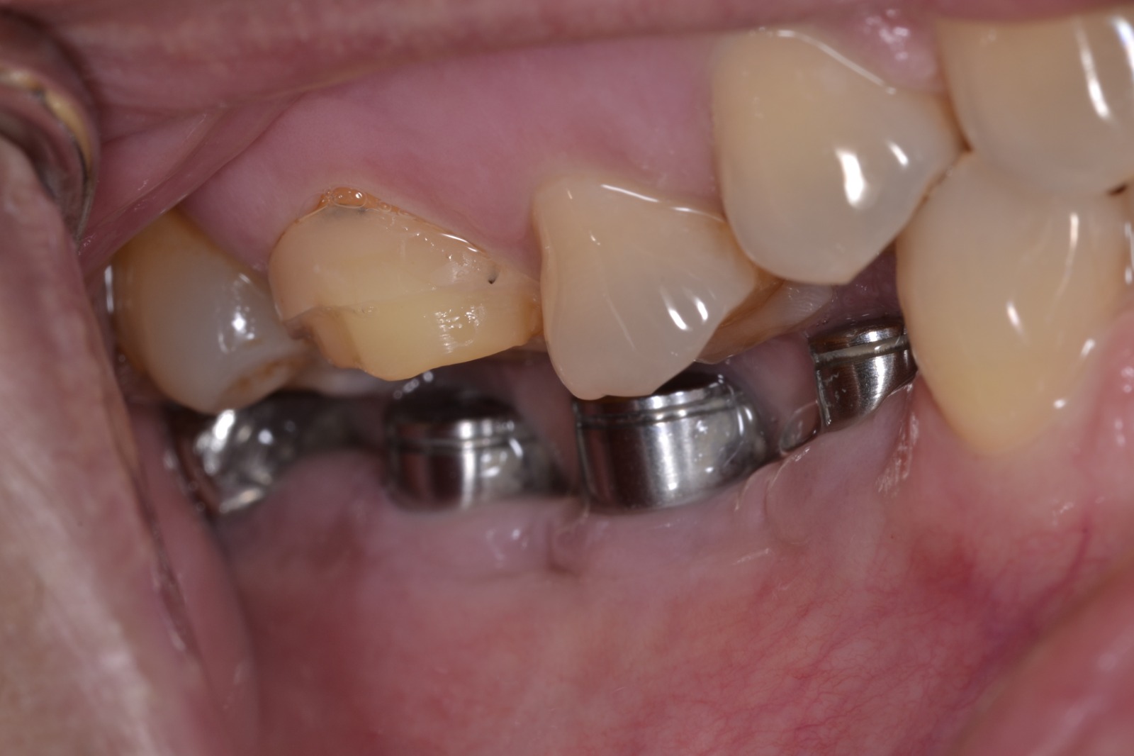
The mistake you made was that you did not advance the flap and suture with a horizontal mattress as your main stitch. I know this because the mucogingival margin is in the same position pre-op and afterwards. Also, the implant cover screw is showing through the gingiva on the post-op picture. Whenever you bulk the area outwards, which is inevitable in the procedure you are showing, you must advance the flap to make sure the tissue will completely cover the site in a tension free manner. If you had advanced the flap and tied it off with a horizontal mattress suture, the crestal wound margin would have an everted appearance at 8 weeks, and the attached gingiva would be significantly coronal to the position indicated by the picture. One other criticism is that you should be more thorough with degranulation prior to grafting, as evidenced by the tag of tissue over the facial aspect of the implant. Use a back action chisel to do this easily. Finally, did you make bleeding holes adjacent to the implant? Just run a 1.2 mm twist drill without irrigation into the cancellous and save the shavings. You must have adequate perforation and bleeding to ensure predictable results. By the way, I don't place implants into defects of this size in the anterior anymore because of the guaranteed insufficient buccal height of bone that usually results. Now, it's always graft first, implant later. The graft is always wider than I think I will need. 
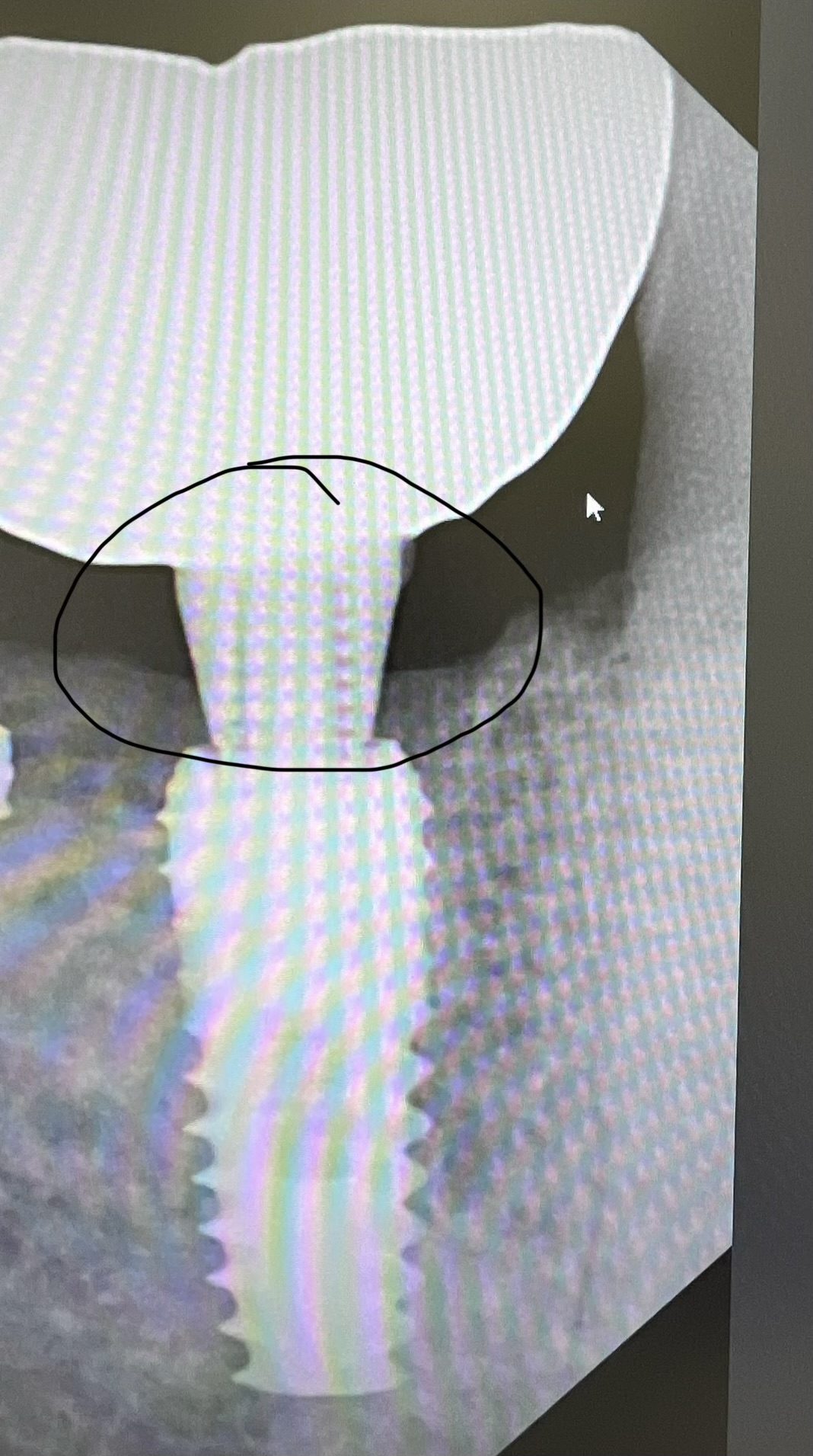
What happened in your case is that the flap margin pulled back and allowed bacteria to enter the wound through the area of the cover screw. This means that you did not achieve passive primary closure of the wound, which resulted in exposed collagen membrane. While it is possible to achieve epithelization over an exposed collagen membrane, it is not guaranteed. Also, with a graft of this size, in a critical area, where there is little blood supply to feed that large area of exposed threads, you needed a lot more autogenous bone, and definitely needed to significantly advance the flap. You can always do an apically repositioned flap at second stage surgery. 
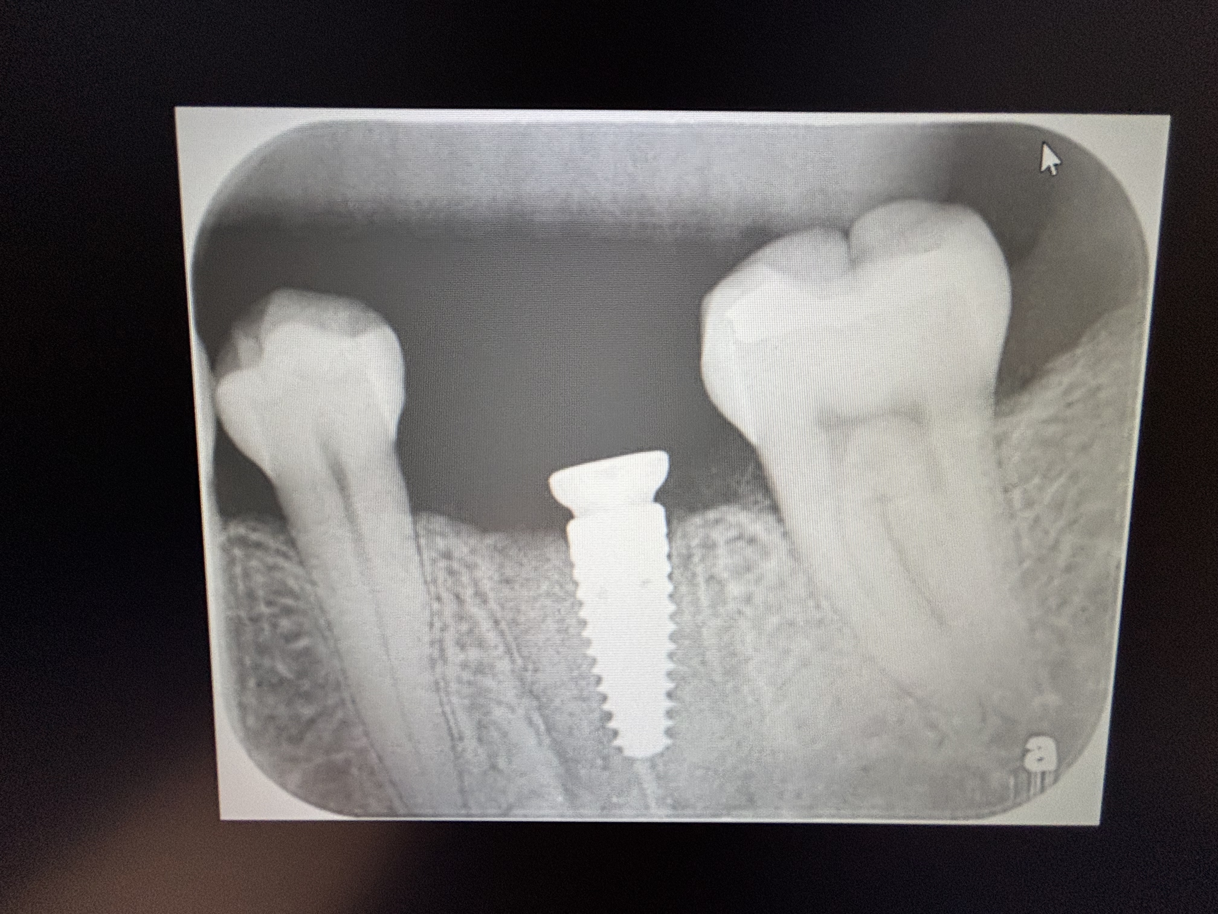
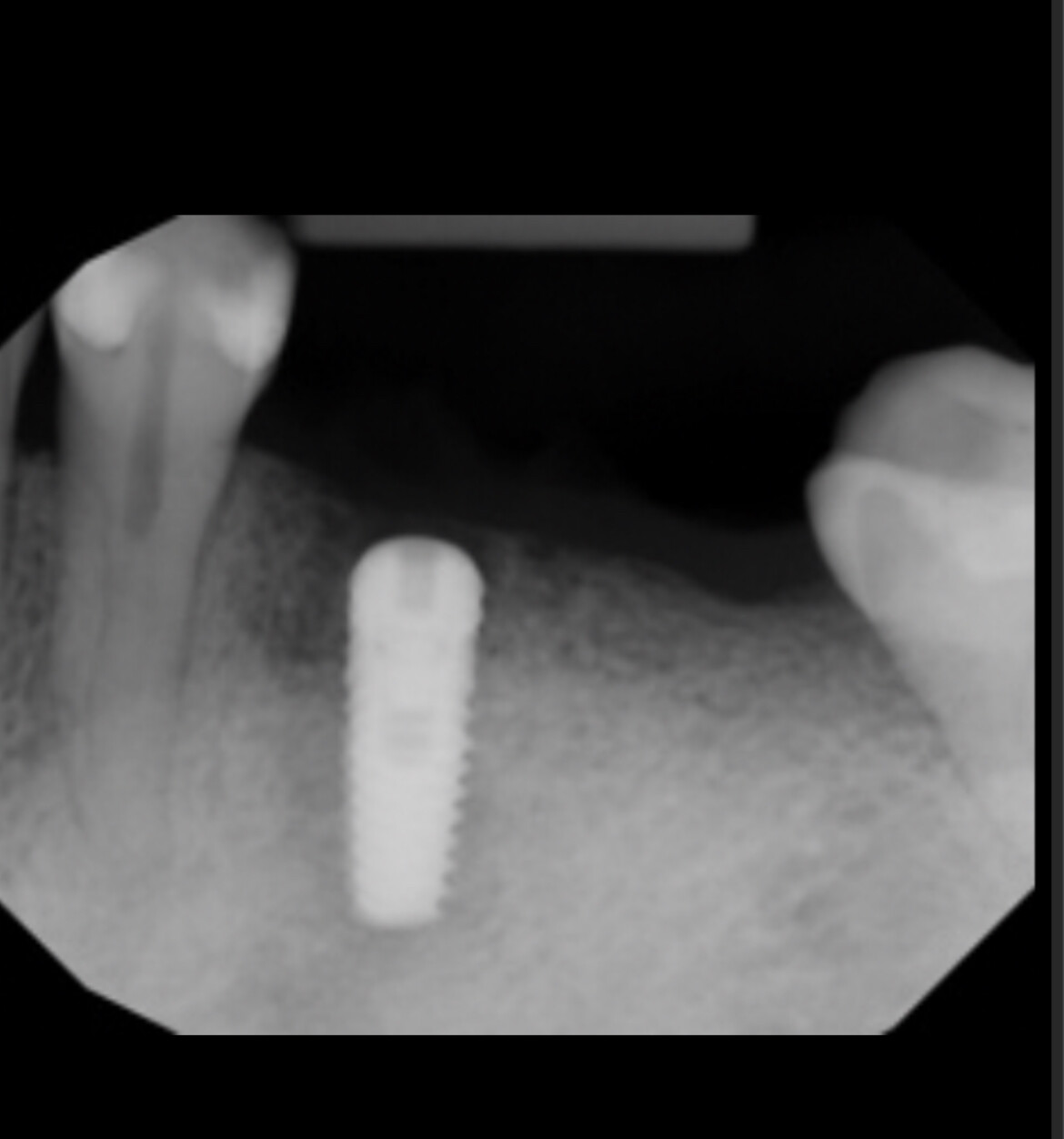
I disagree with the other posters that you placed the implant too deep. In fact, your depth looks perfect. I would remove the graft and the implant, wait 4 weeks for healing, and reenter. When you remove the graft and implant, place your vertical releasing incision (only one) to the distal of the lateral, and intrasulcular to the other lateral. When you reenter, completely degranulate the area, place your bleeding holes first, and overgraft the area. Use a thicker membrane such as Ossix or RCM6, or a titanium reinforced ePTFE or dense PTFE membrane, with BioOss or NuOss. Also, consider the use of PDGF (Gem-21), which will case marked swelling, but is totally worth it is mission critical areas. Make sure your vertical releasing incisions are not so angulated, and PLEASE, no papilla preserving flap. When you close, make sure the membrane margin is 1 mm away from the adjacent teeth (which should have been scaled prior to Chx rinse prior to surgery), and that you use a horizontal mattress suture that has a deep bite (4 mm) into the attached gingiva. The margins should be visibly everted. This is, of course, only possible if you adequately release the periosteum, which is super easy if you use a new blade. I would also advise PTFE sutures, as you do not want this site to become reinfected. Vertical incisions should be closed at base, then ever 1 mm. Modified interrupted on crest at either end of the horizontal mattress suture. Gently press on the wound after suturing to make sure no bubbles come out. This will indicate a complete closure of the wound. Amoxicillin 500 mg, 22 tabs, 2 tab stat, 1 tab tid until gone. Chlorhexidine rinse, bid, until suture removal in 2 weeks. NO pressure on wound, so use an Essix retainer with facial painted with acrylic. Since I started this protocol for wound closure after grafting, I have had ZERO catastrophic failures. If you're not comfortable with the above, refer to an oral surgeon with block graft experience or a periodontist that you know has great success with particulates. God bless.
Shirley A . Colby
9/29/2010
It would be a good measure to evaluate the status of #10. M-cervical margin of the retainer is highly deficient, with ensuing radiolucency underneath. It could be the source of the contamination. Know thyself! If you feel you can measure up to the challenge, as it is definitely going to be a challenge, go for it! Otherwise, Dr sb. had presented a sound advice.
K. F. Chow BDS., FDSRCS
9/29/2010
Hi there. Cut your losses and remove the implant and let the site heal and try again. Trying to salvage the situation is difficult and uncertain, and the effort will result in more trauma to the patient and to yourself 'cos you have to wait and see whether it will work or not. Chances are high that it will not, seeing that it is infected already and there is inadequate healthy living tissue surrounding the fixture. Joseph Kim has given excellent, detailed advice based on the sound surgical principles of thorough debridement of all infected and diseased tissue, ensuring adequate blood supply, and protecting the healing site by good primary closure. To ensure adequate blood supply and therefore nutrients and sufficient defence cells.....use a smaller diameter implant .......3.5mm or even a mini. This will allow plenty of living tissue around the implant and ensure osseointegration and soft tissue protection around the implant. Cheers.
Guilherme Röhe
9/29/2010
Hello Dr.Z Could you provide your Cone Beam CT performed in its planning. I wonder if this bony plate vestibular been displaced during the placement of the implant or if have a history the defect. If the defect was pre-existing consideration should be made graft block and only after 8 / 12 months you can implant. The best approach to the case would be defined by Cone Beam CT, from incision to closure. Without these images we will be in the field of hypotheses. Anyway, I recommend the removal of this implant, wait a period of approximately 2 months and put a new implant BL 3.3 mm. Always approach the palate! But, examine your Cone Beam CT the height of peak bone on the adjacent teeth and, consider the block graft! I await your pictures! Good luck! Excuse my English!
Luis Berumen oral surgeon
9/30/2010
Hi I think then your case has several mistakes 1.- When you have used a graft, the vertical sections of mucosa must to have more ampliation between them for to obtain healthy support of tissue healing and this cuts most be divergent in its base for to obtain more blood irrigation ah! you do not doing cuts on papillas. When the vertical incisions are made over healthy bone and no close of graft is better because you dont have infections 2.- I do not if you have preparation bone surface for reception of graft and also you need make more elongation of mucosa from periosteo, it is very easy. May be you need to learn more about surgical conditions.
Dr Z
10/2/2010
Thank you all for you opinion. I have learnt so much from your view from this failure, which will improve my treatment of future cases to treat. You all have confirmed my suspicions of my poor relivieving of the the periosteum, and inaddequate primary closure. i will be seeing this lady in 2 weeks time..will discuss the failure, and plan for removal of the implant. Will keep you all informed Thank You
Dr Dimitrov
10/4/2010
Cheer up, a complication is not a failure, if you learn from it. This way it won`t happen to you twice. Excellent comment from Dr Kim. Totally agree with him. Plus: 1. Avoid grafting and implant placement at the same time (especially in the esthetic zone) 2. Never place the incision borders next to the graft and the collagene membrane. In my modest oppinion that`s where it all came from, apart from patients not showing after surgery. Next time try to extend the flap releasing incisions distally to the central and distally to the lateral incisor. Don`t worry, you`ll not be too traumatic. 3. ALWAYS puncture the cortical bone to the bone marrow (until it bleeds). Make plenty of "VENT HOLES". This way the graft will receive plenty of blood, which will develop it`s own vascular system and finally transform into new bone. 4. Release well the periosteum from submucosa, until you are able to cover all that bulk. 5. As Dr Kim perfectly stated the HORIZONTAL MATTRESS suture is your main weapon. Sometimes I`ve heard colleagues call it "structural" suture. Interrupted sutures are just for adaptation and additional flap stability. 6.Keep the wound margins FREE from the collagen membrane and biooss particles. If Biooss is present in the wound margins, be sure to expect dehiscence. 7. ALWAYS suture with monofilament sutures (e.g GORE PTFE. What good is charging a pot of money for graft, membrane, surgical time, when you close your wound with simple polyester suture (or even worse-SILK)? PTFE sutures should be the standart for care in these cases. Bottom line- you can`t rewind what happened. Remove implant, remove graft, let the nature repair the site and reenter DE NOVO. This time one step at a time. I liked Dr Kim`s expression- Graft first, implant later Best of luck, Dr Dimitrov
Dr Dimitrov
10/4/2010
One more thing: Are you sure the frenulum was not also pulling the attached gingiva prior to your surgery?
Dott.vdl
10/11/2010
Hi, I am a dentist from Italy. My question to Dr Z. is: which kind of temporary restoration has had this patient? Thank you for your answer.

Featured Products

OsteoGen Bone Grafting Plug
Combines bone graft with a collagen plug to yield the easiest and most affordable way to clinically deliver bone graft for socket preservation.
CevOss Bovine Bone Graft
Make the switch to a better xenograft! High volume of interconnected pores promotes new bone. Substantially equivalent to BioOss and NuOss.