Bringing down impacted canine: prognosis?
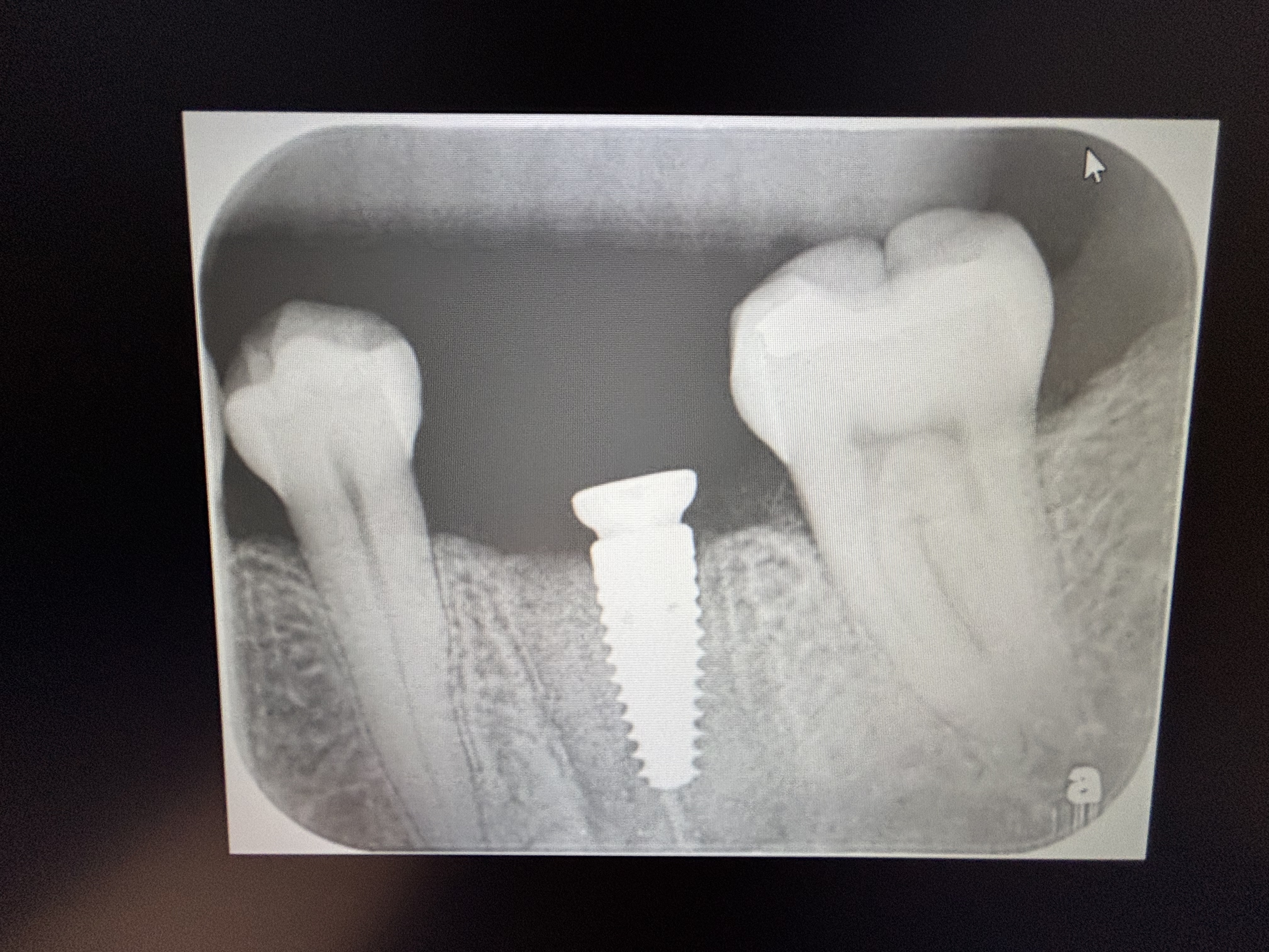
I’ve been trying to bring down this impacted canine since April, 2014. Noticed a radiolucency after the last CT was taken. My question is, considering laceration at the root tip, does this canine have a good prognosis to be put back into occlusion? Thanks.


10 Comments on Bringing down impacted canine: prognosis?
New comments are currently closed for this post.
CRS
9/20/2015
If I don't see movement after six months or sooner if SFOT used then I would remove ,graft and make space for the implant. The root is dilacerated which can affect the path of eruption but most likely in this patient it is anklylosed. Now your ortho is causing pathology. I think this needs to go to the Oral Surgeon. Not sure what this post has to do with implants. Are you an orthodontist and are you working with an OMS?
OMS
9/21/2015
I'm with CRS on this one.. Take it out, graft, wait and implant.
Alex Zavyalov
9/22/2015
Probably, you might achieve your goal but only after root apex resection because of its curvature.
Katona Tamás Dr.
9/22/2015
Just do the ortho. It will be fine.
Dr. Katona Tamás orthodontist.
Larry Forehand D.D.S.
9/22/2015
I am a general dentist who has done ortho for 30 years. You may have done all these things, but I'll give you a "checklist" of things I concern myself with when doing retrievals: Make sure there is adequate if not excessive space to move the impaction into. I keep an open coil spring in place to maintain and increase the available space until the retrieval is complete. Make sure the original exposure was adequate, I use a laser to make sure I can visualize the entire crown. Use steady, LIGHT, forces. I commonly wire ligate the retrieval chain to a 016 archwire and only slightly deflect the wire. I try to erupt the impaction via the path of least resistance, meaning that I will erupt the tooth to the buccal or lingual if necessary and put it in position AFTER eruption. Yes, this can create gingival contour issues, but I feel it gives me better success at retrieval. Above all, discuss with the patient/family the fact that in spite of everything, some impactions can not be successfully retrieved. As previously mentioned, I do use the 6 month rule, BUT if I continue to see movement, I will extend the time as long as I feel I have a chance of success.
Miguel Martinez
9/23/2015
thank you for all feedback given. what would you suggest radiolucency on most recent CT?
Andy K
9/23/2015
This tooth is not ankylose. It's almost erupted. Usually it takes 12-18 months to bring upper canine to go down completely.
The curvature of the apex of the root is normal for some canine and has nothing to do with Ortho movement. The radiolucency is also normal, because of osteoclasts and osteoblasts activity.
You do your patient a lot of favor by not extracting and bring this canine to proper function.
Once you complete this case you will be more confidence to treat similar case in the future.
Please don't take the canine out. You will have the satisfaction that money cannot buy by doing the right thing to this patient.
It will be good for your case presentation for other patient with impacted canine that will come to your office in the future. Give it 6 more month, the tooth will be erupted fully. You can bring it down with 0.014 niti once the tip of the cuspid break the gum. Make sure you get correct orientation of buccal and lingual aspect. I got one case that I need to rotate almost 180 degree. Your work is more than half way done.
Having a healthy natural tooth is much better than having implant that can have risk of perimplantitis, loose crown, etc. Also it is hard to match the color of single anterior implant or crown with the neighboring teeth.
CRS
9/23/2015
After reading your post I had a thought, the first bicuspid may need to be removed eventually but that is a leap of faith commitment. I have been exposing canines a long time and honestly eighteen months to two years of ortho is a very long time. Braces are not great for teeth. what is your opinion on SFOT? I would definitely do it here. I am confused, what was done at exposure? That radio opacity is an odontoma? Usually those are removed at exposure along with the baby tooth my first answer was based on the premise that adequate space and obstructions removed at exposure. The dilaceration at the root tip may have been created by navigating the canine around the odontoma which I missed on the cbct. Remove the odontoma that will be as good as SFOT. You may be seeing the odontoma coming into view on the panorex. Remove the obstruction, if no movement then extract.
Andy K
9/23/2015
The radiolucency in the apex is normal activity of osteoblasts / osteoclasts. But the radiolucency around the crown could be the beginning development of supernumerary tooth (which is pretty common to have it between upper canine and first premolar region ).
You don't need to remove the supernumerary tooth at this point.
I have also one case similar to this. As in bring # 9 down the supernumerary tooth between 8 & 9 started to develop. I ended up to remove the supernumerary tooth 2 years later after finishing up the Ortho for #8. How old is the patient?
The supernumerary is pretty deep, lingually positioned to the canine crown. It's more risky to deal with it now.
Igor
9/25/2015
Use NiTi wire .016 - .018, and do just ortho. Curvature of the root tip is not restriction at all. Finally natural tooth is better then any implant.