Coronal Bone Loss on #7 Implant: Any Suggestions?

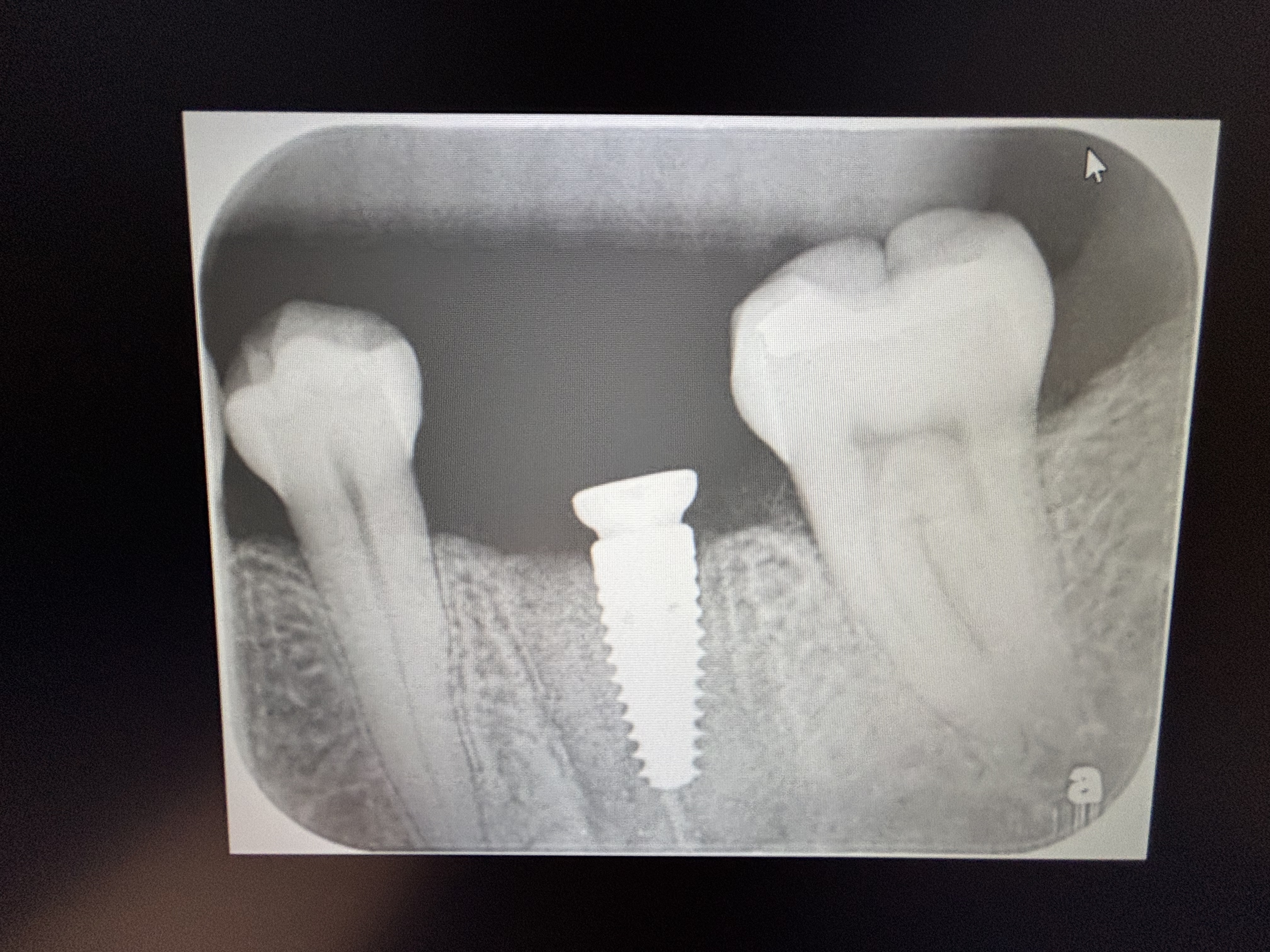
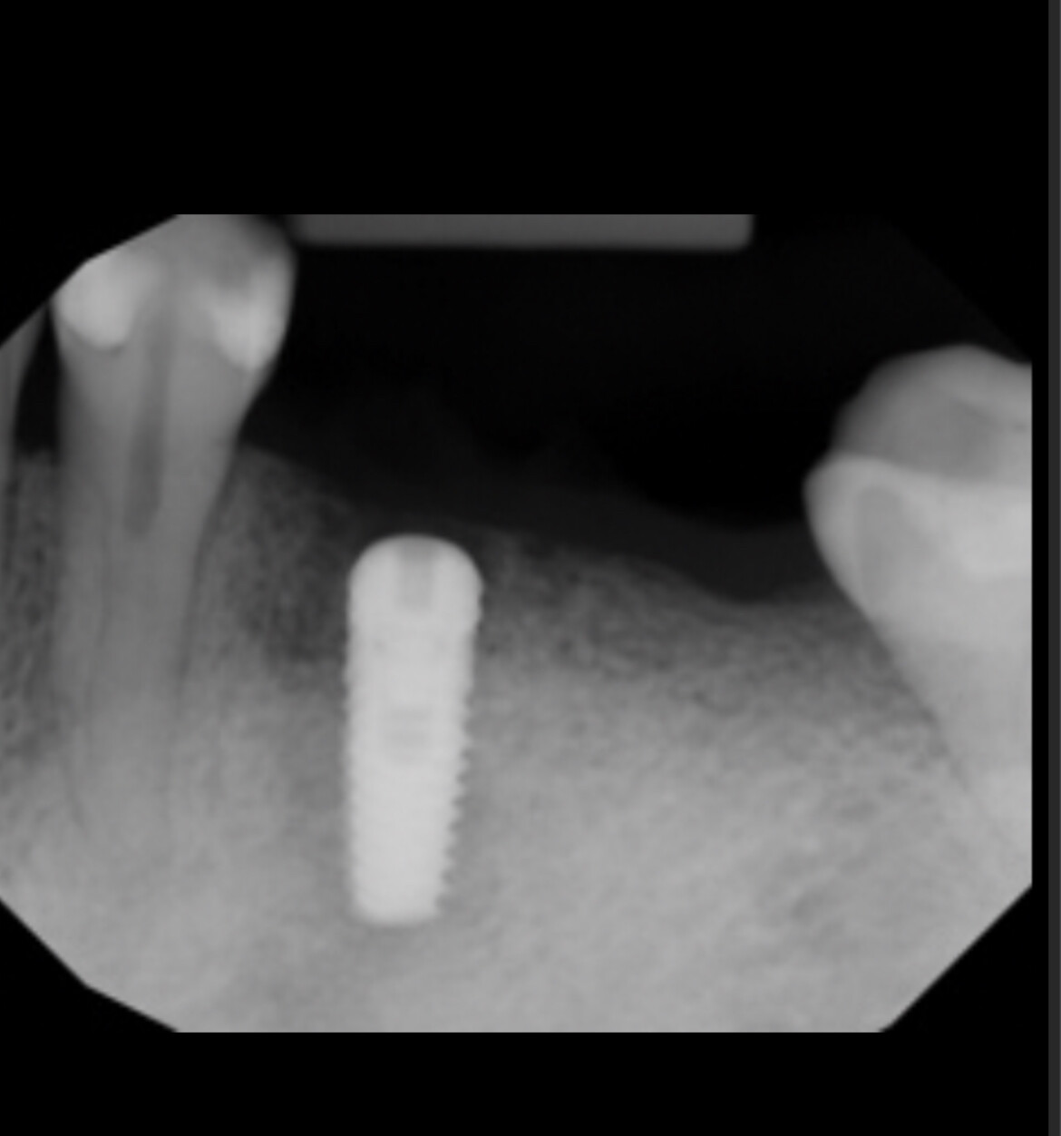
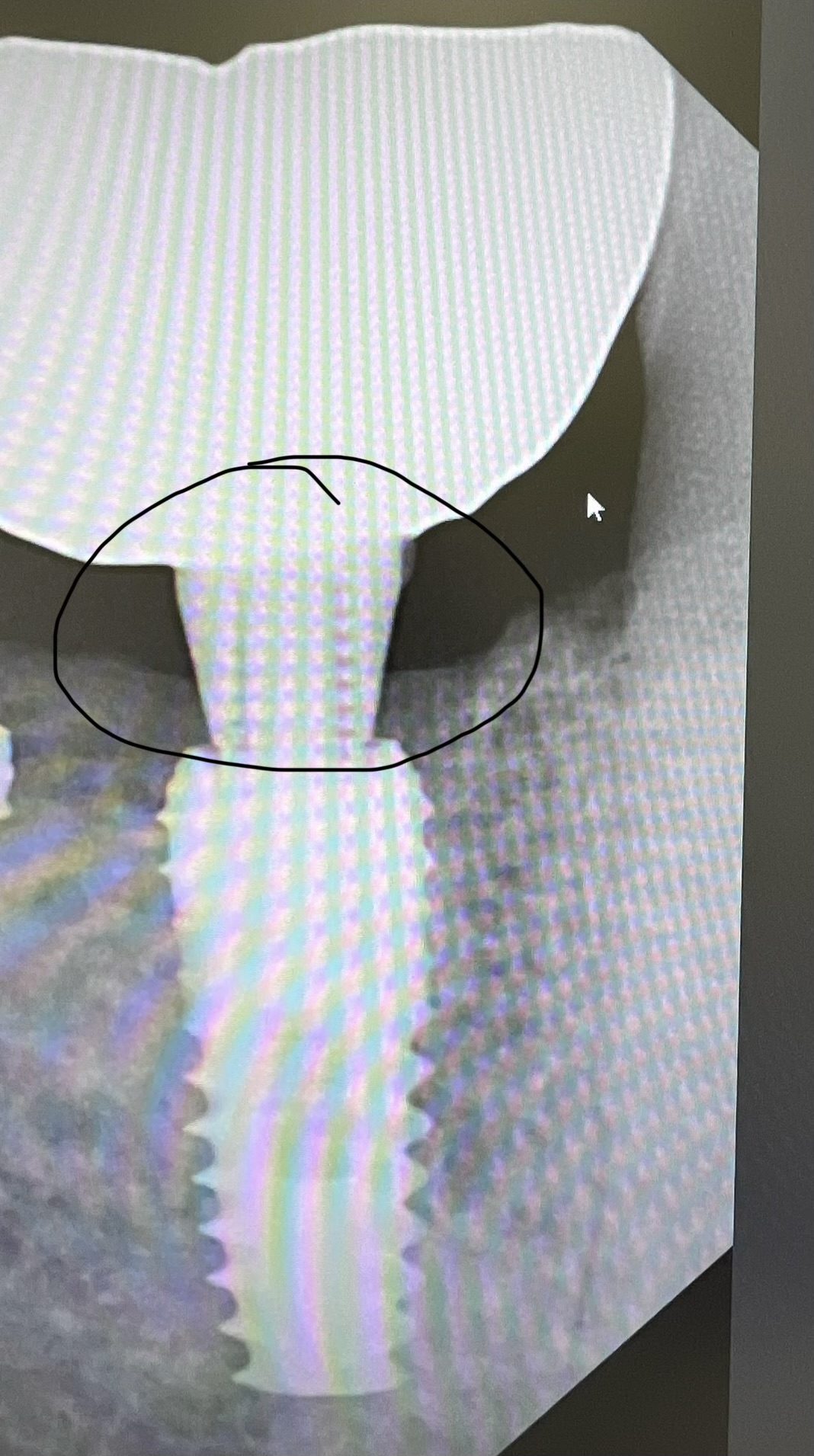
#7 was extracted in April/2013 and an immediate implant was placed (MIS 3.75×11.5) into the socket with a bone graft and collagen membrane. It was allowed to heal unloaded until Nov/2013 when it was uncovered and fitted with an angulated abutment. In Dec/2013, the abutment was torqued and the crown was cemented. In June/2014, the patient returned for follow-up and an x-ray was taken which showed some coronal bone loss. The implant was solid with no mobility. Patient stated that she was taking Fosamax.

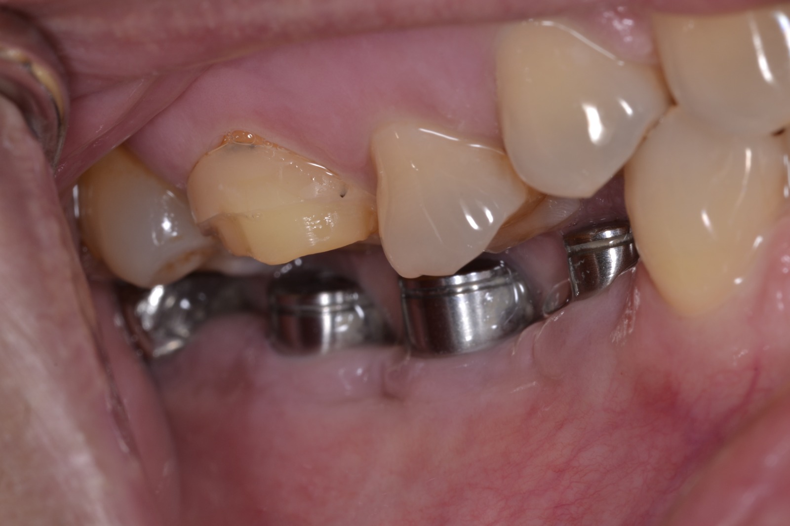
Subsequently, a flap was raised, the area debrided, and a reparative bone graft was placed and covered with a collagen membrane and sutured shut. One month later, the patient was seen for a post-op and appeared to be healing fine. One month later, the patient was seen for another post-op and had some exudate present. She was given a syringe and advised to irrigate the area with Peridex, but was not improving over the next month. In Sept/2014, since the implant was still about 60% anchored in bone and not mobile at all, it was decided to just trim the gingiva mildly and smooth off the threads that were exposed above the level of bone.

The patient was seen for follow up in Sept/2014 and again in Dec/2014 and again in March/2015. In all instances, the implant and the surrounding tissues appeared fine and asymptomatic. However, in June/2015, the patient returned complaining of exudate. She was placed on Doxycycline for 10 days and we reviewed oral hygiene, and stressed the need to irrigate the gingival pocket daily with Hydrogen Peroxide 3%. The patient was seen again in July. The implant is still solid with no mobility and has maintained 5 – 6 mm pockets. The gingiva appears pink and firm but still has some exudate present.

Any suggestions for this case?


10 Comments on Coronal Bone Loss on #7 Implant: Any Suggestions?

New comments are currently closed for this post.
CRS
7/29/2015
Let the patient know the implant is failing. I would remove and disenfect the bone and graft then place the implant. Tooth had internal resorption and was fractured, bacteria had a head start. These are not great immediate cases, I have been burned also with this type of case. The Nd- Yag seems to help kill the pigmented bacteria deep in the bone.
Nathan Vassiliades
7/30/2015
Looks like the implant is failing. I would be upfront with the patient. Bone graft the area and place another implant in 6 months.
doc mcsurgery
8/4/2015
Sadly, the entire implant site is/was compromised from the getgo. Anytime you deal with external or internal resorption, extensive and clearly evident on the preop xray, you are asking for trouble in placing an immediate implant--no matter the "bone graft". That said, this implant has been "failing under direct supervision" for over 24 months and should have been removed long ago. As for the implant being "solid with no mobility", it is better than even money (ie highly likely) that the implant can be "unscrewed" without any resistance whatsoever. As an aside, the defect which is now present to nearly the apex of the central will not be easy to graft.
dr d
8/4/2015
I agree with previous comments. Take implant out asap and graft site before you continue losing bone #8. Anticipate papillary loss #7-8.
EM
8/4/2015
From what can be seen in one of xrays crown appears not fully seated. This could well be the reason for initial bone loss and once rough implant surface was contaminated bone loss kept going. To me it's a classic peri-implantitis case. It's unlikely for fosamax to be the culprit here. You don't normally get cretal bone loss with bisphosphonates. Instead you get bone necrosis and sequestration. With regards to management be aware that in someone who takes bisphosphonates bone grafting is risky. Body cannot remodel it's own bone let alone dead bone graft particles.
andrew
8/5/2015
I haven't done many implants (about 70) and my only failure so far was in a tooth similar to this, external root resorption - and I use MIS implants. Highly likely due to the cells causing the external root resorption having something to do with bone loss here.
Dennis Flanagan DDS MSc
8/5/2015
Post extraction debridement is crucial to prevent this, although that is not a guarantee. E. faecalis is the usual culprit (my opinion). Is this pt a nail biter. These pts usually have E. faecalis as oral residents. Dennis Flanagan DDS MSc
Dean tanaka
8/5/2015
I agree with all the above. another guess is that the graft on the coronal third did not solidify and threads got exposed. Should be easy to reverse it out. At that time, feel for any dehiscence, or perfs. If the bone looks perfect i would put one in, but threads has to be completely in bone. I actually stopped using MiS/Nobel active type aggressive thread implants since if those threads ever get exposed, it's trouble. I like less threads on the coronal part. Since they say up to 2mm resorption is with in normal limits...
PeterFairbairn
8/6/2015
I agree with Dennis , anyway 1 in 70 about right statistically , so good going things happen . Not so called peri-implantitis , which is a more long term mucocitis with some associated bone loss due to poor host response to inflammation . This time scale is generally associated with granulation ingrowth and poor integration as Dennis alluded to . Peter
Saju Korothan
8/27/2017
Hi Doc In such cases try to place NucleOSS T6 implants, much superior, the design of implant for resisting unexpected coronal bone loss.

Featured Products

OsteoGen Bone Grafting Plug
Combines bone graft with a collagen plug to yield the easiest and most affordable way to clinically deliver bone graft for socket preservation.
CevOss Bovine Bone Graft
Make the switch to a better xenograft! High volume of interconnected pores promotes new bone. Substantially equivalent to BioOss and NuOss.