Delayed bone grafting of an extraction site: how to go about it?

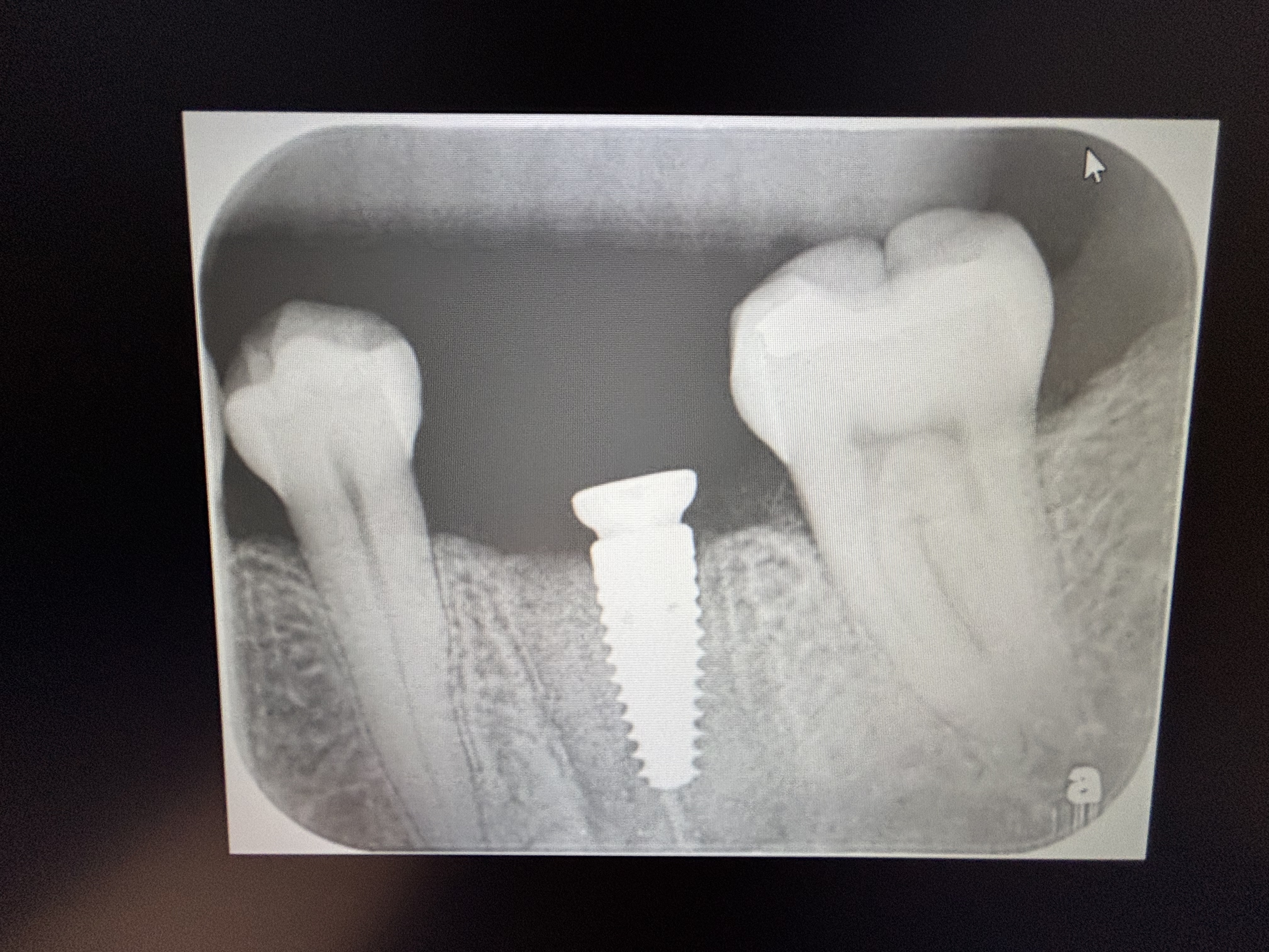
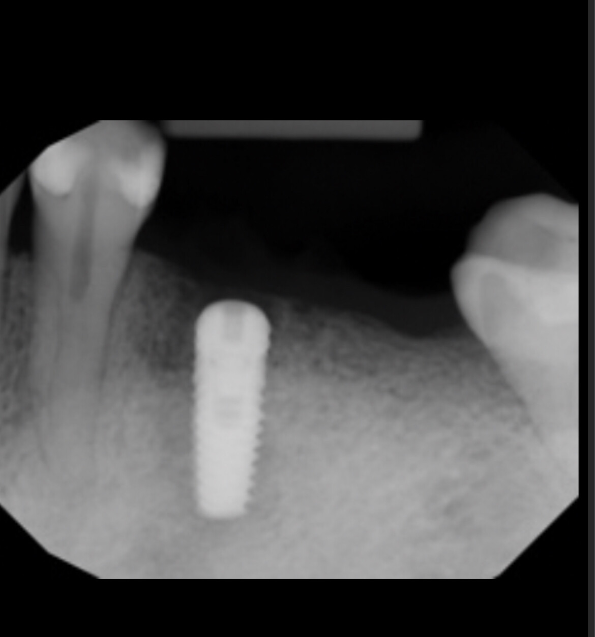
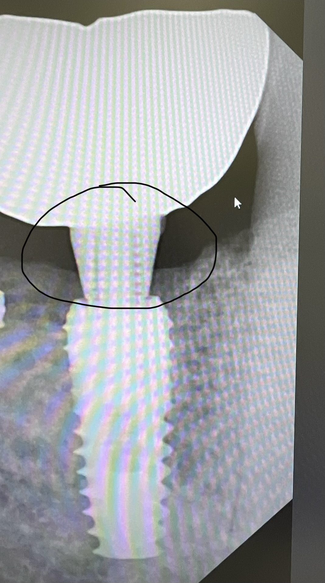
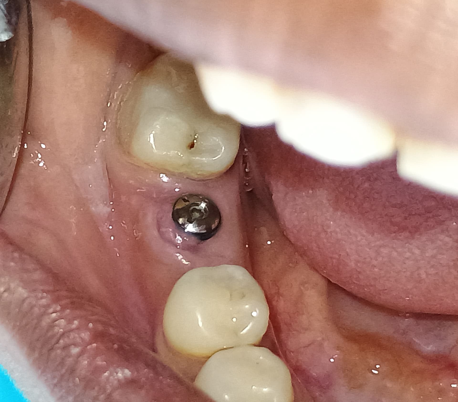
Recently I was presented with a mesial to distal fractured mandibular first molar that had bone loss in the furcation and enlarged periapical lesion. Suppurating fistulas were present buccal and lingual in the furcation region. Tooth was mobile. I surgically removed the fractured tooth by sectioning through the furcation, and was able to remove the tooth remnants in four pieces. I curretted out the granulation and inflammatory tissue as best I can without removing what was left of the interseptal bone. I decided not to bone graft at this point.

My question is on a subsequent visit, I would like to bone graft this site in preparation for a dental implant, but I am not sure exactly how to go about it. Do I flap, remove and discard the granulation tissue, decorticate, bone graft place a membrane and suture (wide area and doubt if I can get primary closure), or do I use the granulation tissue in some fashion? Or is there a different approach I should follow?

8 Comments on Delayed bone grafting of an extraction site: how to go about it?

New comments are currently closed for this post.
Peter Fairbairn
3/11/2014
This depends on your experience and skill set , But my routine for these case over the last 12 years or so is to allow for soft tissue healing of 3 weeks then carefully raise a site specific flap retaining the adjacent papillae. Then comprehensive curretage of the site is performed and you can keep some of the granulation tissue attached ( if possible ) flapping it lingually to use later for improved soft tissue healing . You can use CHX irrigation as well ( the eveidence is very contradictory as to effect on fibroblasts ) then create the osteotomy into the bifurcation residual bone . Done with care this will provide a reasonable primary stabilty for the Implant . Finally graft both the mesial and distal root socket sites with a fully bio-absorbable patriculate graft material ( preferrably bonded with CaSo4 ) . Suture closed ( if you have granulation tissue you replace over the graft material ) careully with Vicryl and leave to heal for 10 weeks . At that time load early checking with Osstell to benefit from another increased host bone metabolism as a result . Do generally 2 cases a week like this , works well to preserve not only the hard tissue dimansions but the attached gingiva as well . Peter
CRS
3/11/2014
I see this unfortunately on a regular basis and I have to retreat these cases. These cases usually have no buccal plate with bacteria present from the failed endo. The buccal plate needs to be regenerated with a barrier membrane usually non resorbable or long standing resorbable and the area disinfected with a 1064 laser to kill the buried pigmented bacteria. The flap needs to be primary. Not a case for a novice. What may happen is if the site preparation is not done optimally the implant will develop a Periimplantitis and you will either be continually grafting this or removing the implant within 3-5years. Regenerating the buccal plate is a good idea and perhaps a bridge may be an option. You are starting with several disadvantages to overcome from the failed tooth. If I could see these cases at the extraction phase often this can be avoided in experienced hands. So just remember my advice as this case progresses, see where it goes over the next couple of years.Often posters take offense at this advice but I unfortunately see this happening a lot, my advice is honest based on experience and judgement. Good luck. Also all the granulation tissue needs to be removed if it is not treated with the laser since it is contaminated along with the first few mm of surrounding bone.
Charlie Levy
3/11/2014
CRS is spot on! Listen to him/her and don't kid yourself. The bacteria will win every time.
CRS
3/11/2014
Thanks Charlie you get it!
Rob Smith
3/12/2014
This question has bugged me for some time. My usual protocol is to extract an abscessed tooth, curette and graft the area so that the graft appears dense radiographically. If there is a buccal dehiscence, I lay a flap and use a membrane. If I don't curette completely and leave a small amount of infected tissue, will those bacteria still be lingering 6 months later and will they infect my implant? How common is it for this to happen?
CRS
3/12/2014
Very and it is compounded when you drill the implant osteotomy threads. The implant may become problematic in the future. The key is using the right wavelength it can't be done with a diode and topical treatments don't get into the bone.
Cliff Leachman
3/19/2014
1064 nd/yag? Interesting, which company and what parameters? Certainly sexier than a bur, probably a lot cleaner?
Larry J. Meyer
6/11/2014
I have a similar case. I did an emergency atraumatic extraction yesterday on tooth # 13 and the patient has scheduled for an implant placement in 2 weeks. I didn’t have time to place the implant yesterday and he was swollen considerably from the infected tooth. He was already on antibiotics and there was no suppuration or apical lesion. I simply curetted the socket thoroughly and let the blood clot form with no additional treatment. The socket is a perfect 5 wall with total boney enclosure and intact buccal plate. I will have good soft tissue closure for the placement. My question has to do with the placement appointment. It seems to me that the socket will be closed and the internal will be in the early stages of healing. If I place the implant flapless using tissue punch and simply screwing the implant into the socket will it react the same as if it were an immediate placement? If I waited 3 months it would fill with bone,,,why not place as is? I thank you all in advance for your advice.

Featured Products

OsteoGen Bone Grafting Plug
Combines bone graft with a collagen plug to yield the easiest and most affordable way to clinically deliver bone graft for socket preservation.
CevOss Bovine Bone Graft
Make the switch to a better xenograft! High volume of interconnected pores promotes new bone. Substantially equivalent to BioOss and NuOss.