Draining Sinus Tract Case: Is This a Failed Implant?
Dr. R. asks,
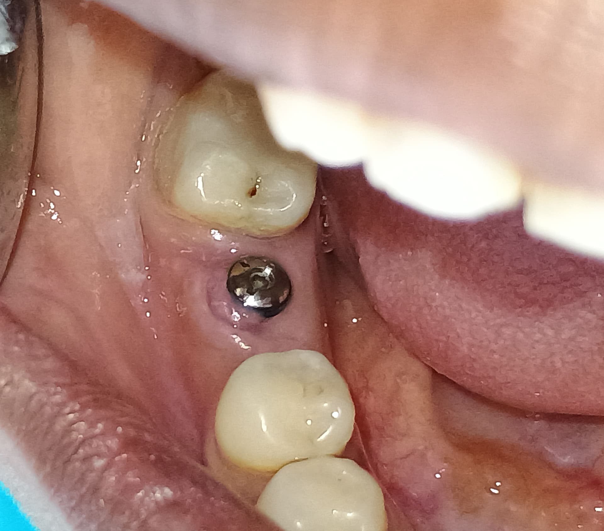
Please see the case photo below. I placed implant(#15) one month ago. Bone gaft was also added on the buccal crest with resorbable membrane. The stability was good. Three days later the wound was opened due to poor primary closure, but i tried to disinfect the wound with chlorexidine ,antibiotic Augmentin(625mg). The wound was responded well. One month post-op, the patient developed a draining sinus tract on the buccal. I drained the area and prescribed Rodogyl. Is this a failed case or is it too close to the natural apex of the neighbouring tooth? I am happy to hear any comments from you all.
25 Comments on Draining Sinus Tract Case: Is This a Failed Implant?
New comments are currently closed for this post.
sb oral surgeon
8/3/2010
multiple issues here:
1. endo / periapical pathology 2nd bicuspid
2. possible crestal infected bone graft
You have had too many issues with this fixture in too short a time.
My advice:
Take out implant, debride, graft.
Endo consult (a high res. CBCT will help evaluate the endodontic status of the bicuspid.)
Re-do implant surgery with confidence that the neighboring teeth are infection free. Wait for absolute resolution of symptoms and good healing (16-20 weeks)of site as well as bicuspid site if treatment is needed
Also remember, if your primary closure breaks down it means that it was under too much tension. If you are grafting over the buccal of an implant it means that some flap release was necessary (periosteal release.)
Did you release your flap?? If not you probably had too much wound tension.
Ron Neff
8/3/2010
An apico for the second bicuspid. Vitality test on first bicuspid, and indicated treatment if any. Clindamycin. Prognosis for implant fair to good, if it quiets great, if not then (after needed tx for the bicuspids) remove and reimplant after a sufficient time for bone healing around 8 months on the maxilla to be on the safe side.
Ron Neff
8/3/2010
An apico for the second bicuspid, probably retreat may have a second canal. Vitality test on first bicuspid, and indicated treatment if any. Clindamycin. Prognosis for implant fair to good, if it quiets great, if not then (after needed tx for the bicuspids) remove and reimplant after a sufficient time for bone healing around 8 months on the maxilla to be on the safe side.
sv
8/3/2010
RCT may be fine. need radiographs with diff angle to ck extra roots or canals.
Was this immediate implant placement after extraction?
what is location of sinus tract on baccal? where does it lead if you take radiograph with GP point?
If it is more occlusally probably it will be fine if you open it up and clean it. Also before you close the flap ck that the cover screw is tight and completely closed.
Best luck.
Dr. T
8/3/2010
I would take a couple of xrays at different angles of #13 to check for additional canals. If there are more canals, retreat and apico #13. The imlant is too close to the root of #13. You need more space between natural tooth and implant body (at least 2mm). I would recommend removing the implant, debride and graft the area. With the angulation of the implant,
Dr. R
8/3/2010
Dear Editor,certainly the sinus tract is occlusally on the implant site only not on the buccal site.
Ed Kusek DDS DABOI FAAID
8/3/2010
Take a CBCT this will show you exactly were the infection is coming from. Then you can use some of the other clinicians sugestions.
Dr.P
8/3/2010
All the advice has been good and hopefully useful. The only thing I would suggest is to get back to us with a follow up when you figure it all out. Very curious to see what we are dealing with here.
My only suggestion is to try and trace the fistula with a gutta percha point. See where it leads to. Try this before CBCT.
Good Luck!
t.v.narayan
8/4/2010
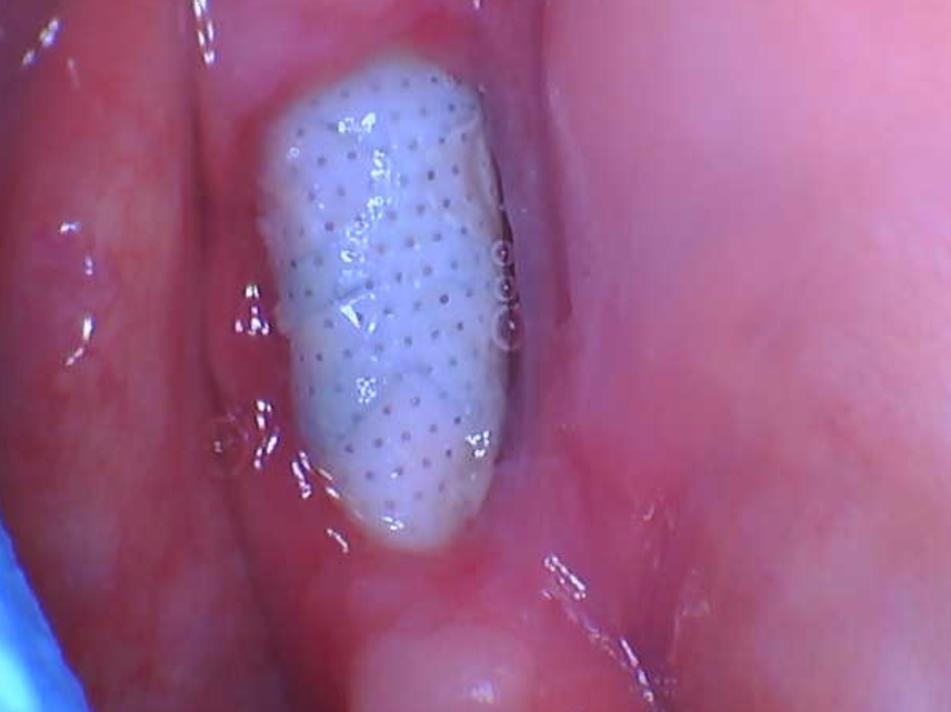
you are most likely dealing with what I term a "Cover screw abscess".could have arisen as a result of some food debris entering the incision due to incomplete closure.A simple remedy would be to expose the implant and place a healing abutment in place of the cover screw.Its very unlikely that its got anything to do with the neighboring tooth.
Dr.Vaziri
8/4/2010
Dr. R. Thanks for X-Ray and let you know this sign is a failed for your case,but I like to added something more. This happening is NOT belonge to poor endodontic 2nd bicuspid or other natural tooth because no sign of infection in neghbouring tooth.The most possible infected came from
- By the sergon
- Infected bone graft
- poor coverage of the sit or tension of the soft tissue with poor blood supply.
However, remove an implant, check expansion of the sinus on labial aspect, Autogenous bone graft the sit and after four mounts place an NEW implant.
Hopefully help.
Dr.Vaziri-from Iran
peter Fairbairn
8/4/2010
All great comments personally although the adjacent tooth could be an issue here I think it is not as there is often associated pain ( not mentioned ) and if draining from this peri-apical are could result in the loss of the implant.
Thus it is probably a spontaneous exposure of the cover screw as tv suggested maybe made worse by the incomplete closure as sb stated.
Then the graft material ( What type of material was it?xenograft?) possibly has become involved in the infection as Viziri stated.
tv suggestion of a healing cap is a good start and then monitor.
Good luck
Dr. R
8/4/2010
i personally agree with Dr naryan who termed it cover screw abcess due to food debris ,and i also agree with open up and place healing cap,but i totally disagree with Dr vaziri from Iran,the stability is good,no pain ,how can you judge it failed?how can you remove implant 11mm in legnth with good stability?
TOBooth BDS Hons MSc
8/4/2010
loose cover screw most likely or microabscess from augmentation material if occlusally positioned.
If the sinus is buccal gp point and pa, most likely an infected second canal. They always track to the buccal of adjacent implants due to path of least resistance. If so isolated second canal and treat.
Enjoy!!
Dr SenGupta
8/4/2010
Bearing in mind we have one x ray and no clinical picture....
Placement and bone approximation to Implant looks good to me.
GP point into sinus tract as far as it goes with gentle pressure
take 2 xrays at different angles remember MBD rule from Endo to confirm source.
very likley to be the endo or sepsis from the flap and graft
I would probably be inclined to clean away that graft ...does not really look necessary and its probably infected
Re enter the site and place healing head
Make sure you flush out the internal aspect of implant when cover screw is off.
i think this case will be fine based on info given
Salangsing DJ
8/5/2010
I agree with Dr.t.v.narayan, I think sinus tract dueto bone cover screw area ,it's call “Cover screw abscessâ€., not from bone graft.
Dr. Danesh from Iran
8/5/2010
Dear Dr. R ;
What to do step by step is;
1-check the adjacent bicuspid w/ percussion, if it's tender then CBCT can be helpful, and if not tender,
2-trace the fistula w/ a gutta percha point(30),to find the source ,if it stays at the cervical of the implant, then check the cover screw, if it is loose , open it and rinse the area thorouly, apply GAP SEAL and replace it w/ healing abutment and prescribe amoxicillin, metronidazole, and chlorehexidine.
3- if the cover screw is not loose, then the GBR is not successful, and should be explanted,careful curetage , and 8 weeks later a new implant ,GBR and periosteal releasing to close the flap(tension free), will solve the problem.
My primary dx. is cover screw abscess.
I hope it helps.
endosch
8/31/2010
Did you trace the sinus tract with a GP pt? Also looks like a missed canal possible.
dr.gurak
9/2/2010
I think your problem is based on the infected graft material. I would take out all the greft material, debride without touching the implant surface, re-graft the area and close the wound with some release incisions on the buccal periosteum. remember that horizontal matris sutures could be quite handy.
Sanglyong Yoon
9/10/2010
Why don't you take couple of X-rays with GP cone inserted or CT in order to rule out the apical infection. For me, the apex of bicuspids just looks fine and infected bone substitues could be the source of infection. Wait for couple of weeks with irrigation and anti. If the tract continues, remove the grafted material and try it again. I believe most implat doctor could differentiate the cover screw infection from the draining tract forming infection. Good lick.
Shirley A . Colby
9/13/2010
Do I see the shadow of a second root, in #13? This
I can not be certain. Occlusal radiolucency may indicate the presence of carious lesion and/or improper
seal. Obturated root shows over extension of gp, which may require re-treatment or surgical intervention.
As many had suggested, use an appropriately sized
gp and insert it into the sinus tract. A simple pa would lead you to the culprit. Using different angulations would certainly help.
Tooth #12 needs to be included in your evaluation.
The concerns with #14 may be independent, but given that it is newly installed and everything is not
established yet, it could be a case of contamination.
Definitely, other possibilities merit consideration as others had pointed out.
We can only speculate from our vantage point. You,
my dear doctor are in control: to investigate, evaluate
and arrive at a definitive diagnosis and treatment plan
Please, keep us posted...
vinga
9/13/2010
it most probably looks like a infected graft material to me....remove graft material, curettage, control infection with antibiotics...at a later date, repeat bone grafting under sterile conditions...implant doesnt look like a failure....removal of implant should b ur last option...if 2nd , missed canal of adj tooth is present then it should b taken care of.
Dr MILAN KUMAR
10/8/2010
well thanks for ur radiogrph. but u'd not shoot any clinical pic.,and u aint mention of the load type. so 1---as per radio .it's not an endo -perio case at all, nor a pathological involment from neibouring tooth.
2---u must have 2 radiographic shooting by G.P. 30 inside the draining fistula to acess the clinical condition properly.
3-- it's not tender at all as lysosomal activiteis is high on draing area, but a CBCT is a must.
4--now clinicaly there is relesing area on appoclosure from ur side , not the tissue tension rather other way round.
5-- so a marginal seeping of disoluted debris n alba causing infected
6--- graft in fection is ruled out as from radiograph stability is nice.
7---still its a clinicaly failed case, so do re grafting, wait for sufficent time of 6,7 month.
8---then do perform placment n load.
Dr. H.
10/16/2010
Simple remove the cover screw and replace a healing screw.
Dr.nader
11/11/2010
hi dear dr, in my opinion,first of all do root canal retreatment, there is no doubt that the second bicuspid has an extra root. more over, there is an obvious widening in PDL of this tooth as well.after that, look for any other complicated treatments.
Dr. M
11/13/2010
First of all isn't the implant in #14 area. As for treatment, I agree with those who say the rct on #13 is not the problem......now, but obviously confirm number of canals and treat appropriately. As for the implant, might you be able to repair the problem? Yes, but the most predictable path is to just remove it and re-graft. I think the periosteal releasing incision is poorly taught or just mentioned in passing. This technique is KEY to anyone placing implants. The easiest way for one tooth, like # 14, is to create two releasing incisions to the buccal, getting a bit wider at the base and leaving a bit of gingiva attached to the bone mesial of 15 and distal of 13. Now release your flap making sure it's full thickness, don't leave the periosteum. Once reflected, get a brand new blade, and while gently pulling flap "up" (incisally) and out make an incision below the MGJ on the periosteum. Hold blade about a 75 degree angle, so more parallel to flap than perpendicular. If your flap is nice and clean one should be able to see the periosteum splitting while beginning to feel the flap "give" or release. Make sure you go all the way across and but don't perforate the flap. Now see how much release you have gained. If your flap didn't give, then you're doing it wrong. If you got some release but need more, open the space between the periosteum and the flap some then place the closed end of a hemostat into the space created. Now open the hemostat creating a pouch between the periosteum and flap. This will give you more release. Getting good at this technique is extremely important, especially if you're going to do bigger grafting procedures or bigger cases in general. Also remember BLOOD SUPPLY is the key to it all. Design flaps with this in mind, any time you can design a flap with your initial incision toward the blood rich palate do so. But as far as your specific case goes, a repair might work, but if it doesn't work, the patient may begin to lose trust. Just be right up front and say, "You are not healing as expected, I could try to go in and save things, but the most predictable path is to clean things up and come back to place the implant later". Or if the patient wants to try to salvage things, at least he/she knows that you told them it wasn't the most predictable approach, so if it doesn't work out you are in a better position.