Gap Between Implant and Bone: Is this Just a Time Issue?
Dr. H. asks:
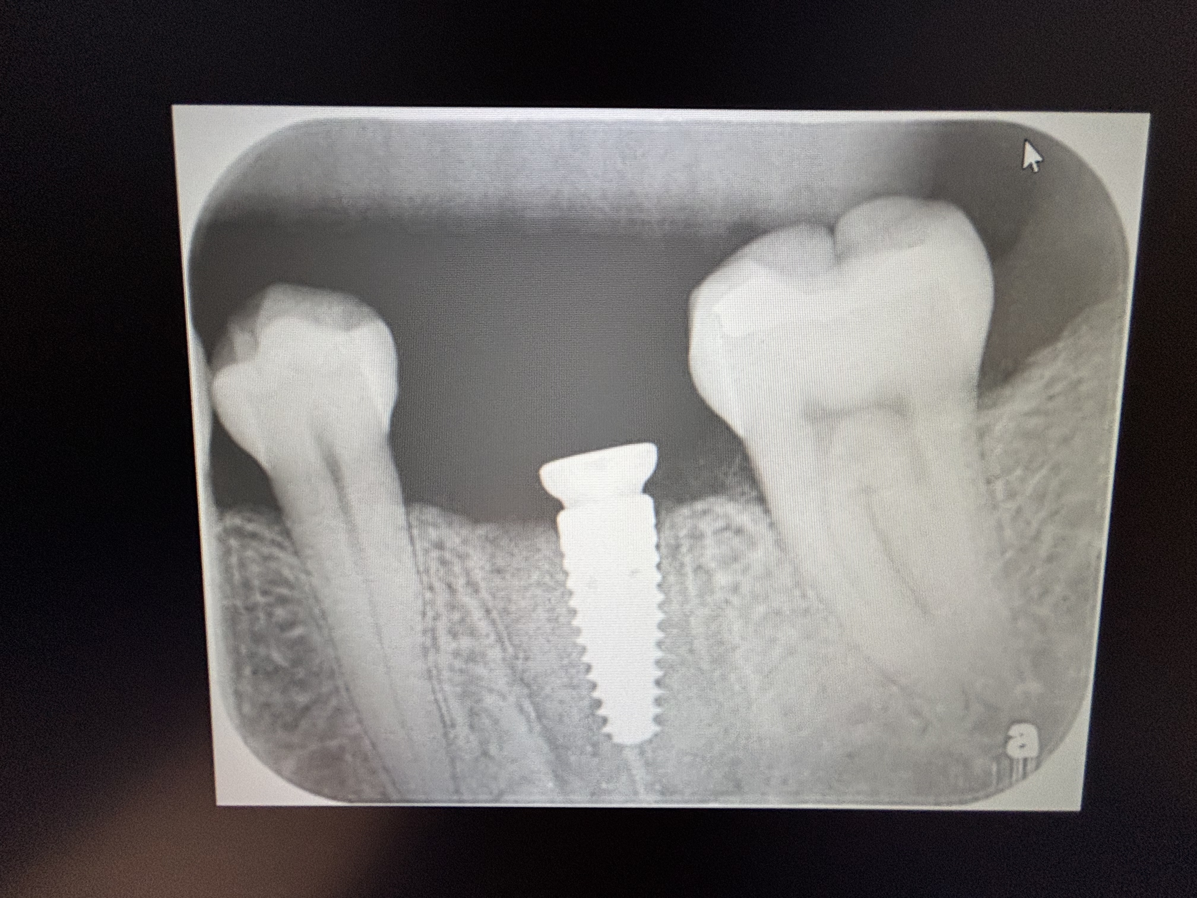
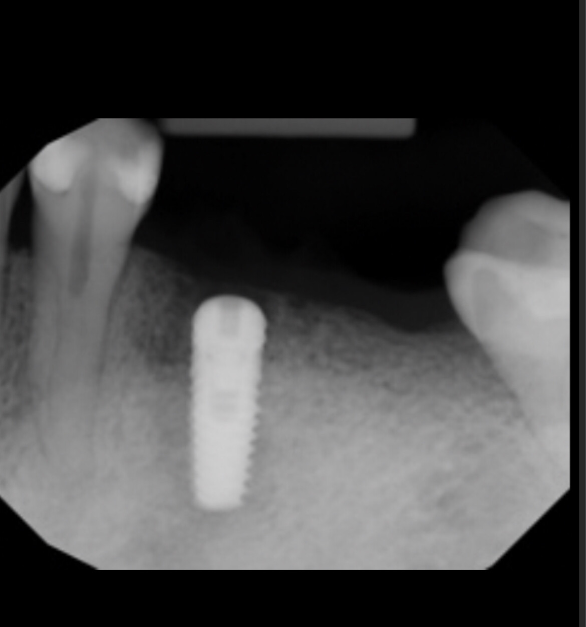
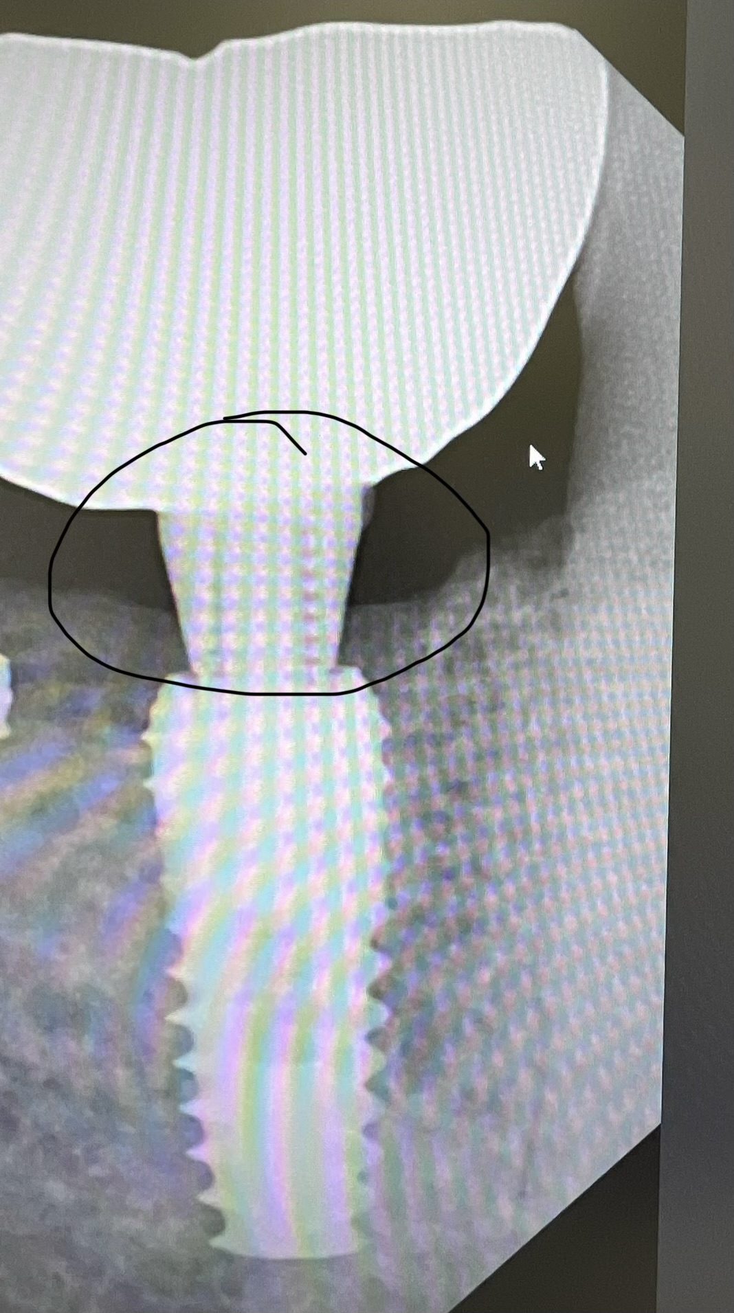
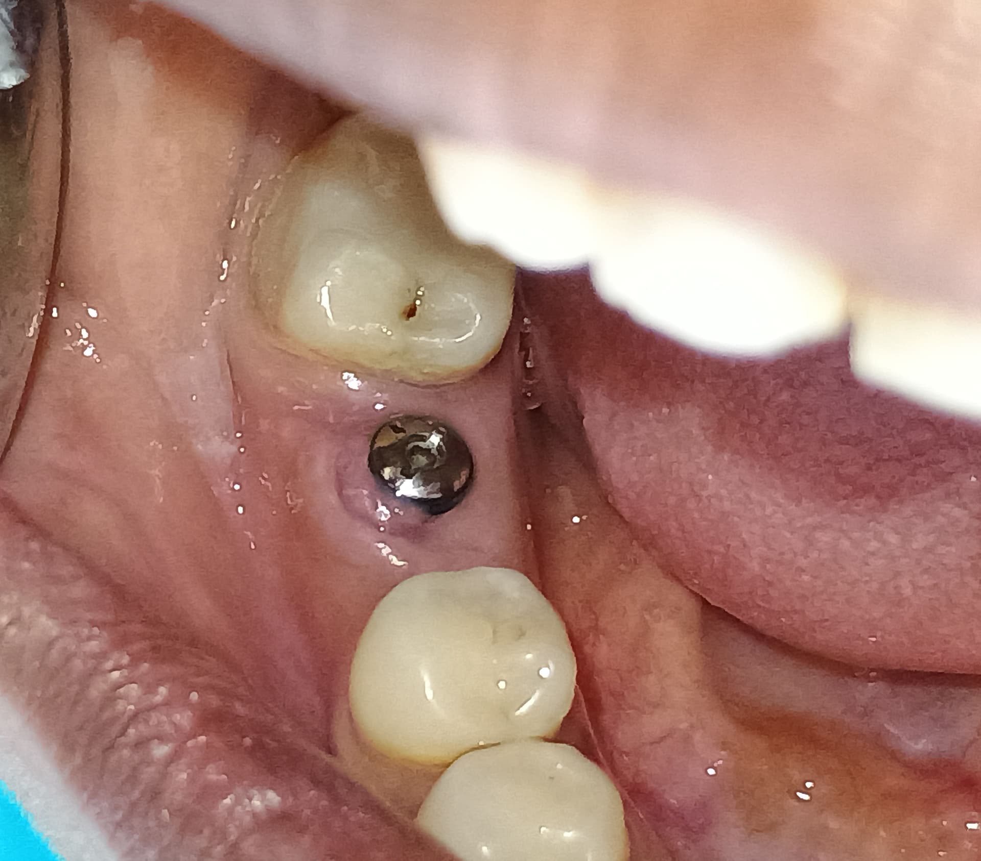
I placed an MIS implant fixture into the socket of #7 [maxillary right lateral incisor; 12] immediately after an uneventful extraction. I packed autologous bone from the bone trap around the implant. There was no pre-existing infection in the periapical area. At 5 months post-operative, I recalled the patient and laid a full thickness flap. Bone volume was adequate with normal dieback. I found that I could insert a scalpel blade between the implant fixture and the surrounding bone in the coronal 3mm. Do you think it is requiring more time for the bone to grow into contact with the implant and that over time the bone to implant contact will increase? Is this just a matter of time?
13 Comments on Gap Between Implant and Bone: Is this Just a Time Issue?
New comments are currently closed for this post.
peter fairbairn
3/7/2011
Dr.H , if it has not by now it will not , is there soft tissue ingrowth in the space? Best to de-granulate , clean implant (Prophy) and re-graft with a particulate. The bone chips may have washed out .
Peter
dental implants
3/9/2011
The Gap Between Implant and Bone is very hard to tolerate the pain. Thanks for advising about Gap Between Implant and Bone. Can You briefly explain me that by how we can make it the normal.
_____________
Paul
Dr H - the question poste
3/9/2011
Thanks for comments, I don't under stand to second post.
To Dr. Fairbairn, the gap is so small it could barely fit for example a bio-oss partible, but non the less it is a soft gap.
I could not clean the tissue out even with a micro-sickel etc, I could only access it with the end of a scalpel blade and tease some of the tissue out leaving strands behind etc. I don't have a implant tester machine....but can purchase on..should I just do a reverse torque test? Also do you need L.A for a reverse torque test?
Thanks
Andrew
peter fairbairn
3/9/2011
Hi Andrew , try reverse torque 15 Nm or so without local as if pain then you have an issue with duboius osseointegration.
If not you can use a soft tissue laser to remove the sof t tissue or the prophy can help as well then use a Caso4 or BTcp ( 100 micron particle size ) and pack in the area.
Peter
Dr H - the question poste
3/10/2011
Thanks Peter,
Sorry for the ignorance, perhaps different terminology in different parts of the world but, what do you mean by "prophy".
Thanks Again.
Andrew
Dr.Bülent Zeytinoğlu
3/10/2011
Dear Dr.H
Was the quality and quantity of bone enough for immediate implantation? Have you got initial fixation at the end of the operation?Researches show that especially at the coronal part if the gap between imp
lant surface and bone is three mm or less there was no need for filling the space with any material it will be filled by bone any way so I what you did was not very necessary.The thing I have diffuculty to understand is have you had controlled the place by an apıcal x ray before opening a full thickness flap.I thing you have disdurbed the healing tissue by the scalpel.If I were you I would wait for a few months more before loading the implant.Good luck.
mike ainsworth
3/12/2011
Hi Andrew, using a prophyjet or air abrasion unit (lightly!) will get the surface clean.
peter fairbairn
3/14/2011
Hi Mike thanks for that , just possibly the best way toclean the implant surface but beware of using pressured intruments on the adjacent bone hence protect as much as you can.
By the way I always peri-implant graft when there is a space even only 1 mm a ssoft tissue will ge there first.
Peter
Robert J. Miller
3/14/2011
A little disconnect here with regard to the clinical consequences of the "gap" in extraction/immediate placement. The object of placing a graft in the facial gap is to prevent epithelial cells from migrating into the space between the implant and facial plate. But there are two ways of doing this. Either graft with a bone substitute or allow a fibrin clot to form which is also cell occlusive. But the degree to which you get a good fibrin clot to directly related to the amount of bleeding observed following extraction and debridement. Our decision to graft a narrow defect at the facial plate is directly dependent on blood filling that space. If there is no bleeding subsequent to implant placement, mix a graft with venous blood or, even better, place a PRF membrane into the site. This will prevent soft tissue invagination and ensure ultimate bone regeneration with little resorption.
RJM
dream dds
3/15/2011
Hi Dr. H: this is my thought process when reading your post. first I would measure depth and width of the socket either by MD & BL dimesions of root or the socket itself. Then I would determine the implant width from this. The width should allow no pressure on the thin buccal plate ie: rule of thumb: lateral incisor replaced by 3.5mm wide implant and socket was 5mm wide. This leaves a 1.5mm gap. I would not go to a 4.0 wide in lateral unless unusually wide socket. A blood clot can "jump" about 2mm. Less "jump" is more predictable. Extrapolate this theory as to your socket width and implant width. In my experience, lateral uppers are the most predictable to fail due to too much pressure on the buccal plate. Always err to the palatal for placement. I like to use a "gel" bone matrix that will ooze out the socket as the implant goes in. Particulate graft can "close you out" at the apex and get a spinner.
I assume you got primary close with a cover screw and that is why you flapped open. My experience would be to make an incision over the implant and use a molt curette to push the tissue away buccal and lingual and then placed a healing abutment. This will keep attached gingiva and also create and/or maintain the papilla for good emergence and esthetics. Why did you full flap past the implant platform? I would not do this unless I had a reason such as inflammed, suppurative tissue. I would have allowed the cul to heal and then evaluate if the "gap" is a problem. It doesnt seem like it was a problem and any intervention could make it a problem. I like the idea of the PRF graft though. But why was this a problem, a scalpal blade width is what? I have had many anterior immediate placements that probably had a fibrous attachement at the body/platform interface, but the companies use this as a positive! Just my thoughts.
Sincerely
Leonard
dream dds
3/15/2011
Sorry I got off point there, to complete this: to check implant integration: you need to reverse torque up to 50ncm. If pain starts, then stop. Note at what level ie: 25ncm, 30,40. If there is pain, the implant is not integrated and from my experienc, at 5 months, it will not get any better. Remember the healing cascade: 3-7 days clot organization, osteoblasts and loose collogen formation. Woven bone matrix up to 6 weeks and then composite bone where lamellar bone starts to lay down. After 3-4 months, the formation has already happened , it is not going to change, if there is fibrous attachment at this point it will stay fibrous. If there is integration, it will get stronger as more lamellar bone is layed down. So, I would not do anything if the implant is integrated. If there is pain, take it out, do an implant "switch", this means remove fibrous membrane and either place larger implant if that seems wise, depends on bone width.....or bone graft and close and wait 4 months to place a new implant. Again, maxillary laterals that start to go bad can get very ugly with severe esthetic defects very possible. Implantology is a very complex area when things do not go ideally.
Sincerely
Leonard
Greg Steiner
3/22/2011
Has anyone shown histologically that bone will integrate to the implant surface in these gaps when not grafted? If so please post the reference so I can rethink my understanding of the biology of this healing wound. Greg Steiner
Pieter Linssen
4/6/2011
agreed good post. Citric acid bath for the implant x3 then get some bone from the pt pack into voids with an endo plugger real tight. It will be fine