Infection and Bone Loss around implant: best course of action?

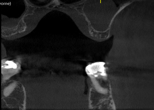
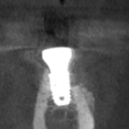
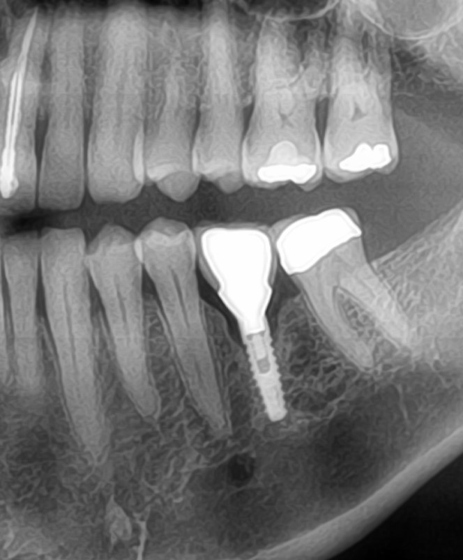
I installed 2 Nobel Biocare Replace Select implants in #30, 29 sites [madibular right first molar and second premolar; 46, 45; LR6, LR5] in a 58 year old male without medical complications. At the 2-month recall I noted some bone loss around #30 but the implant was osseointegrated, soft tissue was normal and it was asymptomatic. I restored the implants with splinted metal ceramic crowns. The patient returned for the 1-year follow up and I noted significant bone loss around the implant in #30 site. Palpation of the buccal cortical plate there yielded copious purulent discharge. The implant in #29 site is healthy. Can the implant in #30 site be salvaged? If so, how do I proceed? If I cannot salvage the implant in #30 site I plan to trephine this out. I am willing to remove and redo if that’s the best course of action, but would love to hear advice about the best protocol/strategy for dealing with this.


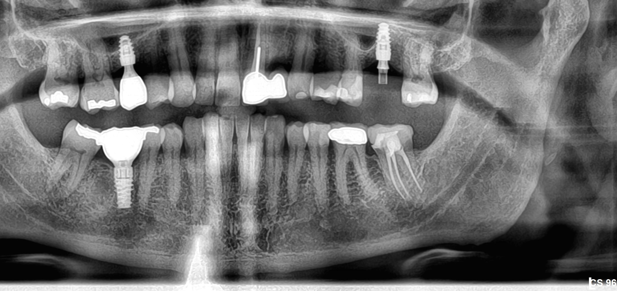
R side in 2012

Lower right today

29 Comments on Infection and Bone Loss around implant: best course of action?

New comments are currently closed for this post.
CRS
10/21/2013
Check the occlusion on the X-ray #30looks high and the trauma may have caused the loss. Section the crowns graft and replace #30 after it has had a chance to heal. Don't splint the new crowns. Thirty needs to go. Plunger cusp on #3?
Sb oms
10/21/2013
Are these screw retained? I think I see cement on the second x-ray. If that's the case, this could be the problem. Why did you splint these, just curious? You created a hygiene challenge when it wasn't necessary. Occlusion a possible big issue as well, but less likely with splinted crowns. Your boney defect is vertical and has walls. If it's in you're skill set, I would try and detoxify and graft. This one could be salvaged from what I see here.
CRS
10/22/2013
That's a lot of bone loss in just one year, copious pus is another red flag. I agree next time screw retained to be sure.
nick b
10/22/2013
thanks for input guys - appreciate the comments. Both crowns are screw-retained and splinted. Other than the possible hygiene challenge (although it doesn't look it from x-ray, he can easily use tepes between crowns) - is there any contra-indication to splinting?
Dr. Alex Zavyalov
10/22/2013
The occlusion is not the cause of problem, because the dentist noted some bone loss before prosthetic treatment. It’s a typical peri-implantitis, which requires a surgical approach with grafting and antibiotics. I would not separate the crowns. It will simply debilitate both implants.
CRS
10/22/2013
I just think it is a lot of bone loss in a short period more than just peri implantitis. The occlusion must be considered.
peter Fairbairn
10/22/2013
This is an great case to show why I try to advocate not splinting .... it can lead to co-axial forces on 1 implant and this can happen with associated bacterial host issues . Host physiology variation may be the primary issue in why some cases show more loss than others like every thing there are too many variables . I would clean then separate them and screw retain , then monitor . If you have experience in cleaning and grafting it can be done but is unreliable . Looking at the pre x-ray there was a small buccal issue mesially . Alex , Ti is very good at coping with Bio-mechanical forces and possibly best left to cope independently. Regards Peter
Simon Milbauer
10/22/2013
Peter great comment as always. It was fantastic to meet you in Dublin Simon Milbauer
Dime Sapundziev
10/22/2013
I agree with Peter the main problem is splinting the crowns. The other thing is is this a foreign body reaction? It might be but the main issue in this particulate case is inappropriate access for cleaning. My advice is remove the crowns, open flap, remove granulations around both implants, treat the implant surface ether with laser or chlorhexidine, do implantoplasty on the implant in #30 place gingiva formers and after 1 month make new separate crowns. Good luck. Dime
Jihad Joseph AKL
10/22/2013
I personally don't see any problem in connecting the two crowns as long as the fit is good. I would like to check whether there is any loosening in the screws and I wouldn't be surprised to see that the screw holding the crown on implant on site 29 is loose indicating a misfit. In addition, I have the feeling that there is an over contour on the posterior crown which can lead to inflammatory process, coupled with the occlusal factor caused by the misfit (would need to be confirmed obviously), all these would have contributed to the peri-implantitis. Course of action: check the fit by checking any loosening screw, and attempt bone regeneration with gd implant surface conditioning ( avoid the over contour). And good luck !!
Marty
10/22/2013
I have seen this before in the same sites, I think it is a Nobelbiocare issue. Personally I think that failure of the distal implant is inevitable, and the sooner it is removed the better for the chance of re-placing after healing. It has already lost half of the integration. I found it more common in the tapered groovy Nobel implants. I could post almost identical radiographs, with teeth unsplinted. I don't believe that splinting makes a button of difference. Hope my personal experience helps. Happy to post my unsplinted Radiographs showing failing (identical) implants. The radiographs were used by ADI(uk) when advertising recent "failing implants" day course. Marty
Eric Katch
10/22/2013
The most predictable approach is to remove #30, graft the site, and wait and replace the implant. In my practice, I have not noticed an increase in problems with splinting.
Dr Bob
10/22/2013
Could be a problem with some foreign material subgingival. I would advise, if my patient, that the implant will probably need to be removed, site grafted, and then the implant replaced. Then tell the patient that the implant might be salvaged and this can be evaluated when the surgical appointment for the removal is done. If the implant is stable after the crowns are removed, and if the defect I see after opening the flap is a well that provides walls with a hole to fill, and if the implant surface is such that it can be cleaned ( a lot of ifs) then a graft may work. I would not promise anything. Even if the site looks great after the graft I would caution my patient that the implant might be lost. I would not charge for the salvage procedure or for a new implant. I would involve the patient in desiding as to how to proceed.
Mahnaz Pears
10/22/2013
I would check the passivity of fit if they are splinted if there is a non passive fit the distal screw may have loosened causing the peri implant problem. If the fit is passive then you may consider removal of the bridge. Flap surgery to debride and remove infected threads. I think the implant will be still partially integrated so explanting would be destructive. Warn the patient of metal showing at the gingival margin but you need to be prepared to clean and remove as many if the threads as possible. You may wish to refer to a periodontist. Good luck
Richard Hughes, DDS, FAAI
10/23/2013
Good point.
Vipul G Shukla
10/22/2013
Dr. Nick, My advice is that both implants can be salvaged. The #30 site clearly shows circumferential bone loss due to bacterial by-products from the micro-gap that I am almost 100% sure exists because the abutment screw is loose. Why is it loose? Because the angle of both implants is such that when you tighten one abutment screw, it lifts the other one slightly, so even when you get 30 Ncm torque on #30, you are never going to get a tight seal between crown and implant platform, and all the bacteria in the screw channel eventually find a way to reach bone and start an inflammatory process that causes the bone loss you see now. Splinting is a good technique, if you actually need it, I don't see the need here at all. Splinting adjacent implants in the direct-crown-on-implant technique [UCLA method] requires an extremely good lab that will have extremely passive fit of the cylinders on both implants at the same time, a difficult task when you see that both are angled towards each other. Here is how I would handle this case: 1) Tell patient that you will try to salvage the #30 implant, and there is a 50/50 chance. 2) Start patient on Amoxicillin or Clindamycin at least a day before exploratory surgery. I would also add Metronidazole to the mix. 3) Under full aseptic technique, access both screw channels, remove composite and then the cotton plugs. Smell each cotton plug. If you smell a necrotic plug on the distal implant, don't be surprised, you have anaerobic bugs that exploded in an unsterile cotton plug and just found the micro-gap to reach your tissue and kept multiplying. 4) Cut away or have lab separate the two crowns such that you can screw back the good crown [use STERILIZED cotton plugs on screw head please]. 5) Raise a flap on both sides, visualize the defect, if you only have pus, then it is easier to handle that tenacious granulation that needs to be curetted till your back hurts and you expose fresh bone that bleeds well. You may have to use a round surgical bone bur for this. Make a solution with sterile saline with whatever antibiotic patient is on and flush the area really well. Some people advise to drill out the screw threads, but I feel that a rough surface like Ti-Unite does have advantages for osteoblast propagation. 6) Mix graft with patient's fresh blood or sterile saline and pack the area well. I suggest a graft like Dynablast paste from CITAGENIX (here in Toronto, Canada), which has bone chips in a matrix of growth factors. [osteogenic and osteoinductive potential] 7) Place a sterile healing abutment for the next 2-3 months. 8) Suture tightly around the graft with a sterile teflon suture. 9) Pray 10) CHX rinse for 4 weeks, remove sutures after 10 days. Check-up and X-rays every month, till you see good bone levels high up. 11) Make a fresh crown, and do not splint this time. Use X-ray to make sure that it was seated all the way down. Sterile cotton pellets over screw heads. 12) Have a cold beer and maybe email me if this works out.
nick b
10/24/2013
Wow - that's very comprehensive and clear advice - thank you. Questions - you suggested making a solution with sterile saline & antibiotic. How does that work please?- i.e how to go through the process & manage the concentration? I will prob have him on Amoxycillin/metronidazole tabs pre op. Also when preparing/cleaning the implant surface - what technique d you recommend? i.e hand scalers/curettes/ultrasonics etc (I don't have a laser) thanks!
Vipul G Shukla
10/25/2013
Hello again Dr. Nick, After the defect is visualised, curette well, this should take at least a good 10 minutes. Meanwhile in a sterile container, dissolve maybe a gram of the amoxicillin capsule content in say about 250 ml saline solution. I have no scientific basis for this, just helping to beef up local concentrations where it is needed most, especially since the patient is on it already. Remember that systemically administered antibiotics can only reach where the blood and plasma circulates, and this necrotic zone has neither, so when you make it bleed afresh and remove the persistent source of bacteria, the body wants to heal anyways, you are just providing the right scaffolding and a little boost with the graft. I have used a sterile endo irrigating syringe filled with this solution and direct a jet at the implant from all sides. Do it till you exhaust the solution you made. If you have a laser, it may help at this juncture, but just gently debriding the surface with a standard titanium scaler should be sufficient, since you mentioned that you don't have a laser. An ultrasonic is designed to loosen away tartar/calculus, here you will find neither (pus/infection is very acidic), so why add scratches when you don't need any. Remove the cause/source of the bacteria, and see how quickly the body heals itself. Good Luck!
Sam Jain
10/22/2013
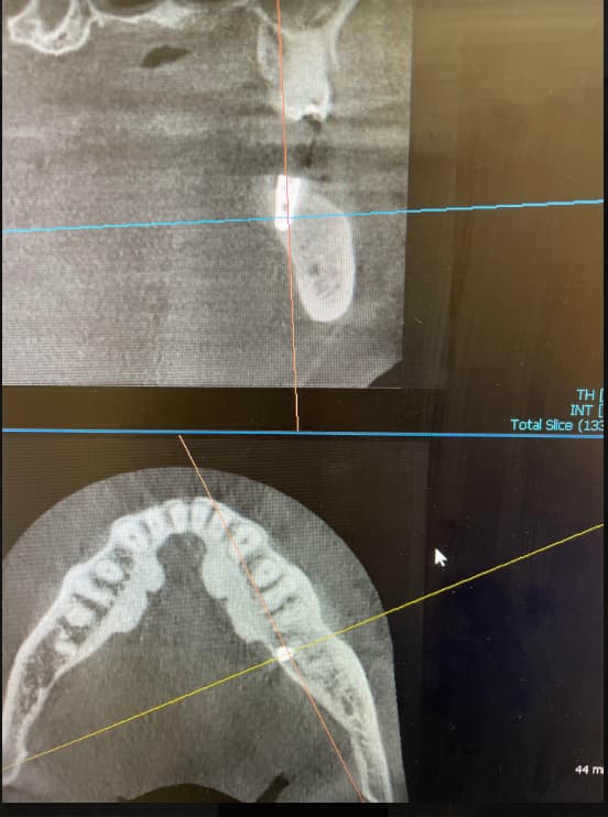
These are the things that don't have good fixes, unfortunately. Seeing a case come out like this turns your good day into a bad day. Any way, pl take a CT scan first, to be able to do a proper diagnosis. What u find will decide what to do next. That's where I would start from. May be there was thin bone left after placement of implant.
Rand
10/22/2013
I have nothing to say about splinting for or against. Occlusion should be evaluated and adjusted as needed. I have had much success over the years with flapping, decontaminating with tetracycline scrub. Bone loss has stopped in most cases. Now I use an Er:Yag laser to the same effect. Does not work always, but is non invasive and more invasive procedures can be done later if needed.
Dr ârra
10/22/2013
leave a least 30 microns of space, the direction of the implant to the antagonist tooth is important, tetracycline cleanse, laser, PRGF and gralth with membran is okay, but i would give the patient vitamins c,d3 ,magnesium, boron to improve bone formation and 5 different fruit 1 every 3 hr
Peter Fairbairn
10/23/2013
HI Simon and Dime great to see you in rainy Dublin . As with all these kind of cases they are multi-factorial and the host can be the biggest issue . Marty I agree with you but do not like to be critical of any product per se . I know you have had a similar issue but again there are vaiations as well as here it is on the lower arch where differnt issues come to play , such as the modelling which can lead to reduced bone on the buccal ( exposed threads ) and a more dense cortical plate. There is also the issue of attached gingiva in this area which can as you know cause issues. The issue with splinting is as I said a co-axial force placed on of the implants when the other is in function which may be one of the problems . These problems in the lower 6 area are one of the real challenges in Implant dentistry as this is the area where we most often see bone loss . I removed ( WITH Neobiotech ) a lower left 6 area Implant I placed 17 years ago a few weeks ago it had lost bone to the second last thread , but had been stable like that for 10 years . It required 400 NM of reverse torque to remove as the bone had remoddelled to becone very dense cortical bone . This is the highest torque I have had to use on an implant held in by only 2 threads ! Often when bone is lost at a point it loses no more for years and we feel this is due to stress micro management by the Ti ( flexing ) which is denied in splinting. Regards Peter
CRS
10/24/2013
The most obvious problem is the Periimplantitis which in this case is rapid. There is a 50% chance of success with surgical debridement, most methods work. I would advise removal since this is so early, in hindsite perhaps regrafting and waiting a bit prior to restoring. I recommend screw retention and no splinting with careful consideration of occlusion. Nobody really knows why implants fail but one needs to know how to manage th problems. Welcome to the world of surgical placement since you placed it now you have to fix it for free. I like a team approach to share the responsibility, good luck this stuff happens. You will be doing either two procedures debridement and or removal replacement, I would just replace it.
Richard Hughes, DDS, FAAI
10/24/2013
CRS, there are several reasons why implants fail. There have been books written that explain why they fail. I do agree that there are times when it is not clear why they fail. The influencing factors are bone physiology and it's factors that influence it, occlusal issues, infection, host immunology, host habits and life style, iatrogenic issues and poor implant design to name a few.
Vipul G Shukla
10/25/2013
Nicely explained Dr. Hughes. Osseointegration fails due to a complex interplay of clearly demonstrable individual factors. Today, we simply cannot say that we do not know why implants fail. We do know, and we are all learning every single day. I, personally, am grateful to all the experienced stalwarts in the field of implant dentistry that help each of us out on forums like these. Without prejudice. Thank You.
Peter Fairbairn
10/25/2013
Well said Richard and that sums it up.... Peter
CRS
11/7/2013
So can you tell me exactly why this particular implant failed? That is my point. There are multifactoral reasons and claiming to know exactly what happens is just speculation. Did not require the treatise on the factors which are obvious and replete in the literature. You missed the point in your learned discussion trying to educate me, sometimes doctors can hide behind their one upmanship. I like to refer to this blog as the "Implant Sports Page" since it is filled with opinion, speculation and arm chair quarterbacking. Occasionally there is useful imput for learning if we can get past the pride and pile ups. Ah human nature!
DrT
11/7/2013
I find opinions and even a bit of speculation very interesting and I often learn from this type of brainstorming. As long as we can put our egos aside, this can be a very educational forum...
Richard Hughes, DDS, FAAI
11/8/2013
CRS, I did not intent to offend you. Many things could cause this failure. We don't have first hand knowledge of the insertion or prosthetic phase or occlusion.

Featured Products

OsteoGen Bone Grafting Plug
Combines bone graft with a collagen plug to yield the easiest and most affordable way to clinically deliver bone graft for socket preservation.
CevOss Bovine Bone Graft
Make the switch to a better xenograft! High volume of interconnected pores promotes new bone. Substantially equivalent to BioOss and NuOss.