Is this more than periodontal disease?
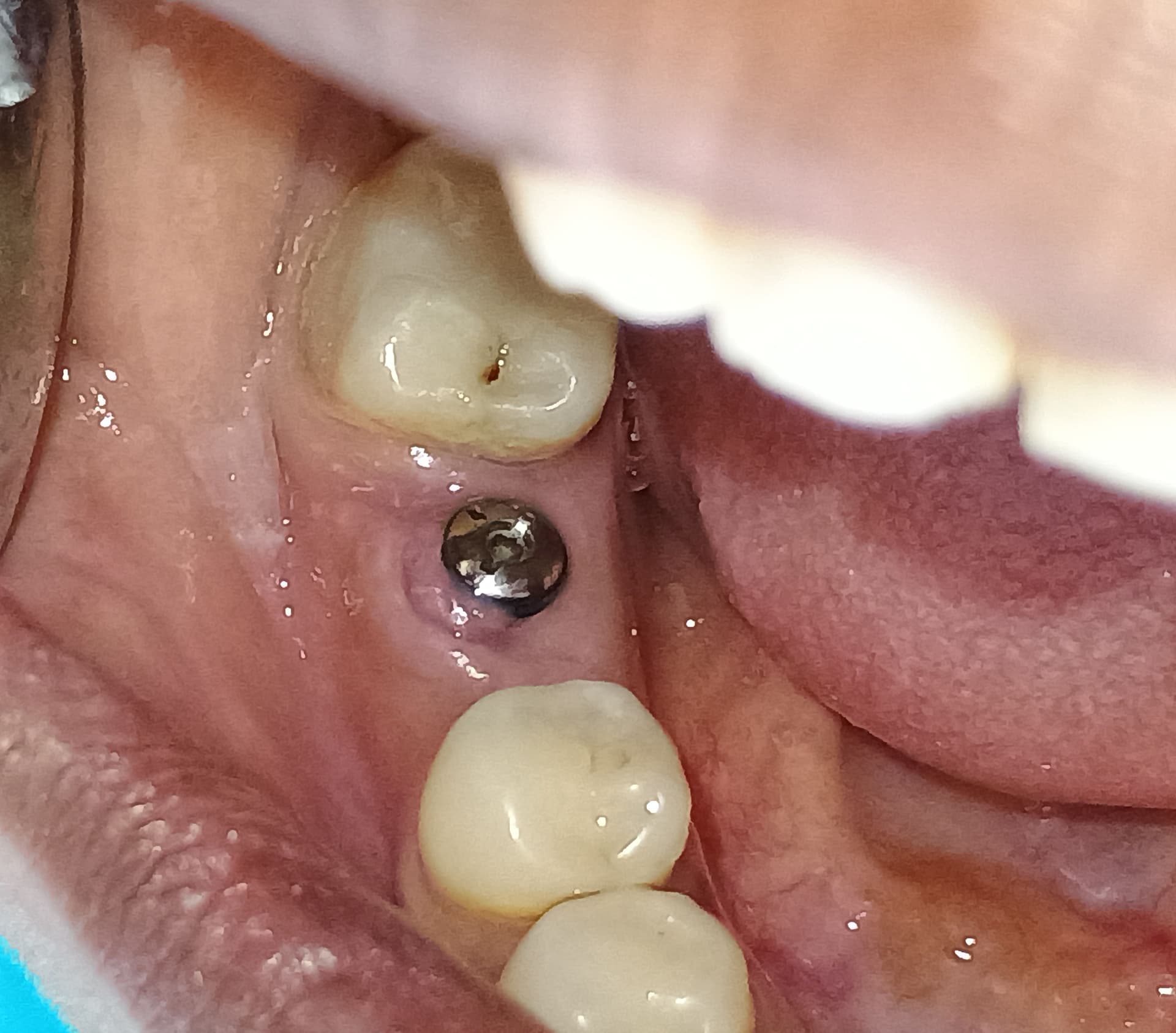
During a new patient exam of a female patient with asthma and arthritis, and no history of diabetes, I found peri-implantitis, general periodontal problems and an unsual bright redness to the gingival all around. Is there more going on here than periodontal disease? Is there more going on here than periodontal disease? Your thoughts would be welcome.
![]](https://osseonews.nyc3.cdn.digitaloceanspaces.com/wp-content/uploads/2016/01/case-6892-8ace232064ce63932f68c25c50aeb41d.jpg)
18 Comments on Is this more than periodontal disease?
New comments are currently closed for this post.
CRS
1/13/2016
Pocket depths? That is what perio pockets look like clinically sometimes with mouth breathing. See how patient responds to treatment. Perfect LANAP case all that granulation tissue responds well to that particular laser wavelength.
Justin
1/13/2016
Thinking out loud...
Probings, fmx, historical data if possible from previous office (probings, previous x-rays) would probably help. Periodontitis as a manifestation of a systemic disease? Certain autoimmune diseases and burning mouth syndrome are a few things that come to mind with this case. Magic mouth rinse in addition to periodontal therapy?
Thanks for posting!
Justin
DrT
1/19/2016
CRS: How can you recommend a treatment...LNAP..without making a proper diagnosis. Periio
pockets do not typically look like this, even in a mouth breather (which has not been established).
My "guess" is desquamative gingivitis related to hormonal etiology. I cannot see how
LNAP is going to manage this problem. Biopsy and more thorough medical work up
BEFORE any treatment, please
CRS
1/20/2016
I read the information in the post which perhaps you may have missed. This patient has significant systemic inflammation from here periodontal, arthritis and asthma. Start with what you know clear the oral inflammation, the Nd-Yad laser wavelength targets inflamed tissue very well. The desquamative portion will be be helped by controlling the inflammation. The periodontal inflammation and any systemic inflammation such as arthritis, heart disease, diabetes are linked. Her physician will be able to better manage the medical issues and the dentist will be able to control the dental issues. It is all linked. The medical diagnosis is made by working in tandem with the physician. Inflammation affects the entire body it is all linked. So hopefully now you understand how LANAP will help this problem by controlling the inflammation. There is probably some mouth breathing going on. That's my initial diagnosis and treatment. That's what I think is going on to answer the posters question. I have treated similar cases with good results. Giving the patient hormonal therapy or a mouthwash is ignoring the inflammation, treat the cause.
DrG
1/20/2016
I'd be concerned about your approach CRS. While temporarily the Laser might create a surface healing the underlying cause would still create an environment on a cellular level to make the signs of the patients autoimmune disease return.
Personally I'd try the following
1. Biopsy - r/o pemphigoid, pemphigus
2. Treat topically as needed lidex, dexamethsone rinse(in concert w/MD's tmt)
3. Treat Peri-implantitis lesions locally (LANAP) keeping in mind it's the combination of the stepped abutment and open contact causing the bone loss and food impaction in the #30 area.
The desquamative lesions can be from a variety of issues, everything from meds the patient takes to environmental local issues (toothpaste allergy etc). Going ahead with surgery seems a little risky from a medico-legal standpoint without a little background homework first.
CRS
1/21/2016
I don't feel that cleaning up the mouth is risky vs how long do you want to wait to treat this. You do know that periodontal disease is managed not cured. The bacteria load can be monitored with salivary testing and observing the tissue response, the important part is no bleeding on probing. Treating a patient with topical steroids is risky. If I still did not get a tissue response then biopsy. This clinically based on the photos does not appear to be autoimmune but coordination with the MD as I stated before is a good approach. Autoimmune disease is aggravated by periodontal inflammation I would start treating the perio. If the patient is placed on systemic steroids or immune suppressive meds it will complicate the picture since the immune system is altered so eliminate the dental factior and help the MD.Decrease the bacteria many ways including LANAP to do this as a teamed approach with the MD. If you don't have the right laser, start with oral hygiene, salivary baseline testing, supra gingival scaling to leave some attachment for the LANAP to work. It is about re-establishing the attachment to provide a healthy barrier to oral bacteremia. The systemic issues will be treated by the MD not the dentist putting Lidex on the area. Also figure out why the patient might be mouth breathing. Part of medicine is formulating a differential diagnosis and starting treatment informing the patient what you hope to achieve. Start with what you know and have a differential diagnosis.
DrG
1/19/2016
What did the periodontist you work with say?
Rand
1/20/2016
Possible lichen plannus or errosive desquamative gingivitis.
Gregori Kurtzman, DDS, MA
1/19/2016
I would have her physician do a blood work up to rule out systemic involvement. How often does she use her inhaler? that could be contributing to the redness marginally in the anterior and not connected to the bone loss issues.
The appearance of the gingiva doesnt look typical for perio. I would recommend full mouth probing and scale and root plane her. Use of a diode laser will help. Also consider Perio Protect trays to control the biofilm in the pockets. the perio tx will also help rule out systemic connection because if it responds to the perio tx then most likely no systemic connection if it doesnt then there might be one
CRS
1/20/2016
The diode just burns the tissue, treat the inflamed tissue with the optimal wavelength to get the pigmented bacteria. The perio protect is good for long term after LANAP. Look at the bone loss on the X-rays . Unfortunately scaling and root planing will just remove the tissue which could be disenfected and regenerated.
.
DrT
1/19/2016
I wouldn't jump into scaling and root planing and lasers until you have completed your medical work up. I do agree that her inhaler may be an etiologic factor. I recently looked at the Perio Protect trays and there is no strong
research by qualified clinicians to support any claims that they make
Gregori Kurtzman, DDS, MA
1/19/2016
there is research that supports Perio Protects claims on perioxide gel in biofilm elimination and maintenance. having just completed an article on peroxide use in perio I was able to find 35 lit references to support its use. How do you define "qualified clinicians?"
DrT
1/19/2016
I checked the references on the site...all are too non-specific for me....IMHO...to recommend this
to any of my patients. If you are satisfied by their claims then by all means use it
on your patients.
Dr. Harry Klapper, DDS
1/19/2016
The displayed x-rays do not show the anterior teeth. The posterior x-rays do show evidence of bone loss .First of all , I would explain to the patient how un-natural and un-healthy the gums look and that some bone loss has occurred.
Make sure that she has a thorough work-up and blood test from her physician to help rule out any systemic causes. Have her brush her teeth , at least twice daily, with the combination of baking soda and hydrogen peroxide to make a paste and I would want to see her again in two or three weeks with the results from her doctor and from the brushing and then evaluate the situation.
dr fred gustave
1/19/2016
I agree with JUSTIN AND DR T that this appears to be an autoimmune condition and a diagnosis is needed before initiating any treatment. My first reaction to the photograph was that we are looking at an advanced case of Errosive Lichen Planus. I would recommend a tissue biopsy and if it confirms LP then a referral to her Rheumatologist for treatment.
Rand
1/20/2016
The patient is a mouth breather, which could contribute to her problem. I have referred her to the oral medicine department at the nearest dental school.
Mark Bornfeld DDS
1/19/2016
This displays the non-specific appearance for which in the past the blanket term "desquamative gingivitis" was applied. There are a number of different pathologic entities that present in this way-- mostly falling within a range of autoimmune disorders (e.g., Crohn's, erosive lichen planus, mucous membrane pemphigoid, lupus). These may be identified by a range of blood tests (ANA, rheumatoid factor), as well as routine and direct immunofluorescence histology. Occasionally, more rare birds, like eosinophilic gingivitis, also look like this.
Patient should be referred to oral pathologist/oral medicine specialist.
CRS
1/20/2016
Treating the periodontal disease in tandem with the physician treating the medical issues is a win-win. The bacteria fuels the fire of systemic or autoimmune inflammation. The mouth matters and this inflammation allows oral bacteria to seed the bloodstream and promote any systemic inflammation. This is very simple and what dentists know best. Sure do a biopsy if that makes you comfortable it most likely come back with inflammatory cells. I'm not sure what this has to do with dental implants but happy to give the info.


