Large Nutrient Canal: How to Manage?
Dr. A. asks:
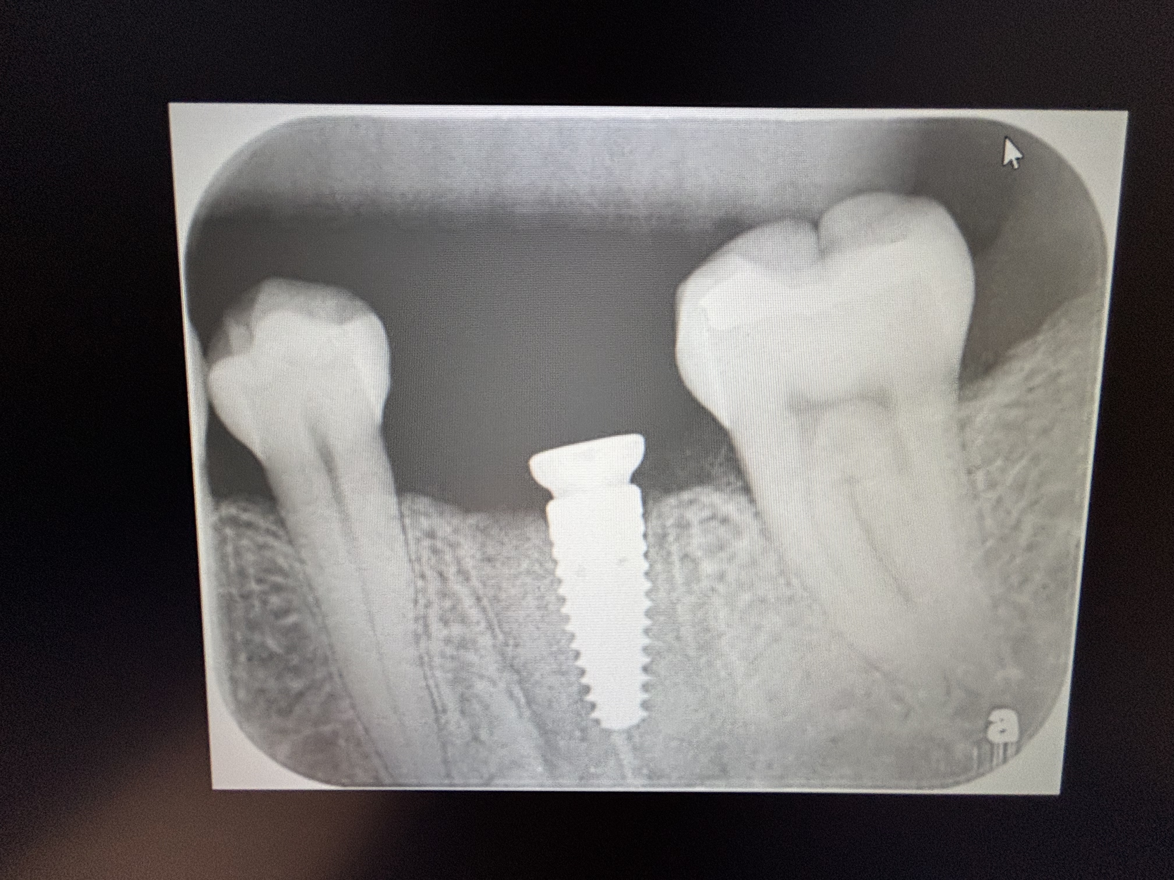
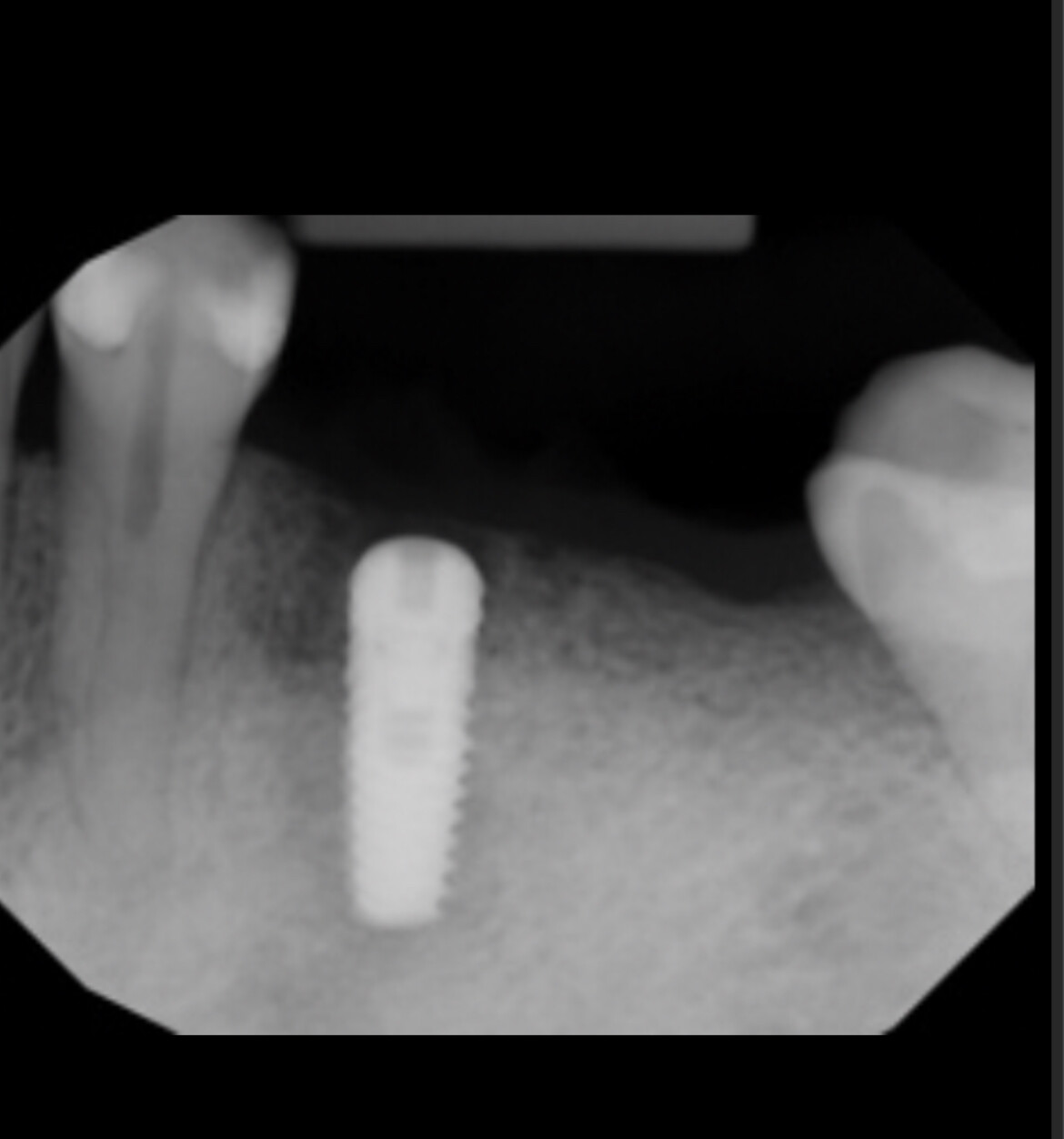
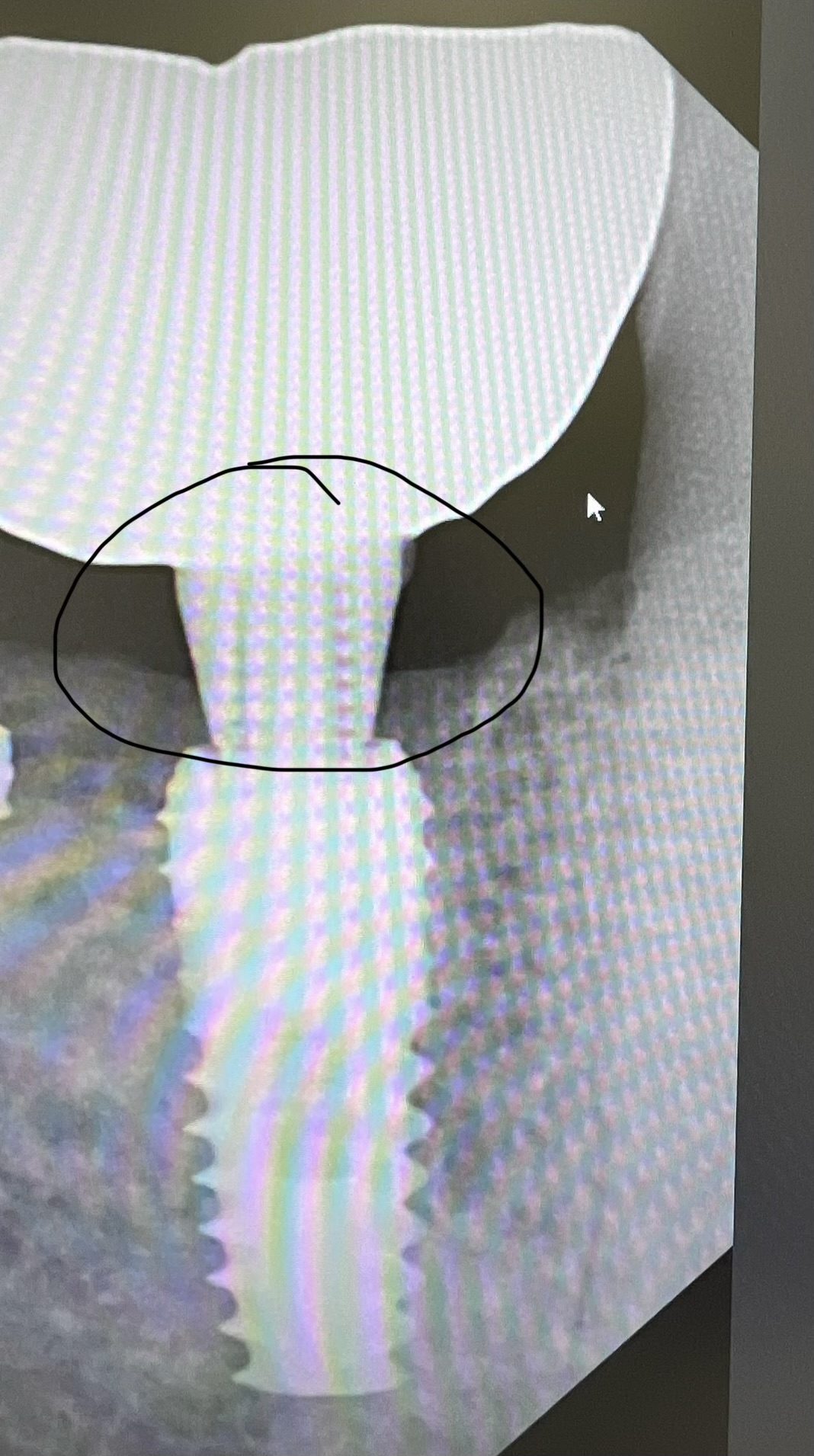
I have treatment planned a patient for extraction of #27-22 and for immediate placement of dental implants to support a fixed partial denture. On the panoramic radiograph I see a large nutrient canal subjacent to the area where I will be placing the implants. If I penetrate this canal with a drill or implant, how severe can the bleeding be? What special precautions should I take? If I get significant hemorrhaging, how do I manage this?
10 Comments on Large Nutrient Canal: How to Manage?
New comments are currently closed for this post.
satish joshi
1/28/2008
Yes there is a very good chance for excessive bleeding from osteotomy.Best way to stop profusely bleeding osteotomy is to place implant right away in osteotomy instead of trying to stop bleeding by other means, in my opinion.a CT scan should be done in this case.
P
1/28/2008
I never thought about using implant to stop hemorrhage.Does it work?
SeaMentum
1/29/2008
My advise is to avoid the area as much as possible. There have been reports about serious complications regarding this anatomical variance and placing an implant may/may not stop the hemorrhage. I agree that a ct is required in this case, which can save you a ton of headaches by helping you avoid the complication.
As far as bleeding, expect much more bleeding than you are used to. Furthermore, make sure you monitor this patient longer than you normally do before discharge. Sometimes the pooling of blood may cause the floor of the mouth to rise, causing difficulty breathing.
This is not a contraindication for treatment, just be prepared.
mike stanley
1/29/2008
(lokingly) One way or another, the bleeding always stops...
I have noticed Dr. Joshi giving consistently well-thought-out advise and I would definately get a CT with someone who can help visualize the nerves & major vessels. You may be able to completely miss them with careful angulation or by using a placement guide.
satish joshi
1/29/2008
Dr.Stanley,
Thanks for your kind words.
Satish Joshi
Larry Duffy
1/30/2008
I think either a ct scan or a new tom volumeteric scan is necessary...especially if the canal is large enough to see on a pano...after the scan you will determine if it is mid-mandible or buccal or lingual and place accordingly...if it is mid mandible I would consider shorter implants in this area to avoid the canal and maybe in that case place two more implants in the mid-symphysis area
Robert J. Miller
2/2/2008
The concept of "nutrient canals" is a means to explain away anatomy that is a variant of normal. Using 12- and 16-bit CBCT technology, we have been able to precisely dissect the intraforaminal area and interpret our findings. At the cuspid positions, in 80% of the scans that we perform, we find secondary anterior foramena known as the superior genial foramen. It is a neurovascular branch of the inferior alveolar nerve anstamosing with the incisive canal and innervates the chin. Surgeons performing symphysis block grafts often find that there is numbness in the chin post-operatively. This is probably caused by inadvertant resection of this branch. While there will not be parasthesia of the lip, patients often complain about this altered sensation in the chin area. If the incisive canal is also resected during block harvesting, you will also deinnervate the anterior teeth. Patients will describe a "wooden sensation" in these teeth post-operatively. In addition, the lingual, submental, and mylohyoid arteries are in this region and should be avoided to prevent potentially severe bleeding episodes. CBCT should be utilized before implant procedures in the intraforaminal area to prevent these untoward consequences.
Dr SDJ
3/30/2008
How come these nutrient canals come up after an extraction in an area where a tooth previously existed? Shouldn't nutrient canals be in bone where there never was a tooth? My humble queries.
R. Hughes
9/12/2008
All good point to consider, this is an interesting and revelant topic.
famouri DDS
7/7/2009
You must not trust to panoramic view for anterior regions because of ghost image!!!.….also you must role out hemangioma and A.V.malformation which are life threatening for example with fine needle aspiration.
Hope thats helpful