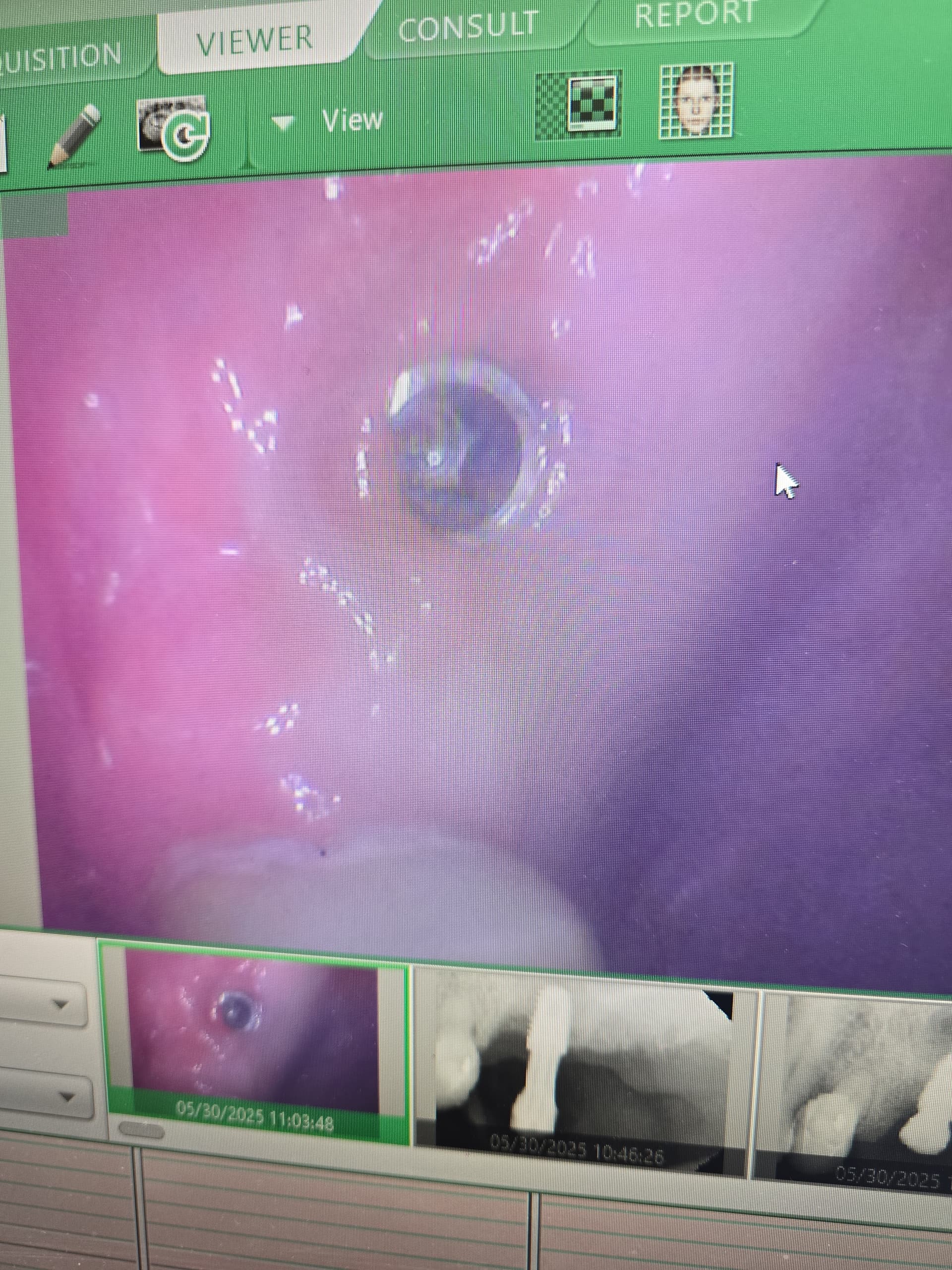
Membrane Dislodged: Recommendations?
Anon. Asks:
I extracted tooth #19. I removed any potential granulomatous or infected tissue. The extraction site looked clean. I performed site preservation at site #19 with BioOss and Collatape coverage and a few days later the Collatape membrane became dislodged, exposing the graft site. Is there anything I can do to save the grafted bone? Should I go back in and remove the graft now? What would you recommend?
As noted below, Periacryl high viscosity is an excellent solution to prevent membrane dislodgement. By covering a resorbable membrane with PeriAcryl, you can stabilize and preserve the membrane and protect the graft by using it as a barrier.
13 Comments on Membrane Dislodged: Recommendations?
New comments are currently closed for this post.
Neda-Moslemi
12/2/2008
I do not beleive in preserving the extraction site in regular cases. It will delay healing rather than accelerate it!
Membranes placement in extraction sockets will result in membrane exposure in more than 50% of cases. The high rate of membrane exposure in such cases is because of the presence of sulcular epithelium in the area. It will prevent flap margins to be approached and dehiscence will occur.
In this case, if the exposure is extensive, remove the membrane to prevent soft tissue loss. If it is minor, daily mouthrinse irrigation may be helpful.
Neda Moslemi
doctorberg
12/2/2008
actually the collatape is not a membrane for grafting, so remove it and yes peroxide solution mouth washes will help.next time use some resorbable collagen membrane or similar and tack it so it wont be dislodged.
Yes this kind of graft has been suggested by some studies to delay the actual healing process
Joseph Kim, DDS
12/2/2008
The BioOss will probably not integrate well due to the use of the collatape, which is not a true membrane. I've found that bovine is great for sockets that have thin buccal plates, but you cannot place an implant for 6 months or else you will find a sea of bovine particles. Another technique that works great for sockets that have a little more thickness to the buccal plate is using a teflon membrane and inserting it into a small lingual and buccal flap, secured with a nylon suture. With this technique, you plan on leaving it exposed, and remove it one month later. By then, there has been enough differentiation to prevent soft tissue invasion of the socket.
John Clark
12/3/2008
As taught to me by Dr John Giblin from Australia, after placing your grafting medium (he tends to use bioss-collegen) the outer part of the graft is covered by a block of Spongostan® (a gelatine based absorbable haemostat- google it) which has been squashed flat and then cut to fit the socket. The spongostan is then fixed in place by a web of 5.0 vycryl, sponged dry with a clean gauze and then immediately wetted with some PeriAcryl™ (cyanoacrylate tissue adhesive) by using a microbrush. Make sure you don't get it anywhere else!!, wait a minute or two and you will see that the graft is nicely sealed with minimal bleeding if at all. Generally, this cover hangs in for 2-3 weeks with keratinised gingiva growing in from the margins underneath.
Of interest I have tended to move away from Bioss, Bioss-Collegen mainly because in Australia once used the patient can no longer be a blood donor and also some patients object to its use. Nowadays, Itend to use synthetics such as cerasorb or strauman bone ceramic ideally mixed 50:50 with some harvested bone chips or when doing immediate implants, with the trapped coagulum obtained from the osteotomy..
Of perhaps even more interest is the use of DR Giblins technique in addressing Oral Antral Communications. I'm trained in fixing these surgically, however, circumstances arose where a referred patient with a large (7 x 5mm) OAC could not be treated with the normal buccal flap due to a large gash through the buccal sulcus area, right where I would have been releasing the periosteum!!!. Anyway, I decided to try a modified 'Giblin" where after suturing the buccal gash with some 5.0, I then sutured in a 'Bio-Guide membrane 'hammock' over and in close contact with the OAC, then place a wedge of Bioss-collegen to entirely fill the socket, then the spongostan and periacryl as described earlier. I then took an alginate impression with a little bit of cling film over the grafted site with which I then immediately fabricated an acrylic obturator worn 24/7 for 2 weeks. Antibiotics therapy was Augmentin duo forte (Amox875/Pot Clav125).
Remembering that this was a huge OAC, the result was perfect - full coverage of the ridge with keratinised gingiva, no dropping of the sinus floor and no loss of the Buccal sulcus. Definiely ok for an implant down the track. With the 'solid form' Bioss-collegen cut as a wedge, I probably could have got away without the membrane, however mainly placed it as a barrier against any epithelial intrusion into the graft from the sinus- would definitely need it to contain a particulate graft though.
John Clark
12/3/2008
I forgot to mention.. in answer to your question... the spongostan/periacryl 'capping' done now may not work quite as well as when used in the first place but it should stop or drastically minimize any further loss of your bioss graft.
John
John Clark
12/3/2008
One other thing I forgot!!, the obturator was taken out at meal time, I actually used flexible mouthguard type material not hard acrylic and a CHX mothwash was used 2 x day for 7 days, commencing the following day.
Peter Fairbairn
12/3/2008
Chlorhexidine mouthwash before surgery but may not be such a good idea post-op.
alexgurin
12/3/2008
to John Clark:
You said that in Australia once used a xenograft the patient can no longer be a blood donor.
please, can You give any reference to that?
John Clark
12/3/2008
The Australian Red Cross administers the blood bank here in oz and my information came directly over the phone from one of the chief administrators here in Brisbane. Nonetheless, I'll try and track down the document he referenced and post the gist of it. Also re the CHX mouth wash, I also tend to not use it immediately after implants preferring to use saltwater for two days before then going onto the CHX for 5. Completely a gut feeling but it seems to work ok. However, with socket preservation and with OAC closures, I use the CHX straight up the next day, figuring that I have a nice little seal over the socket and I'm more concerned about bugs around the surface of the seal and on the obturator than I am on possible effects on fibroblasts. Again, no scientific studies here but so far so good.
anon
12/4/2008
Use a resorbable membrane that can be left exposed without rapidly melting away (Epi-Guide). As it was mentioned earlier, some 50% of these cases will expose the membrane anyways. Synthetic bone has performed better then bovine bone in my hands but make sure it does not have Hydroxylapatite in it as this material does not have a solid track record. Good luck.
Larry J. Meyer DDS
12/6/2008
To John Clark in regard to the oral antral communication. I am somewhat confused about the sequencing you described. You used a collagen membrane as a hammock. Does that mean that it covers the graft, or is it draped into the defect to contain the graft material and keep it from penetrating the sinus? If that is the case, is there direct bone- graft contact? What is the healing sequence?
Last week I extracted an upper second bicuspid, fractured endo-post tooth lying along the sinus floor. It came out in three pieces with the schneiderian membrane attached to the root tips. The defect was 3x4mm. I placed an RTM collagen membrane over the defect, tucking it under the palatal and buccal flaps and sutured with Cytoplast sutures. I wonder what will happen with the bone underlying the membrance and how it will heal.
I am very glad to have seen this post so as to get a discussion going on this subject.
Larry J. Meyer DDS
I can submit the preop x-ray if needed.
Russell
12/30/2008
I have no suggestions to your present situation, but in the future, you might want to try using a membrane that is designed to be exposed to the oral enviornment, eliminating the need for primary closure. Clinicians have been using gortex type membranes exposed for one month, enough time to establish a new blood supply.
Other clinicians have used things such as a cellular dermal matrix, facia lata and pericardium also, intentially leaving them exposed.
Good luck to you and best wishes.
David Arango
2/25/2009
The same thing happened to me a few months back due to a brush stroke from the patient, I just removed the sutures and the remaining collagen tape, applied some CHX gel and re-sealed the socket with Collacote, which is thicker, then sutured it with 5.0 vicryl. Healed uneventfully and the implant is rolling. Some things that work for me:1.Pack the graft really tight. 2. double layer of colla cote or colla plug(cut it in half)it´s thicker. 3. Upper anteriors: cover the graft with a saddled CT Graft, tuck the graft under a little buccal and palatal flap, this gives you more soft tissue for aesthetics.
Best of luck


