Pain after dental implant placement in mandibular posterior region: recommendations?
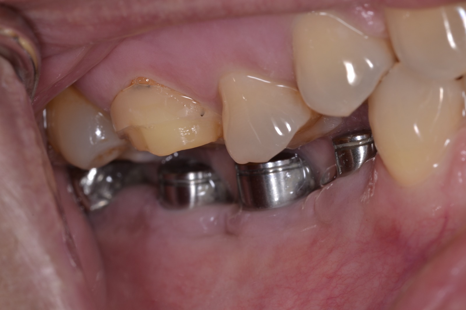
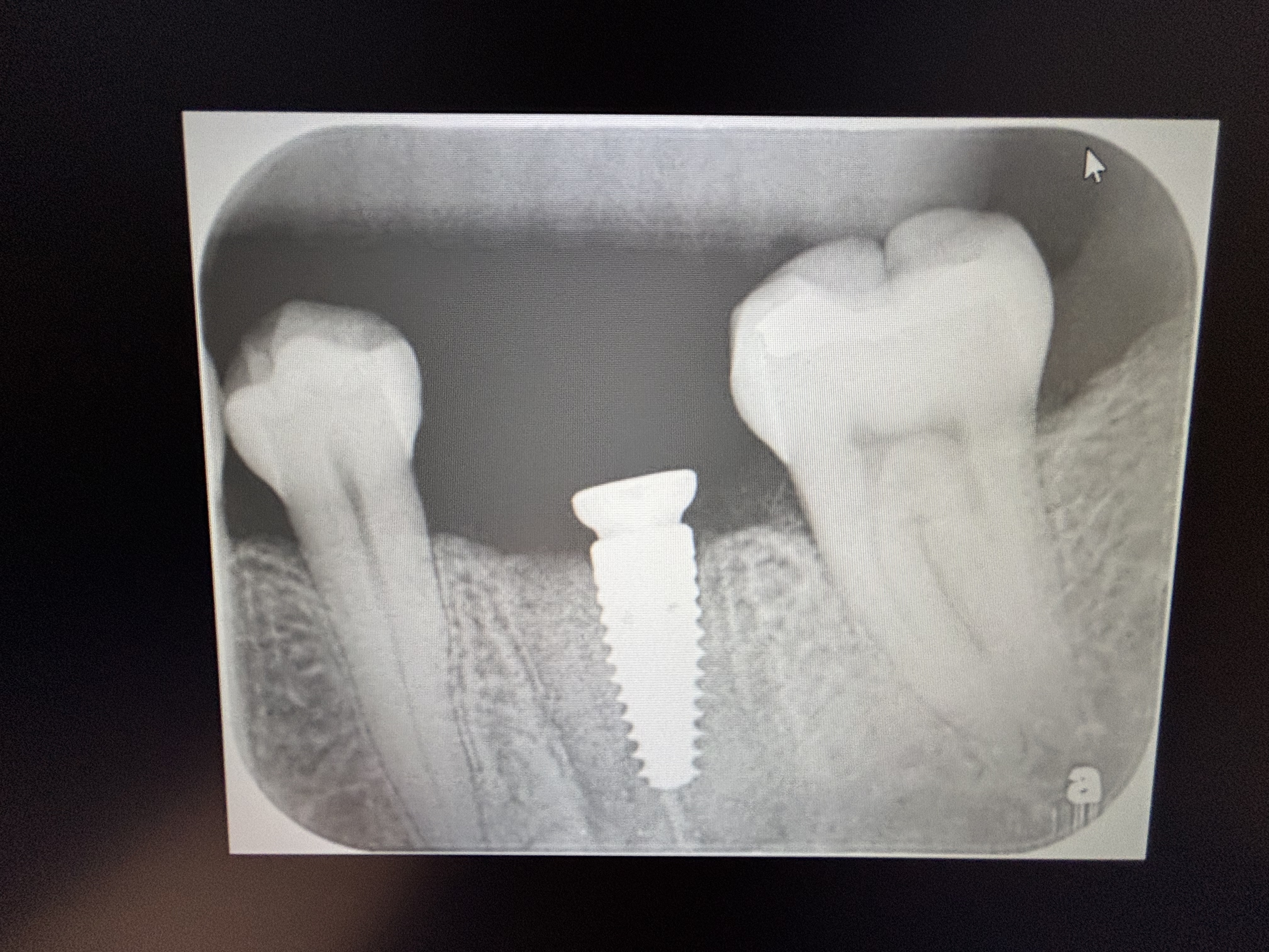
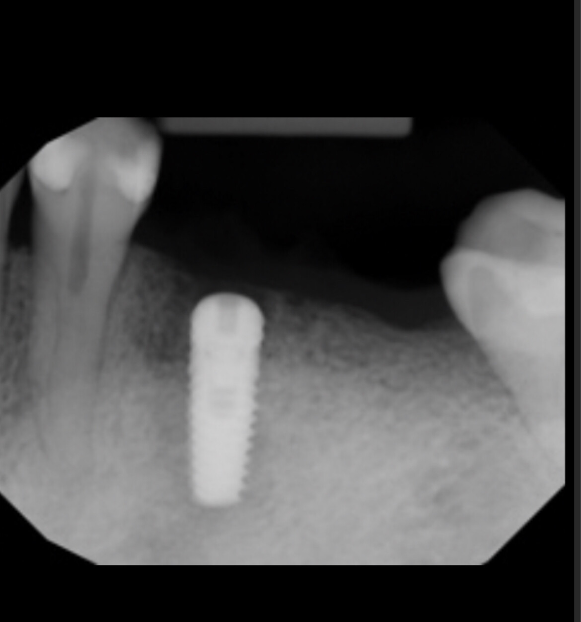
I installed 2 implants in the mandibular posterior region without any apparent complications. The patient immediately began to complain about pain. After 19 days, the pain has decreased but is most intense at night. Anatomically the implants are not near the inferior alveolar nerve. I have the patient on NSAIDS. What could be the reason for pain? Can it be due to extra torque placed while placing the implants? Is this too short a time frame to consider surgical intervention? What do you recommend?
14 Comments on Pain after dental implant placement in mandibular posterior region: recommendations?
New comments are currently closed for this post.
CRS
8/23/2014
Can't really make a working diagnosis without a film. Usually after placement there is very little pain other than the surgical incision. The problem is without a diagnosis even removing the implants may not alleviate the pain, ie trigeminal neuralgia? Could be patient's response to inadequate pain meds? Type of pain throbbing, burning, tingling? Could be a lot of things, welcome to the world of surgery, I feel your pain!
matt watson dmd
8/26/2014
I had same problem when I placed implant in #30 area. This happened on my daughter so I got to witness the discomfort first hand(every day). After 10 days on intense "electrifying" pain, I decided to remove implant. When I torqued in the implant she actually told me it was painful, but I assumed more anesthesia was needed. After profound numbing she still felt the pain/pressure of the implant every time I turned it. No pain when making the osteotomy, just when I turned the last 3rd of the implant into place. When I finally removed the implant, which was no where near the inferior alveolar nerve, it opened a floodgate of blood for 5 seconds and again while she was numb she said she could feel the pressure release. It was instant pain relief, no meds needed. I grafted the site and replaced the implant 4 months later. when replacing the implant I used the final drill with cortical tap drill. Placed the implant to minimal torque and she has been great, 2 years later. This was my first time experiencing this but I do believe the torque was the issue. Good luck with your episode but don't hesitate to remove and wait. The patient will be upset short term, but if you keep them in and hurting, they will remember you long term(in a bad way).
Tuss
8/26/2014
Could there be a compressive effect on the canal if the implants are sitting close to it? High insertion torque compressing bone over the ID canal
KPM
8/26/2014
UGH. Isn't that the worst? I just had that happen to me last week (#6, immediate placement with some GBR). I'm convinced, at least in my case, that the culprit was compression necrosis...aka....my fault! I over torqued the implant and knew I did it. Took it out and replaced it but the damage was done. Three days later I had to remove it. This has happened before and usually, after removal, the symptoms disappear within 24hrs, all things being equal. Alas, this was not the case this time, 48hrs later, but it's heading in the right direction. That said, I, and I'm sure anyone placing implants, has had post operative pain that has subsided over a few weeks after placement and been fine since. The question of course is when to remove and when to leave. Now a days I remove it if there is more than moderate discomfort for more than two days. The odds are it's headed for failure otherwise and if you wait too long you're going to have bone loss and complicate matters when you do replace it. I'd say remove the implant, let the site heal for 4 weeks and replace paying strict attention to the torque applied when placing the implants.
Richard Hughes, DDS, FAAI
8/26/2014
The following may be contributing factors: insertion torque; implant geometry; thread design; how wide and deep did one prepare the osteotomy vs the width and length. All of these contribute to compression necrosis. The other most obvious are possible nerve involvement.
Bone tissue is elastic but only so elastic. It can only stand so much compression.
Ashish Shah
8/26/2014
Pain is due to periosteum insult.It is the most innervated region oof the site..Sometimes the periiosteum is not approximated back on the bone after reflection and hangs on especially in immediate implant placement after extractions,also after placing gingivaformer.This is my humble opinion.
Joy Mandhub
8/27/2014
I perfectly agree with Dr.Shah. Of course we do not have a post operative radiography but my guess would be compression of the periodontium of a neighbouring tooth. I would suggest to remove implant, gbr and replace implant after 3 months.
Anooshah hajiheshmati
8/27/2014
In this case Severe pain after 2 week is because of injury to bone tissue or laceration of gingiva or periosteum and exposing bone tissue or infection each one have a different solution
Anton Andrews
9/2/2014
99% it's torque.
Uzair Luqman
9/3/2014
I have faced such problems as well. And mostly the pain is similar in its character and intensity to alveolar osteitis. My question is that is it possible to have alveolar osteitis around the implant or a similar process?
Marik Guizot
9/5/2014
Is that possible we use the pain killer and wait instead of removing the implants ?
I have case 3 weeks ago put the implants 3 immediate loading with immediate crown and the patient back to his country and dont know when he comeback again. Im sure and did a lot of case with immediate placement. Its quiet bad time if the patient is still present i will remove the implants. The patient also feel the numbness. Before the final reatoration done for 1 week after the implant placement. He dont have any complain but smile.. Is there change any infection occured ?
Robert J. Miller
9/13/2014
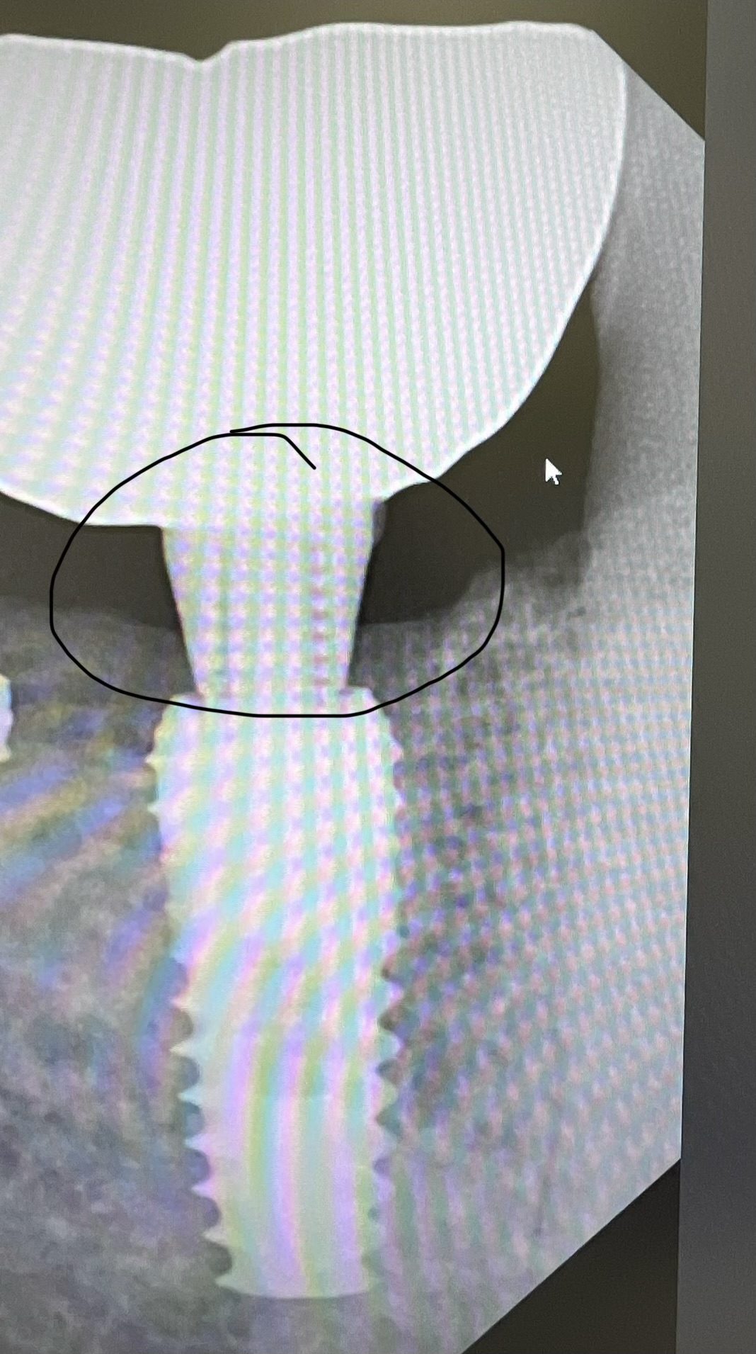
As CRS said initially, it's hard to diagnose without a radiograph. However, there are several reasons for this untoward event. First, and probably the most common, is a bifurcation or even trifurcation of the inferior alveolar nerve. After reviewing hundreds of CBCT scans since 2004, we have discovered that there is about a 10% chance for a bifurcated IAN. While you may see the main branch on a periapical or panorex, you will most often miss the coronal branch, or dismiss the appearance of this branch as artifact. We are currently finishing a paper on anatomy of the poserior mandible which will highlight these anatomical variations. This is why we recommend a CBCT scan prior to surgery in this area. These branches are easily diagnosed on cross sectionals and can be avoided during surgery. The second most common issue is drilling an osteotomy to depth in a ramped ridge. Then, during implant placement, if the implant body is slightly exposed in the ramped area upon seating, we turn the implant slightly farther to seat it more apically. However, the apical part of the osteomy is not prepared to this final depth. As a result, the apical bone is compressed downward and you get a compression injury of the IAN. There are many other reasons that we can elucidate, but we'll save them for another post.
RJM
Richard Hughes, DDS, FAAI
9/14/2014
Bob,
Will your article appear in the JOI?
You are making significant cobtributions to this field.
Robert J. Miller
9/14/2014
Richard; This is the second paper of a four part series we are publishing in JOI. The previous paper was on the anatomy of the intraforaminal zone and was published in 2012. The second paper is posterior mandible, followed by anterior and posterior maxilla. These are in between some other papers we are working on, so it takes awhile to sequence them.
RJM