Perforated the Lingual Cortical Plate: Expect Complications?
Dr. O. asks:
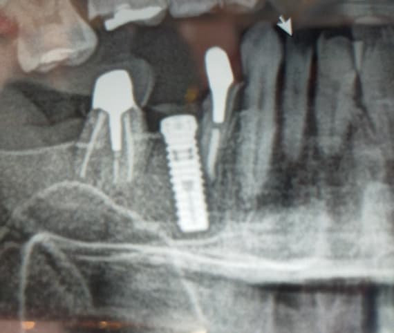
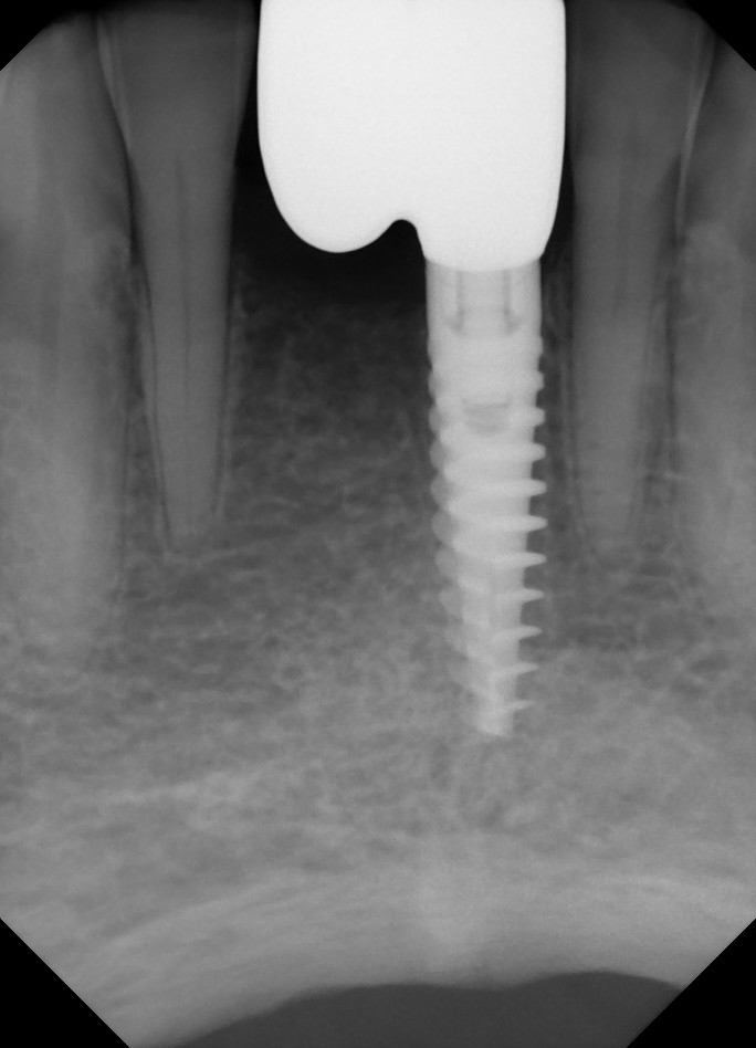
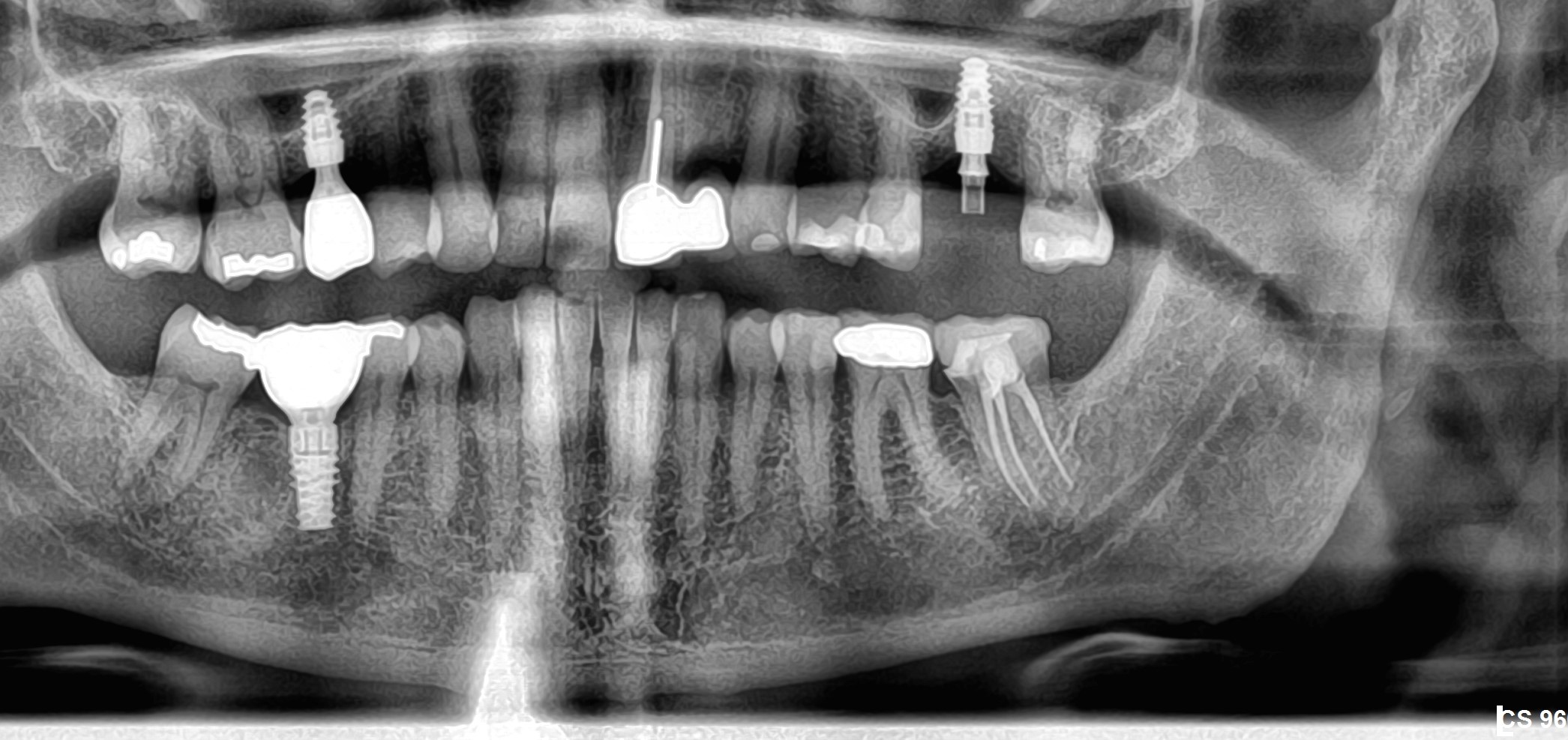
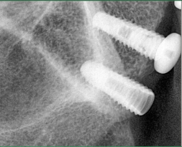
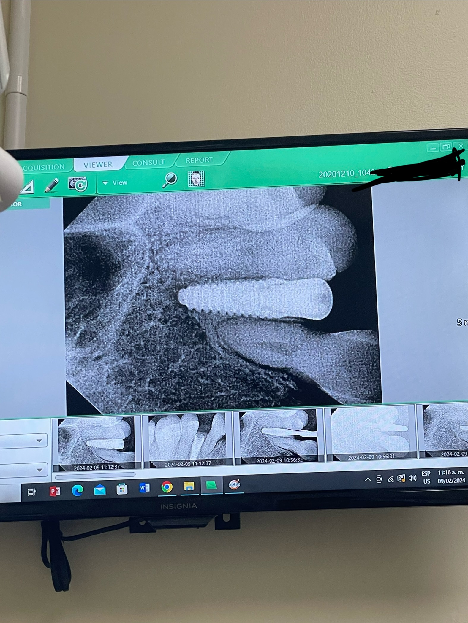
While placing a dental implant fixture in #19 area I perforated the lingual cortical plate. The lingual half of the last few threads are showing through the lingual bone. The buccal half of these threads are firmly engaged in bone. Should I have unscrewed the implant and drilled the hole oriented more towards the buccal and then re-inserted the implant to its new buccal orientation? I left the implant as is and closed the flap. Should I expect complications in osseointegration? Could I have placed a bone graft over the exposed threads?
29 Comments on Perforated the Lingual Cortical Plate: Expect Complications?
New comments are currently closed for this post.
DRMA
11/17/2008
There can be a periosteal pain by tong movements.
I think bone graft will not help in this case.
(if you are lucky, there will be no symptoms)
I would wait one month before reimplant and use gelatin foam after explantation.
eric-sb oms
11/18/2008
Obviously the key here is prevention. The lingual undercut is different in each patient, many times surprisingly severe. It is even worse in the second molar region. I think it is a bad idea to leave an implant in that can be felt under the periosteum. Although it does not represent pathology within itself, it may become a source of irritation for the patient. If the mucosa ulcerates, you could be in big trouble. It would be unlikely to heal and could cause the patient and you a lot of grief. There is no realistic way to fix this with a bone graft. The anatomy would not allow it, plus it would be quite an invasive surgery. Another thought is the concept of bacterial invasion. The implant osteotomy is a contaminated hole, as much as we think it is sterile. By introducing bacteria into the floor of the mouth or submandibular space and then sealing it in with an implant, you could cause trouble.
I would have taken the implant out and re-drilled. You may have had to delay your re-entry depending on the thickness of the mandible.
You can never be faulted by removing a fixture and trying to do it better. By leaving this one in, you will certainly spend a lot of your time thinking about it. Again, early implant removal is always easier than 2 or 3 months down the road. The important lesson here is not letting it happen again.
Jeffery B. Wheaton DDS,MD
11/18/2008
I think the implant will integrate and there will be no symptoms and you'll be fine. You can go a couple mm into the sinus with the apical portion of an implant and be O.K., likewise you can go through the cortical plate a little and be fine. We do it all the time with bi-cortical rigid fixation plates for trauma. Infection is a possibilty but the osteotomy is now plugged with an implant, as long as you were covered peri-operatively with antibiotics infection is a low risk. Just follow the patient, if asymptomatic in a couple of months- restore.
Dr Frank Moloney
11/18/2008
I am assuming the #19 area is the lower left first molar? I Australia and most other parts of the world we use a different numbering system. How do you know you have perforated? Perforation of the lingual plate will probably not compromise osseointegration, but you might get a nasty surprise one day if you do that again, as I did this year, when I hit the lingual artery and watched an "oil well" coming up the socket of tooth#28 (your system), which was easily tamponaded by immediate insertion of the implant BUT was followed by serious swelling in the neck and floor of mouth. Luckily she was having a GA so we kept her intubated and sent her to ICU for 3 days until CT scans showed that the trachea had returned to a central position and was no longer obstructed. She was able to have her intubation tube removed on day 04 and she went home. At stage 2 implant surgery the implant was osseointegrated and my Dentist has gone on to successful crown preparation and finish---but that was a potentially life-threatening situation. Cheers Frank Moloney MD DDS Oral and Maxillofacial Surgeon, Brisbane, Australia.
Joseph Kim, DDS
11/18/2008
Next time, make sure to feel the type of bone you are penetrating. Cortical bone has a distinct feel to it, especially when following cancellous bone. Don't push the drill in areas where significant undercuts are present without being sure about the angulation of the bone and drill relative to it.
Dr. Bill Woods
11/18/2008
How do you know you perferated the plate? How could you see it, under the tissue? Im not a specialist, but I think if I perferd, I would take it out, period. Cant be at fault for doing so. Dr. Maloney's comments are pretty graphic as to what can happen. Measure twice, cut once. Did you have an implant length in mind and if so, how did you determine how long to go? I know how long my implants are going to be before I do the osteotomy. It doesnt sound like you knew how long to go and when to stop. Do you have depth markers or guards on you surgical burs? I have backed up an implant when I thought I was in the safety zone (
Dr. Gerald Rudick
11/18/2008
This is a very interesting situation, and quite unpredictable, if only a 2 dimensional panorex or periapical film were taken in the initial diagnostic stage.
When installing implants in the lower posterior region, our main focus is to be sure that the implant lies safely between the crest of the bone and 2-3 mm short of the inferior dental canal.
It is rare that a less experienced implantologist would consider the possibility of a lingual perforation, when the concentrated effort is to stay within the boundaries for safety.
Years ago, I used the open flap approach, visualized a sufficently wide crestal ridge,to place the implant in a comfortable position and had plenty of room to avoid the mandibular canal.
I inserted the implant, which was an HA coated press fit Calcitek Integral, which fit snugly into the osteotomy up to 60% of its length, and then had to be gently tapped into its final resting place.
Radiographs were taken throughout the creation of the osteotomy, and everything seemed fine.
Upon placing two gentle taps on the implant, the entire implant virtually disappeared from my sight!!!
A radiograph was taken immediately, which showed the implant lying horizontal and directly in the mandibular canal!! I was not sure if I should call my cardiologist for the heart attack I was sure I was about to have, or call my lawyer!!!
Instead, I called the dental expert at Calcitek, who calmed me down and told me that I had in fact perforated the lingual plate, and not to worry.
I extended the lingual flap apically with a periosteal elevator and was able to see the implant lying on the outside of the cortical bone, secured in place by the lingual mucosa. I fished out the implant with a college plyers, and was able to place the implant back into the osteotomy....only this time, instead of tapping....I just gently pressed the implant into place so the cover screw appeared at the crest of the ridge.The implant fit securely into place, and would not be affected by forces of chewing.
The advise of the Calcitek implant expert was to suture the area, keep the patient on the medication originally prescribed for a couple of days longer.... and leave it for six months.
This story is 25 years old and this implant has been supporting a fixed bridge all this time,without any problems and minimal bone loss.
The message here is that before considering placing a mandibular implant without the benefit of a CT scan.... run your index finger along the Mylohyoid line with your thumb on the buccal plate and try to feel if there is a deep concavity on the lingual side below the mylohyoid ridge.
When drilling the final depth, keep a finger on the lingual plate and you will feel the vibration of the drill if it is close to the cortical platebefore it has the chance to perforate the cortex.
Occasionally in endodontic therapy, a strip perforation may occur through a wall of a root without the operator being aware of it, because the radiograph will not pick it up, unless there is a huge excess of the sealant, and pain and bleeding occurs.
By feeling the vibration of the drill on the lingual, the perforation can be avoided, and a shorter implant can be substituted.
Best advise is to wait, as you say a very small portion of the apical end of the implant is actually coming through.
Good luck Dr. O.
Gerald Rudick dds Montreal
R. Hughes
11/18/2008
Dr. Wheaton is correct. However, be careful.
Leopoldo Bozzi, MD Italy
11/18/2008
Dr. Rudick wrote:
"The message here is that before considering placing a mandibular implant without the benefit of a CT scan…. run your index finger along the Mylohyoid line with your thumb on the buccal plate and try to feel if there is a deep concavity on the lingual side below the mylohyoid ridge.
When drilling the final depth, keep a finger on the lingual plate and you will feel the vibration of the drill if it is close to the cortical platebefore it has the chance to perforate the cortex."
While agreeing with the first part of dr. Rudick comment and suggestion, because your's was an incomplete diagnosis before surgery, and the one suggested by dr. Rudick it is the easest way to check the mandibular lingual anatomy if you're using only a bidimesional x-ray examination, You should never place your finger below the inferior border of the mandibula "to feel the bur apex" perforating the plate, because of the higher risk to provoke this way a laceration of the eventually lying there blood vessels, kept against the bur by your fingers. So in any case you may want to place an implant in the mandible or in the full thickness of the mandible, and ovbiously here I'm thinking of an athophic, seriously atrophic mandible, it is far better never place your finger below the expected exit point of the bur. Use ever a new bur to have the best cutting capacity with the lowest pressure you may place on it, and be careful. Mandible is a completely different implant situation, respect to the maxillary sinus.
So, in absence of any image of you clinical case, my advice is to take out the implant, you may place a shorter one in the same position if the available bone housing is sufficient to host -say- a 7mm long implant, or let it heal undisturbed (no gelatin sponge into it) wait three month and redo the surgery after a in-depth preliminary examination.
Don Callan
11/19/2008
This can happen to any of us, learn what not to do so this will not happen again, there is some good advice above. TAKE THE IMPLANT OUT, GRAFT, WAIT 3 MONTHS AND PLACE ANOTHER IMPLANT. If the implant is left there, it will be a problem--I have been there.
Dr. Kimsey
11/19/2008
I agree with Dr Callan to remove this implant if for no other reason that some other dentist will one day say how you placed it poorly and you could have killed the patient.
Jeffery B. Wheaton DDS,MD
11/19/2008
I think it is irresponsible to just say "take out the implant." Dr. Callan, exactly what problem have you experienced like this? That is a rather vague statement. As for subjecting this patient to more trauma because you are worried what some other dentist may say in the future, I think that is a little silly. I think this all depends on to what degree of perforation you are talking about. Get a CT, if it is barley through, bone will form over the apex and you will be fine.
Dr S.
11/19/2008
Amazing thread going on here, with the most experienced implantologists giving their valuable inputs based on experience.
However I think the question here is mis-understood. The dentist here tipped the last drill too lingually and the lingual cortical plate perforation that is being mentioned is obviously in the gingival 1/3rd of the implant not the apical 1/3rd. I haven't done too many implants to qualify to comment here, but I guess it is going to make the prosthesis construction challenging probably needing special abutments. Secondly the patient is going to complain about the prosthesis encroaching on the tongue space.
Dr S.
11/19/2008
On a lighter note.
A lifeguard on the beach got frantic SOS call from a group of hyperventilating youngsters who wanted him to help their friend get out of the quicksand on the beach.
"How much deep is your friend stuck in?" asked the lifeguard.
"About ankle deep" retorted the victim's friend.
"Then you don't need my help, you can pull him out yourself if he is just stuck in ankle deep" said the life guard.
" No he is stuck ankle deep UPSIDE DOWN! His ankles are sticking OUT of the quick sand and not sticking IN the quick sand, Please rush!"!!!!!!!!
It helps to understand the question first.
Jeffery B. Wheaton DDS,MD
11/19/2008
Dr. S. Good point, I was thinking ( as I think a few others were as well) about apical perforation, mainly because that is something I have experienced before. I agree with you, it does not sound like this implant is ideally placed restoratively. If that is the case then I would remove, graft, and replace in a few months...my point is I think alot of the concerns about periosteal inflammation, infection, etc. are unfounded.
JAV
11/19/2008
The problem with this case is that it was not treatment planned. If you have exposed threads on the lingual your osteotomy is off. You probably did not have a stent and tried to free hand it. If you take it out and try to redrill the osteotomy you could easily perforate the lingual more apically. Take it out, graft the site, and then treatment plan the implant with a surgical stent or guide.
Dr. Bill Woods
11/19/2008
I think that perfing the lingual plate, whether immediate or later consequence, is setting you up for some very unwanted misadventures in clinic or court. Period. Neither will be fun. No CT + later infection that is now sublingual with a fixed connection that would be an extremely difficult surgical situation to resolve, I just cant see it. Maybe that would get some bone over the apical portion of the implant. Havent seen that as "what to do" in any CE I have participated in, including AAID courses or Pikos or Callan. There isnt any schnederian membrane there as a boundary for a bone graft. Would you do a Summers procedure and just graft there? I dont think so. Not me. I have seen many lingual cortical plates that I intentionally place the threads for initial stability. Others thin and something to avoid. Sublingual is just somewhere I dont want to be with final placement. I completely agree with those that share the philosophy to take the implant out and place another later. Perfing a lingual plate is not a "no balls...no blue chips" situation. I'll go by what I was sensibly taught and sleep better. "Surgeons whistle in the graveyard way too soon". JM2C. Bill
Dr S
11/20/2008
I have posted a case in osseonews cases section. Please opine urgently
R. Hughes
11/20/2008
You can prevent this by careful planning, ample surgical exposure etc. You can determine if there is a perf by palpation, and probing.
ddsyoon
11/22/2008
I'd better remove it unless I am free of being charged misplacing it at the court.
Dr. Scott Kareth
11/26/2008
Dr. Moloney,
Here in the U.S. I assure you that we all know the rest of the world uses the International Charting System. I not only assume, but know that you are fully aware that #19 is in fact the lower left first molar (or rather 36) but in addition you feel the need, due to an Ausie or OMFS derived insecurity,to point out this obvious fact. The reason that I feel the need to call this out, is that I truely enjoy this site but believe many including myself are distracted by observations, such as your "lower left molar" revelation. Please leave the ego at home and let us all focus on the spirit of this site LEARNING.
satish joshi
11/26/2008
Dr.Rudick's advice to place finger under lingual plate during drilling is very dangerous and irresponsible.
I would rather face the consequences of being sued for malpractice than placing my finger in the path of blood ridden drills and subject myself to be a victim of HIV,hepatitis B, hepatitis C,or other dangerous diseases.
We must understand that all patients are not honest in revealing their medical history.
toothdoc
12/5/2008
I agree with Satish Joshi.Is it worth to take a chance on your safety or well being?
R. Hughes
12/7/2008
If you learn the bone expansion techniques as per Dr. O. Hilt Tatun, you will be disinclined to perforate the lingual plate.
Chan Joon Yee
12/28/2008
Taking the implant out is the last thing that the dentist should do in this situation. Regardless of whether you try to graft the osteotomy or not, your re-entry will find a situation many times more challenging with higher risks of perforation than the first attempt.
Personally, I would, as far as possible, avoid removing the implant. If there is primary stability and the perforation is small, there should be no problems with integration. Even with significant perforation, the mucosa is unlikely to be stretched to such an extent that dehiscence will occur. I would place a rigid membrane covering the perforation and close up for at least 4 months.
I've found that grafting is always more predictatble with the implant in place. I have managed to achieve 3mm vertical ridge augmentation grafting over super-crestally placed implants. Doing it without implants in place has seen little success.
Yes, a little bump will be visible on the lingual mucosa, but the fact that most people don't complain about their tori should be an indication that it doesn't really matter.
As far as ideal positioning is concerned, most dentists perforate precisely because they want ideal placement and drilled straight down, right beneath the upper arch. If the dentist had drilled parallel to the concavity, he may not perforate but the implant would indeed be difficult to restore.
I just had a case like that recently. I advised the patient to take a chance and she's now glad that she did.
Dr. Guy Levi
12/30/2008
I'd better remove it and manage the case like
Dr Callan said. The main reason is extremely high risk
of prosthetic complications and prosthetically -related surgical complications,as well. I am sure , in such cases, implant position is far away out of opposite dentition. Getting back the buccal bone volume and
creation of surgical stent prior to implant placement is mandatory, especially in a such situation.
Dr. Guy Levi
12/30/2008
... not saying about a highest risk of lingual cortical
plate resorption with consequent soft tissue and other
problems.
Dr. Gerald Rudick
12/30/2008
I left an earlier comment on November 18th 2008 about preventing a lingual perforation by placing thumb and index finger over the buccal and lingual sides of the mandible while drilling; when not having the advantage of a CT scan.
I described a situation of an event 25 years ago, which has turned out to be completely successful.
I thought I was perfectly clear that when drilling the depth of the osteotomy, when your index finger is placed below the mylohyoid ridge, the vibration of the rotating drill can be felt....thus avoiding the possiblity of perforating the lingual cortical plate.
Dr.Leopoldo Bozzi did not fully understand what I said....in that he commented " you should never place your finger below the inferior of the mandible "to feel the bur apex" perforating the plate.
I said that the index finger should be placed on the lingual plate below the mylohyoid ridge... certainly not the inferior border of the mandible, and certainly before it has a chance to perforate.....if too close, you will feel only the tip of the pointed drill before it has a chance to do any damage.
Dr. Satish Joshi also criticized my comment by saying "Dr. Rudick's advice to place finger under the lingual plate during drilling is very dangerous and irresponsible...etc."
Once again, this clinician read through the lines too quickly and without thinking, as I said " when drilling the final depth,keep a finger on the lingual plate and you will feel the vibration of the drill if it is too close to the cortical plate before it has a chance to perforate the cortex"
So my advice is to be respectful of the anatomy, and feel the vibration of the rotating drill before you can do any damage.
Remember the case I referred to was 25 years ago when CT scans were not used as frequently or as routinely as they are today.
I certainly take no offence to my collegues Dr. Bozzi & Dr. Joshi, and am glad they took the time to read what I had to say.
Gerald Rudick dds Montreal
Kim Bradbury
4/11/2018
To help you dentists; I had 2 right mandibular dental implants placed in 2009, both pierced the lingual cortex. The periodontist had told me I was an ideal candidate for dental implants, even after the preliminary scan reported there was a sclerotic lesion in which needed careful assessment before I was considered for dental implants. He was morbidly obese, with a huge stomach which required all his concentration to prevent him from smothering me in the chair with it; his stomach made the whole procedure truly uncomfortable, it was almost like a nightmare being pinned to the chair by that huge soft, heavy mound. He free drilled; no guides, no thought, no care, then when I repeatedly returned because of the pain I was experiencing in my neck, he repeatedly told me I was over reacting to the feeling of having dental implants placed in my jaw. After a year of horrific pain and many problems in my neck, he wrote to my GP telling him I had emotional problems because the implants had been placed perfectly; I managed to get my records through the FOI act and discovered he'd known the implants had pierced the lingual cortex for all the time he'd been writing letters to other specialists to advise them I had serious mental and emotional problems. The 4/6 had also gone right through the floor of my mouth and the second implant in the most posterior position of my right mandible had hit the sclerotic lesion and deviated so it was lying alongside and almost parallel to the angle of my jaw, "osseointegrated onto the lingual surface of the right mandible" the threaded end pointed past the far end of my jaw, into my neck. Over 8 years, those dental implants tore holes in my upper oesophagus, allowing food and fluids to escape into my skull base, the point of my spine where it connects to my head and into my neck from where it leaked down into my shoulder, chest, into a big bubble between fascia down my back, into my abdomen and further down into my right leg and right foot. Because of that periodontist's refusal to acknowledge his mistakes and rectify them, because of his deceitful letters to colleagues I desperately saw to try to find out what was wrong, in which he said I had emotional problems and mental disturbance, because I was 50, single and female, I was unable to obtain medical assistance from any Queensland specialist, I became too ill to do anything except exist, from day to day. In 2016 I packed up everything I owned and drove to Victoria to see and say goodbye to my daughter, son-in-law and 3 grandsons, because I knew I was dying. I do not know how I managed to drive that far and do not remember any of it. My daughter had found a little cottage I could rent while down there, so I moved in and after a month, something in my right jaw burst. Pus and rotten debris poured out of my right mandible for days; I was sick, vomiting, severe diarrhoea, dizziness etc etc. I went to a doctor who referred me to a maxillofacial surgeon who luckily, didn't know the periodontist in Brisbane who'd placed the implants. He ordered scans to see if any damage had occurred and showed me the position of the implants; the Victorian maxillofacial surgeon refused to remove the dental implants, he wrote to the periodontist and enclosed copies of the scans to show him the placement of the implants and the damage to my right mandible, which was paper thin in places. Up until 2017 I had believed the mandibular implants had been correctly placed; several Queensland maxillofacial surgeons, periodontists, dentists and other specialists had repeatedly told me so. The Victorian maxillofacial surgeon referred me back to that Queensland periodontist, telling me it was the periodontist's responsibility to remove the implants and organise medical help for all the injuries they'd caused. The periodontist did not respond to him, the periodontist refused to accept my phone calls when I tried to make an appointment. I was prepared to borrow money to fly to Queensland so he could remove the implants, but even after hundreds of phone calls and dozens of emails, the periodontist will not respond and has continued to ignore me. I sought assistance at a Victorian, then a NSW hospital, but they told me I had to be treated in Queensland, so I eventually managed to drive back to Queensland. Before I left Victoria, the implant right at the back of my mandible fell out, then a few weeks ago, the 4/6 implant came out bringing with it about half of my right mandible, now less than 1 cm thick. Ever since my right mandible abscess burst, fluid has been seeping out of my throat and right jaw. I now have huge holes in my jaw and on a liquids only diet, sucked through a straw down the left side of my throat. I am very, very ill, living in a caravan park after 9 years of horrific illness and only managing to just get through one more day, while food and fluid have been jammed up into my brain stem, skull base, neck, shoulder and been forced down into my chest, body and right leg. Now I am back in Queensland, terrified of going to a Queensland hospital after being refused treatment so many times by so many different Queensland hospital specialists who had all been influenced by the periodontist who placed the dental implants; it's appallingly easy for a male periodontist who is also a professor of periodontics at a large Queensland University to bully, slander, malign and intimidate a single female grandmother of 50 years old, now 59 years old after the past 9 years of horrific pain and illness. It's very easy for a well known male periodontist to ensure that no colleagues provide that female patient he has grievously injured, with any medical examinations or treatment of any type. Because of all those Brisbane dental and other specialist's horrific misogyny, I am now going to die,


