Peri – Implantitis: What are the Indications?
Dr. B. from California asks:
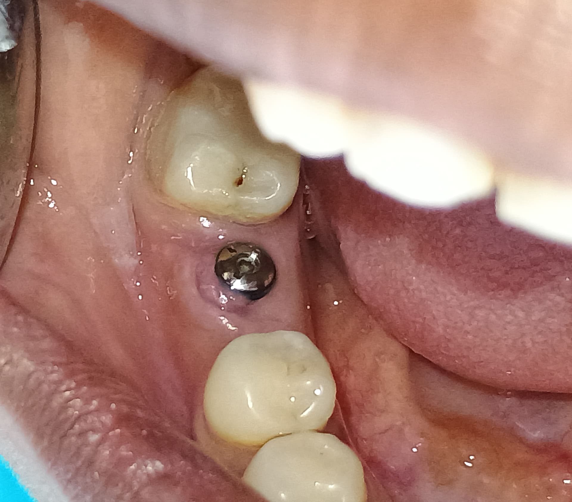
I placed a regular platform dental implant in #9 area. Bone volume and quality was excellent. I let the implant integrate for 3 months. No signs of purulence or any other problem. Follow-up radiographs showed excellent healing. But when I went to torque down the abutment, it produced pain and I could feel slight movement. Is this an indication of peri-implantitis? What should be my next steps here? Thanks.
9 Comments on Peri – Implantitis: What are the Indications?
New comments are currently closed for this post.
Dr. Jim
9/17/2007
Dr. B,
Unfortunately there is a good chance this implant will not be around long enough to even develope peri-implantitis. Based on what you have described, it does not sound like the implant completely integrated.
Dr. J
9/18/2007
The implant has failed and should be removed if it really did move. You could give it more time to integrate but, most likely, it has failed and no amount of time will change this. I would remove the implant, degranulate the area, perform a ridge augmentation procedure, allow 4 months of healing time, and replace the implant. Then it should most likely be successful.
Rand
9/18/2007
It sounds like a failure. Nonetheless, are you sure that the abutment was down all the way when it "moved?" If tissue was caught under the abutment, it would cause pain upon torquing the abutement and might discourage you from torquing it down all the way and until fully seated it would seem like the implant was moving, when in fact it could be just the abutment moving.
If the abutment was fully seated then the mobility indicates failure. With failure you have two retreatment options:
1) remove, decorticate the osteotomy and graft and place another implant some months later.
2) remove the implant and score the bone to make it bleed and immediately place another implant that is wider.
One sure test, if the implant is mobile, then you should be able to remove it easily by "unscrewing" it out of the bone.
Good luck to you and best wishes.
Dr R Bryant
9/19/2007
I have had about two implants with this problem. I have retorqued the implants and left alone for a further few months and they have both integrated.
Andy Howard
9/19/2007
3 months of healing in the premaxilla may not have been adequate time for integration. How much torque did you apply? If it exceeded 30-35Ncm, you may have sheared the bone. However, this does not usually cause pain. A complete fibrous encapsulment of the implant can definitely cause pain. IMO: flap it and inspect for vertical bone defect. If no significant bone loss and no purulence, aggressively currette the osteotomy and place a slightly larger diameter (and longer if possible) implant. Wait 5-6 months before exposure. If you remove implant and graft the socket only you may loose some ridge width. PRP or rPDGF may be of some benefit here also
Dr. Bill Woods
9/19/2007
I agree with removal if it isnt a tissue issue. There should be zero movement. Wait around for what? No waiting or watching will solve anything. Remove, regraft and replace. If there is pathology there, it could worsen the situation and end up creating a mess for the future. Ask me how I know. Bill
Don Callan
9/23/2007
Bill is 100% correct.
jerry Drury
9/23/2007
It is possible that there was not sufficient bone to implant contact to prevent pain on when you torqued the implant. You can wait longer and see if re-integrates, or if indeed there was movement then you can remove and start over. If you are dealing with type 4 bone you may want to consider using a rougher surface and,or somewhat less torque next time. This is one of the instances I would consider an HA screw design on the second round as long as the patient understands some of the potential drawbacks.
Dr R.Shah
9/26/2007
I have recently placed two Straumann implants with a view to provide an overdenture arrangement with two retentive anchors.
The implants were placed in the anterior maxilla four months ago, in what was quite a thin buccal ridge.
One of the implants has developed a buccal sinus but appears integrated and is solid. There is an area of bone loss aound 5mm radiograpically which corresponds to the area which was grafted with autogenous bone at placement.
As the patient is a smoker I am pretty sure that the graft has become infected at this stage as a result of this.
I am considering lifting a flap, curretting the implant and regrafting the area with Bioss and Biogide to fix the defect. The other option is to remove the implant, graft and start again four months later.
In view of the fact that the implant is solid and clinically appears well integrated, it also had good primary stablity at the time of placement I am tempted just to go for the first option.
What would other people do in this situation