Placing Implants in the Atrophied Anterior Mandible: How Should I Evaluate This?
Dr. R. asks:
I am a general dentist and place and restore my own implants. I currently am using Camlog and Implant Direct. I am concerned about placing implants in the atrophied anterior mandible with decreased bone height like 10-12mm. What chance is there for fracturing the anterior mandible? What key indicators should I evaluate? What precautions should I take? What signs or symptoms would indicate a jaw fracture? How should I treat a jaw fracture if it occurs?
13 Comments on Placing Implants in the Atrophied Anterior Mandible: How Should I Evaluate This?
New comments are currently closed for this post.
Dr.Alejandro Berg
4/26/2011
I would say that your chances of fracturing a mandible are slight to none. If you think about it, when they find human remains that are thousands of years old they find exactly that... a mandible. This bone is really strong (the only movile bone and i´m not counting the inner ear ones because the have no load bearing work). If you have a good planning and are somewhat of a gentle surgeon you shouldn´t have a problem. If it were to happen.... you will need an experienced OMF surgeon, plaques and screws and so much luck to make it go away that if you have doubts, you should send the patient to be implanted by an experienced crew.
best of luck
Bruce GKnecht
4/26/2011
First of all. Get a Cat Scan and really see what you are dealing with. The trajectory of the bone is a big cancern. If you look at mandibles with resorbed ridges, the Genial tubercle is at the height of the ridge. The bone has an inclanation towards the lingual. The cancellous bone is limited and the cortical bone is thick. I have placed 4.3/8 in the area to prevent causing a fx., but I had a cat scan adn I placed two to hold a bar adn adn lowr denture. I also used a surgical guide so that the implants exited in the middel of the porsthesis and not to the buccal or lingual. The other ooption could be Mini implants. the 2.5/10 MDL is a great implant for this. I place four and use either the enclosed retainers or Saturnos by Zest.
sag
4/26/2011
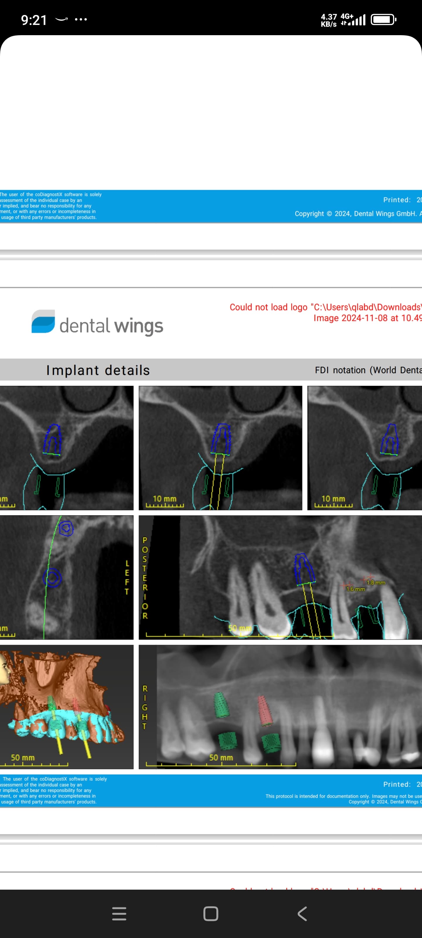
Fractured mandibles can and do occur following placement of dental implants. You should be careful to not penetrate the inferior border of the mandible, since you will already be drilling through the superior border. The labiolingual dimensions must be accurately measured to avoid placing an implant that is too wide and will therefore weaken the mandible further. Cone beam CT is clearly indicated.
If the jaw breaks, you should refer to an oral and maxillofacial surgeon since they have the most experience in treating those fractures.
In the alternative, you can refer the patient to an experienced surgeon for the implant placement and then restore the case as you see fit.
Carlos Boudet, DDS
4/26/2011
Great comments by all, very good information, Bruce!
With the easy availability of a conebeam scan, I agree that it is indicated in these cases. If you have no access to a conebeam ct, you can get useful information with a lateral film of the symphisis area using a radiographic marker attached to the patient's denture. With this you can look at the amount of bone available (height, width, shape) and compare it to an object of known dimensions (the marker on the denture can be a 10 mm long wire) and even the angulation.
In my opinion, narrower diameter implants in cancellous bone is better than wide diameter implants in cortical bone, so don't overdo the diameter.
I just mentioned the lateral film for your benefit, but the cbct is so much nicer!
Good luck!
osurg
4/27/2011
As an aside, if you fracture the mandible be prepared to speak with an attorney. Fractures in attrophied mandibles are not easily treated. They often require hospitalization and open surgical procedures with plates and screws. Patients are not happy campers.I can assure you that these patients will seek legal redress. Dont be fooled that an informed consent will protect you. A smart attorney will know how to get around it. Even if you prevail the experience will not be pleasent.Sag makes a good point about letting someone with more experience place the implants.
Richard Hughes, DDS, FAAI
4/27/2011
To osurg, your comments are spot on. Actualy a subperiosteal implant is an excellent choice.
Dr Rob Dunn
4/27/2011
Whilst cone beam scanning is esential in these cases, why complicate things when you can choose to use Mini Implants. Initial research being carried out at Manchester University and Liverpool University in UK shows no difference in performance between these and conventional implants. The only difference of note is that they are cheaper for the patient, have less side effects post surgery, and in most mandibular cases can be loaded immediately. The 3M Espe MDI's range from 10 to 18mm with the most commonly used sizes being 10 or 13mm. Make life simpler for yourselves, and improve the quality of life for your patients.
CGP (omfs)
4/27/2011
Great comments by all. I find treating the atrophic edentulous mandible really challenging for two reasons: 1) the keratinized tissue usually is very poor and attached tissue is a must in my mind and 2) the bone in these patients a lot of time is not what it appears as there is generally very hollow marrow with a steep inclination making it easy to perforate.
The comments about fractures and the edentulous mandible is spot on! These are very debilitating and to go one step further not only require a bone plate but the AO principles of treatment are a very large 2.4 recon plate.
I think a CT scan (conebeam) is a really good idea. Evaluate the bone but don't forget the soft tissue and if they are in good order, be gentle and place the implants. If it looks more complicated refer to a very good surgeon and ask to watch/assist. I never have a problem with helping out b/c the way I see it when I do I will have a better relationship and will still always get referrals. Remember it is the practice of dentistry for a reason, we should all still be learning no matter what we claim to be....I sure try.
Good luck and no matter what the scenario is, do what is right for your patient not you. If that means you are the most capable then treat the patient. I have heard it said, "every time I have done something that doesn't feel right it turns out it wasn't."
ozzo
4/28/2011
indicating lack of keratinized tissue in posterior mandible is a good point. Wide range of applications from simple implantation to pre- or simultaneous bone-grafting requires passive flap closure, which is closely related to the amount of keratinized tissue and periodontal biotype and flap design as well. Adding to nice comments on IAN damage-avoiding, such an evalution on the so-to-be peri-implant tissue is highly important. By the way, in especially high-cortical low-spongious bone areas, risk of distal nerve compression increases. Although implant looks correctly placed&angulated on a conventional radiography, stress of implantation and compressed peri-implant bone tissue may cause compression on IAN, resulting un-resolving paraesthesia in related area. In short, evaluation through Cone beam CT is essential.
Dr. Dan
5/3/2011
I'm sorry to say, refer to a specialist that you know is good at this. Not worth screwing up. I'm a specialist and when I'm not sure of something, I refer out.
Thomas Cason MFOS
5/9/2011
Hi - all good and fair comments. I wouls agree eithr a lateral ceph or better still a scan to evaluate the anterior mandible. I would also advise you elevate the lingual flap to ensure you havent perforated the lingual cortex - you have more chance of this than causing a fracture. These can lead to significant bleeds and I have knowledge of a patient that needed a trachyostomy due to the severity of a sublingual haematoma and airway compromise.There is often a small vessel in the midline area thats ready to greet you when you dont want it!!
Good luck.
Carlos Medina
5/22/2011
The anterior mandible generally will have bone when there is none in other places...so, take xrays to determine height and measure with a caliper, or take a 3D scan.
ivy oconnor
6/6/2011
what is the danger of taking a 3d ct scan


