Pre-existing Periapical Radiolucent Lesion: Should I Be Concerned?

Dr. T asks:

My patient presented with an edentulous area at #9,10 sites [maxillary left central incisor and maxillary left lateral incisor; 21, 22]. My treatment plan is to place 2-implants and to restore with custom abutments and cemented crowns. The two teeth had been extracted 10-years prior.

A periapical radiograph showed a radiolucent lesion at the approximate apical level of the prior teeth. The area has been asymptomatic, there is no sign of a sinus tract and there is no expansion of cortical plates. How should I approach the placement of implants at this site? Should I be
concerned about the periapical radiolucent lesion or should I just assume it is apical scar tissue and proceed with the implant placement? Should I do a surgical biopsy of the lesion first?

Upper left cental and lateral space

26 Comments on Pre-existing Periapical Radiolucent Lesion: Should I Be Concerned?

New comments are currently closed for this post.
peter fairbairn
5/20/2011
Be very aware here , scan the case first to check position of the area , remnant cyst tissue could cause many issues later and must be removed. Peter
Dr. B
5/20/2011
Try to obtain older radiographs, was the lesion there before? Has it grown? Either way, I think this looks like a cyst and it should be removed prior to placing implants. As Peter said, get a CBCT. Good luck.
ttmillerjr
5/20/2011
The X-Ray may be deceiving but it doesn't look like you have enough room for two implants. I would open a semilunar flap and clean out the area. You will for sure find a lesion in the bone, and in my experience these lesions that may have been associated with endo can be nasty. So I say just clean it out and give it 4-6 weeks before placing implant(s).
DR MILAN KUMAR
5/21/2011
as per radiograph u'd given, there is a significant periapical radiolucency on apical vernix which extends to the cortex.1--- its clearly demanding a CBCT.2----Before placement u'hace to clear the osteogenic debris in form of granulation at the cortex.3--------------------------use dfda for the site4-----------wait for 3 months 5----take cast model again n compare the previous impression n do a dummy6------------in dummy try imp copin too as its a esthetic requirement in ur case7-------------place implant in gradual drill . a routine 2.7 will do
doc
5/21/2011
Hi, In the radiograph the radiolucency is definatly not a cyst , but yes there is an erosion of the cortex. A Dentascan is the best option, clinically you could also try aspirating with a syringe (wide bore needle ) under LA . this will give you a fair idea. All the best.
Carlos Boudet, DDS
5/21/2011
Some suggestions in addition to the great comments above: After enucleating and debriding the cystic cavity you may want to place calcium sulfate in the lesion as suggested in the endodontic literature. No graft also works but I believe using calcium sulfate produces a better repair of the lesion. Take photographs and document the smile and gingival exposure. If the extractions were done 10 years ago, you probably don't have adequate volume of bone to place the implants without grafting. A cbct scan or at least bone mapping is indicated. It is great that you want to place one implant per tooth, but remember that there are recommended spacing between implants and teeth, and if you squeeze two implants too close together or too close to the adjacent teeth, it can become a catastrophe. Good luck!
dr naser
5/24/2011
if the area is asymptomatic and the radiolucency is there for 10 years or so insert one implant distal to the lesion without penetrating it you have no room for 2 implants good luck
howard marshall
5/24/2011
Some very good comments above. Recommendations: Catscan first. Must determine whether this is a cyst, or whether there had originally been an infectious destruction of either the palatal bone or the buccal plate at the apex. Years ago we would have done an exploratory flap on buccal and lingual to check this out, but today the Catscan is state of the art. You can have had an original perforation into the palate, or a failed buccal apicoectomy with subsequent extraction, and either of those can give you the radiolucent circumferential appearance shown on the radiograph. In addition, you could have a residual cyst. Once you determine from the scan which of the above it is, your treatment is simplified. If there is solid bone from buccal to lingual within the marrow, and there is a break in the buccal or lingual plate, but no further pathology, I would place only one implant of adequate diameter, and do a bone graft in the socket base where the defect is before placing the implant. If it is a cyst, it must be enucleated from the buccal, and for faster regeneration, use a cancellous bone graft with a membrane, and sit tight for 5 months minimum. Then place the implant. Hope this helps.
Mario K Garcia
5/24/2011
Doctors, deciding on treatment by looking at a PA! Please, lets move away from that. Get a CBCT scan; then let's discuss treatments options. In addition; get some history: how and why were these teeth lost (trauma, failed root-canal, anodontia, perio?..) Always remember No-roots/no-bone. Have fun...:)
Dr D
5/24/2011
I agree with the great comments above. I do not think there is room for 2 implants. For peace of mind,a CBCT is ideal as most likely there is insufficient ridge width. A good analysis of the case with bone mapping and wax-out could be an alternative route, with the hope you can place on implant mesial to the lesion at #9 site and cantelever #10. Removing the lesion seems a most because your implant is going to be close to the implant. Good luck
Frank Avason
5/24/2011
Nasopalatine duct
Dr. Gerald Rudick
5/24/2011
From the radiograph provided alone, it is difficult to judge if there is adequate space for two implants.As far as the lesion is concerned, CBCT would give additional information, but so would opening a full thickness flap, and with a sharp explorer, pick at the cortical bone to see if it can be perforated. It is not unusual to find the remnants of a globulomaxillary cyst in this area; or a cyst of a another nature or granulation tissue left behind from the previous pathology.Grafting would be a definite indication, as well as orthodontic consultation so as to obtain optimal spacing in the aesthetic zone; before implant placement is consdered.
Dr Mario Marcone
5/24/2011
I do not think it is the naso-palatine duct ... However, the naso-palatine duct is partially visible in the given view at the top left corner, where it is expected to be found, in the area between #8 & #9. The area in question is most likely a residual lesion from previous tooth pathology. May sound a bit exaggerated, but perhaps one may consider CT scanning the area to help arriving at a differential diagnosis before proceding with any kind of surgical intervention. Best Wishes ...
Dr .T
5/24/2011
The site is pretty tight for 2 implants. My other option is to restore the tooth no. 9 with an implant and distal cantilever the 10. Or conventional cantilever bridge off of the 11 which is already crowned. Would you still treat the radiolucent area if it is avoided? There is no obvious change over the last 3 years on radiograph. Unfortunately I do not have records before this. Teeth were lost due to failed post crowns. No recall or surgical intervention other than XLA. Ridge width is more than adequate.
M&M
5/24/2011
If the radiolucency is located next to your maxillary right central incisor, this is likely the nasopalatine canal. Please verify with a CBCT.
Dr. Vipul G Shukla
5/24/2011
Dr. T, Based on the X-ray alone, it does not look like a cyst or the naso-palatal canal opening. It does look like an apical scar from a previous p.a. surgery though. Or p.a. infection that was left to heal on its own after the tooth was removed. You mentioned the teeth failed with cast posts and crowns. Either way, do you have adequate room for two implants here? My money is for a single implant with a cantilever lateral incisor crown. No, I would not worry about the radiolucency if I were not entering the site surgically. Just a rad every year or so. Good Luck!
athina
5/25/2011
I agree with the option that the radiolucency might be the nasopalatine canal. Taking an X-ray with a diffrent angle might help before going for a CBCT.
John Manuel DDS
5/25/2011
Good comments. Better to have models, patient, and other films to better evaluate this situation, but... - This type of lesion is common where the periosteum has been violated by drainage, procedures, etc.. The fibrous tissue has beat the bone into the defect. - 
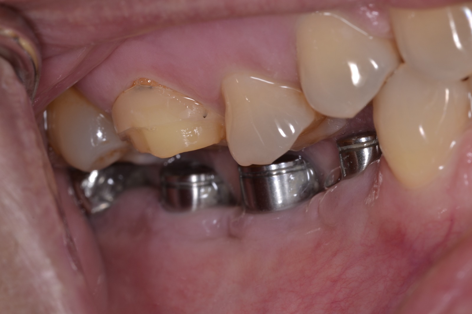
This area can clearly be seen visually upon full flap opening, and the vital structures are easily placed, so exotic 3D films are not critical. - As discussed previously by the experienced surgeons, you most likely do not have adequate ridge width to place a standard implant in this space without bone grafting and ideally tissue revision. - The labial periosteum is programmed to eat away non-loaded bone over the long period since extraction. - 
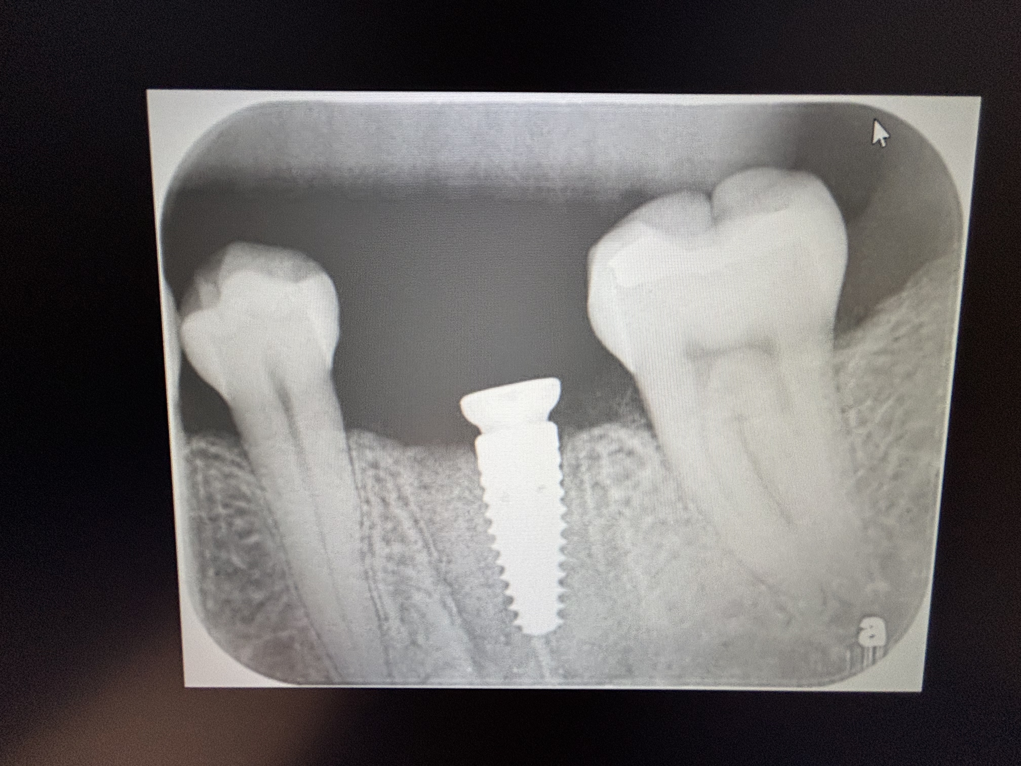
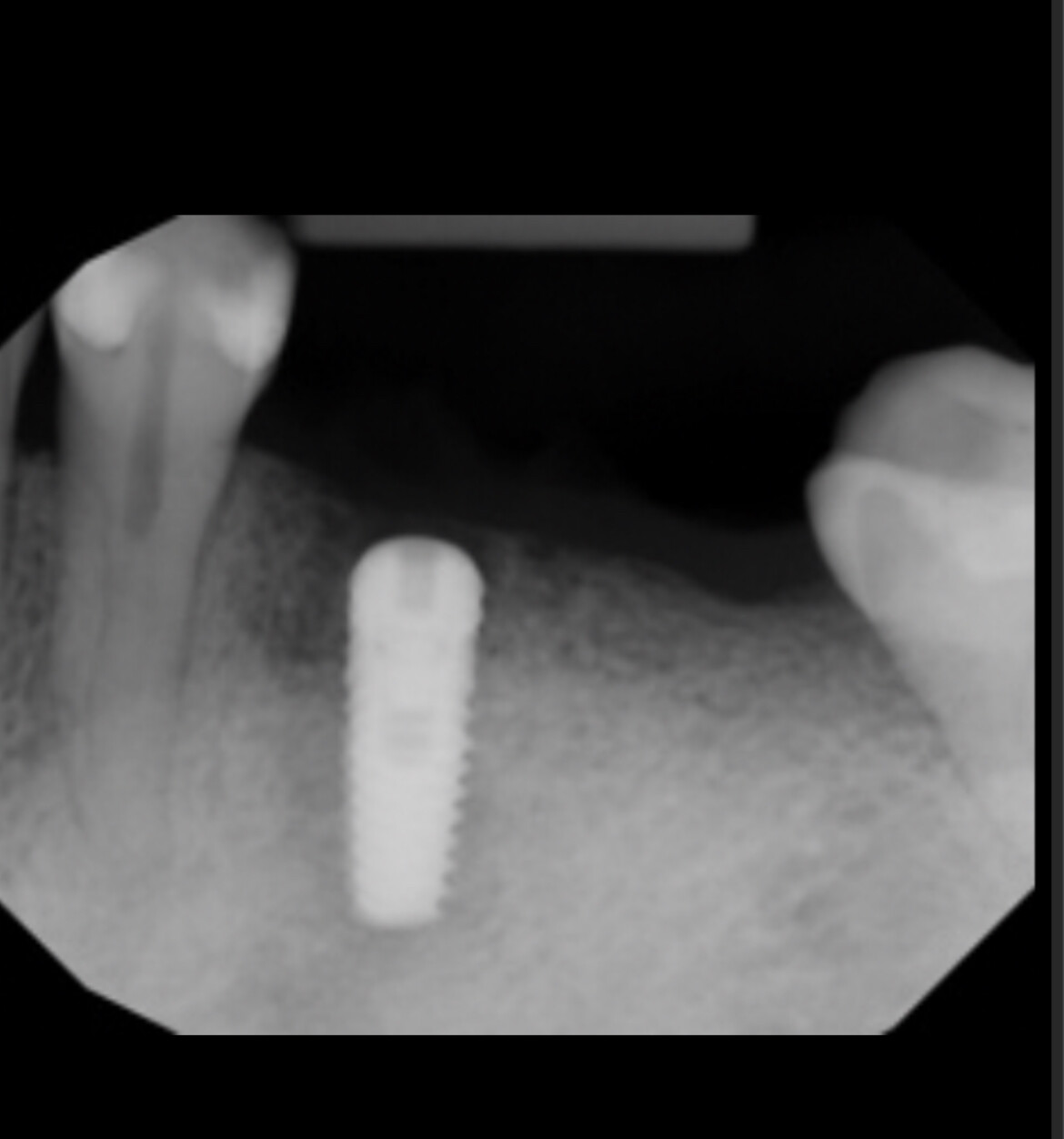
It is a common situation to find a Palatal plate with 2-4 mm bone overhanging the hollow mid and apical areas of the extraction sites. An expected cross section would show a "y" shape: the leg of the "y" is the palatal plate and the top of the "y" is the basal bone. This is not a rare finding, it is the expected finding even before any records. - We cannot see the ridge, but, even if it had a 6 mm bone width at the occlusal surface, you'd likely have too little width more apically. - So, you want to approach this fully armed with knowledge and options: -When you pull the flap you'll find a coarse plug of fibrous tissue from the periosteum , tightly bound into that bone defect. You'll have to scrape it out and thoroughly clean that bone defect. You'll have to use a membrane to keep the fibrous tissue from the periosteum from invading again. You can excise the fibrous lesion and send it in for Path Lab evaluation. - 
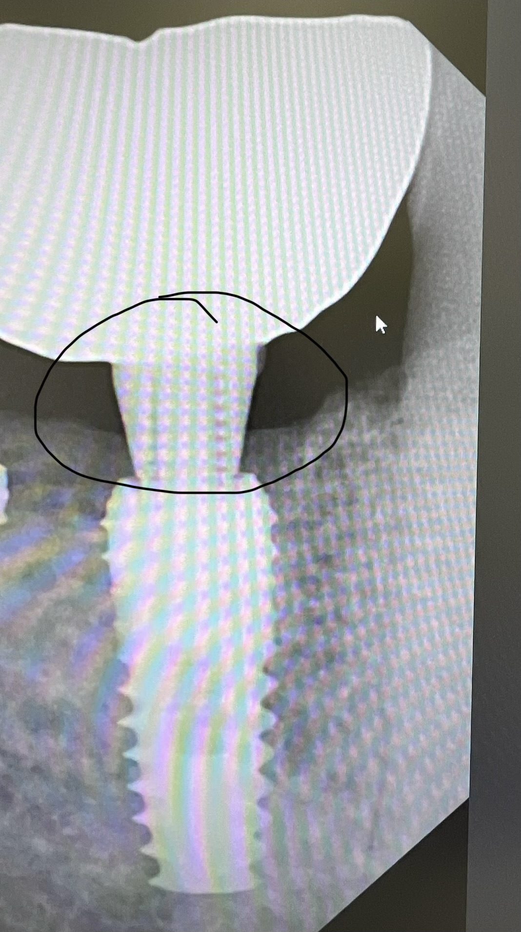
You'll need an implant design that will reliably become bound with laminar bone when immobilized in a sea of graft crystal/blood mix. (Bicon) - You'll need an implant designed to sit 3 mm below the anticipated Facial bone crest with a tapered top and a thin, 2.5 mm abutment leg rising up through the tissue, allowing you room for two implants, maybe 4.5 x 5.0 and 4.5 x 6 or x8. (Bicon 2.5 well series) - You'll need an implant that allows placement with only graft contact around half the surface. (Bicon) - You'll want to design a flap which takes 2-3 mm of the palatal tissue apically repositioned about 5-6 mm with the sutures in a periosteal set of loops and a second surface set. - You may want to put perforations, gently, in the plate vertically next to the implants and adjacent teeth to allow circulation in the graft. - the Membrane, prob. 20x30mm, will come up an over the tops of the implants with the patient's harvested bone atop the implants and be held down with 5-0 Chromic Gut. Then a Colla Plug is placed over that to cover the opening left from sliding the flap apically. - You must cut the incisions on attached tissue vertically parallel so the repositioned flap will close easily on the sides. Some loosening of the side tissues will give you a smoother transition. Then wait 3 to 4 months and you can uncover the implants with a slit. You will have a thick layer of attached tissue on the Facial and another 5 mm of tissue on top of your new, thicker ridge where the gums have grown over the CollaPlugged area. There are many things missing in this scenario, but it is a possible way of approaching your situation. Whatever you do, at some point you must flap the area and look at it. You can clean, place grafts over several appointments or you can inform patient you are prepared to do it all at once if the situation warrants that after you open and clean it. John
dr.danesh
5/25/2011
dear colleague; I think it is more likely a remnant cyst. to be sure you can do a biopsy & cbct. but if i would be the surgeon, due to lack of space mesiodistally,and a good bone buccolingually ,I place only one regular implant at the safe site, w/o entering the questionable site.the other one would be a cantilever. Even I donot bother my pt. to pay extra for CBCT nor the biopsy headache.happy pt. & surgeon w/ minimum discomfort and trauma to the pt. good luck.
Massoud Hosseini
5/25/2011
I think this is just scar tissue ,perhaps at the site of previous apicectomy. Much quicker and more certain to establish a diagnosis is to lift a semilunar flap and biopsy it. Additionally if it looks like scar tissue ,bur away the contents and the walls and bone graft it .
stephen travis
5/25/2011
Really good comments. I agree that treatment planning from PA is fraut. CBCT essential but for anterior cases discussion is made much better by having photographs. This shows smileline, ridge loss need for soft tissue replacement, gingival architecture and biotype- all important in decision of surgical intervention and need for one or two implants. The central lateral replacement is the hardest as it is rarely possible in an edentulous area to have a papilla. Hope this helps.I could not suggest a treatment approach until the photos were seen.
Richard Hughes, DDS, FAAI
5/26/2011
First get a history from the patient.
ktau
5/26/2011
I read many comments on this site, most were good and helpful especially this thread. Perhaps may i suggest that the drs posting questions to also post the final outcome so that we can all learn. Those who posted comments can also have the benefit of verifying if they were right.
Don Cohen
5/28/2011
I agree with most of te previous comments except nasopalatine cyst. It's not well corrugated, but needs to be scanned no matter what for proper diagnosis. Also one implant wig a cantilever is best to preserve the papilla.
John Manuel DDS
5/28/2011
For those fairly new to surgery, here are a few notes: 1- Why did the one surgeon suggest the removing the fibrous lesion before placing an implant? One reason is the scar tissue has little or no circulation. The bone gets is circulation from a healthy periosteal membrane and the medullary bone. Putting a bone graft under that scarred area of periosteum is like puttiing it in the Sahara desert. It is like spitting onto a blood agar plate and then placing it into an incubator. 2 - Why not just do one flap to remove that lesion, one flap to place and bone graft, one flap for tissue revision and another flap for the final implant placement? Every time you pull a flap, there is some cicatrical healing upon replacement, reducing the circulation. Every time you make a cut, you have built a "fence" in that tissue, a fence that will weaken it in future flapping and that will reduce the number and size of blood vessels crossing your slice line. Careful surgeries build barbed wire like fences with better circulation, but still less than that with which you started. Sloppy surgeries can almost completely bock the circulation with the thick band of secondary healing - somewhat like The Great Wall of China. If I already know the patient's general medical history, why would reviewing history make any difference? It is not the magic of the surgeon that makes for successful outcomes. Rather it is the diligence of the surgeon in collecting and evaluating critical information prior to the beginning of the surgery that has the greatest control over outcomes. For example, as mentioned above, every cut and flap in the area from past incursions will reduce the circulation and and weaken the covering tissue. How many times has this area been entered before? Also, does the patient have adequate blood circulation behind the graft (in their medullary bone)? What meeds are inhibiting circulation? Can I use meeds to increase circulation? Would the surgery benefit from no vasoconstrictor in the anesthetic to protect thin, narrow tissue areas? Why not just take each problem and solve it in a separate surgery? As mentioned above, each flap reduces blood supply to the proposed graft. Each incision places a barrier to circulation across it's length. So, one needs to step back and see if there is a safe way to combine procedures within fewer interventions. My suggestion to use an apically repositioned flap in moving the strip of tough palatal tissue to the Facial cervical area with parallel cuts on the attached area, would have those incisions flair widely at the end of the attached tissue, giving more loose tissue to reapproximate the edges of an almost round hole where that periosteal scar tissue was removed. A semilunar incision above that would reduce the available blood supply to the area in later surgeries and not increase the attached tissue. Also, the thin, mucosal tissue found over these F and B undercuts is notoriously fragile. Placing the main supporting sutures of a flap this large in those thin tissues is like suturing through cooked La Sagna noodles . If you take a 2-3 mm strip of tough palatal tissue at the top of the flap, you can put a suture in the under surrface of tough fiber with no risk of splitting and less invasion than going from outside in with the suture needle. Sorry for all the verbeage, but I am thinking some of the newcomers may not be understanding what is behind the suggestions of the surgeons who have been kind enough to share their experience here.
Fabio B
7/29/2011
Without a CBCT a correct treatment planning is not possible. The image could be a cyst, a cortical erosion or simply the nasopalatin foramen. I think it coluld be’ a corticale erosione but it’s only a personal opinion.

Featured Products

OsteoGen Bone Grafting Plug
Combines bone graft with a collagen plug to yield the easiest and most affordable way to clinically deliver bone graft for socket preservation.
CevOss Bovine Bone Graft
Make the switch to a better xenograft! High volume of interconnected pores promotes new bone. Substantially equivalent to BioOss and NuOss.