Refer large apical lesion on #19 to an endodontist or place implant?

This endodontically “virgin,” #19 [mandibular left first molar; 36] was an asymptomatic tooth. In retrospect, it had a very diffuse and essentially, unreadable apical lesion(s) noted on a periapical radiograph taken two years ago.

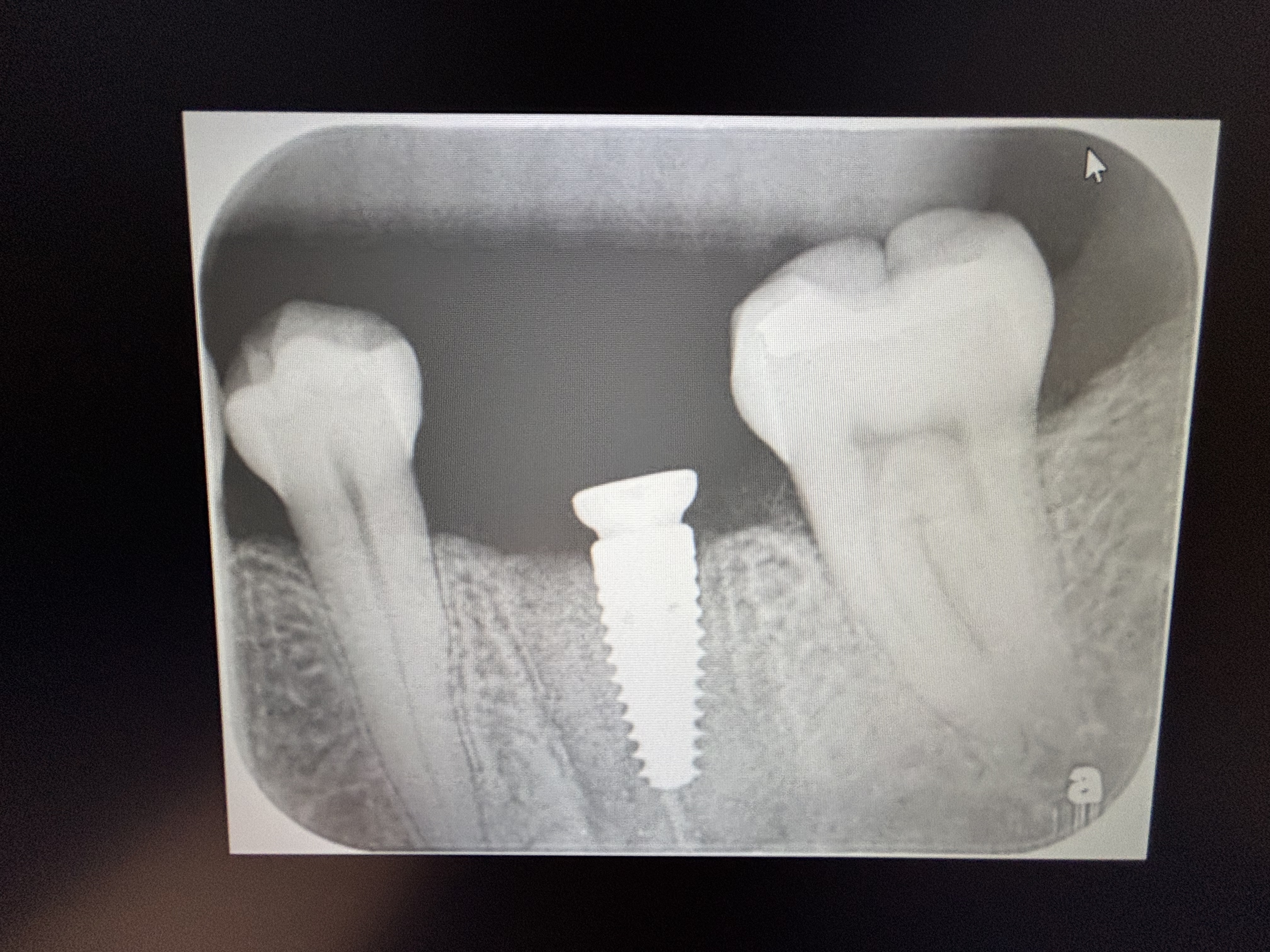
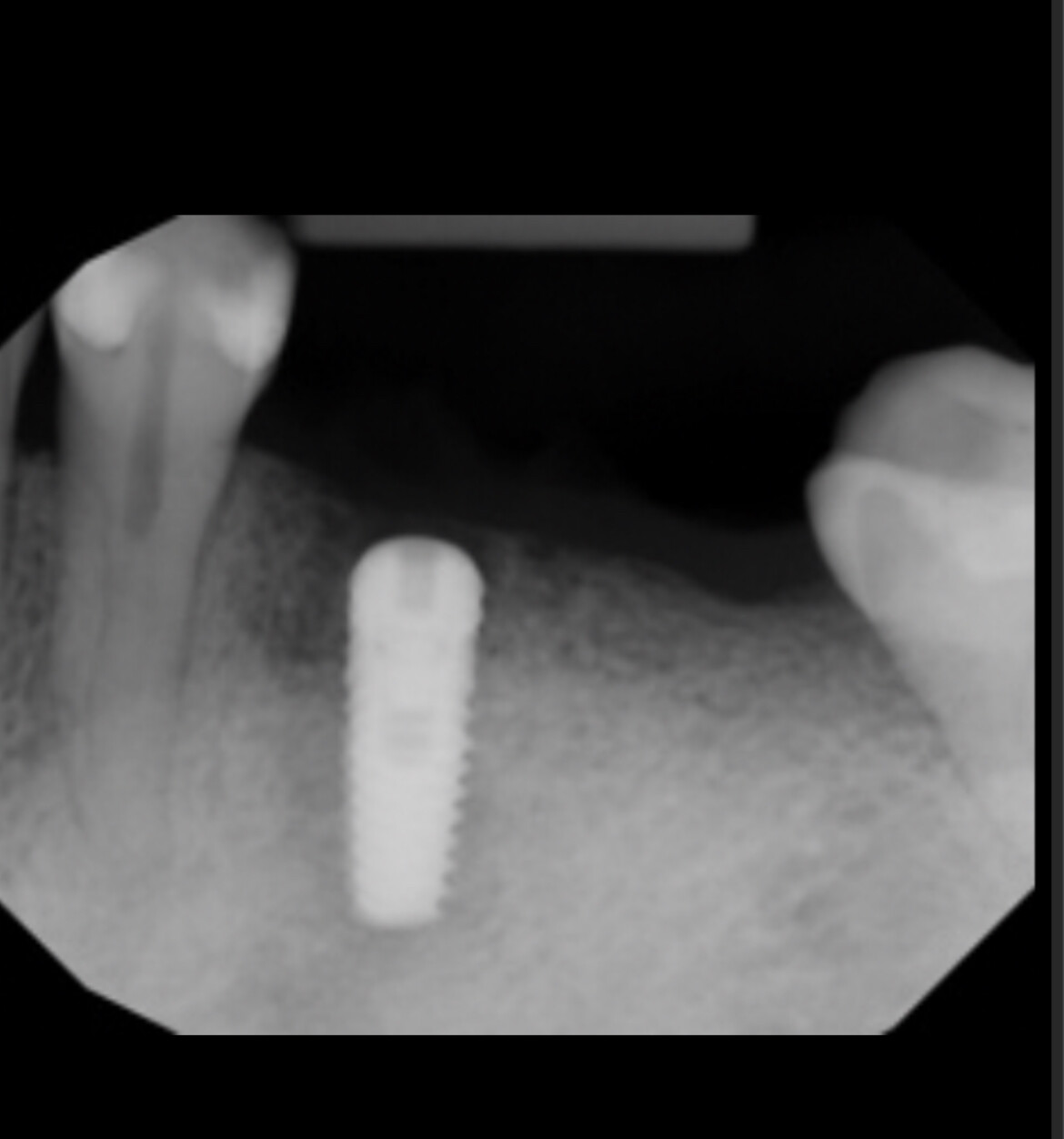
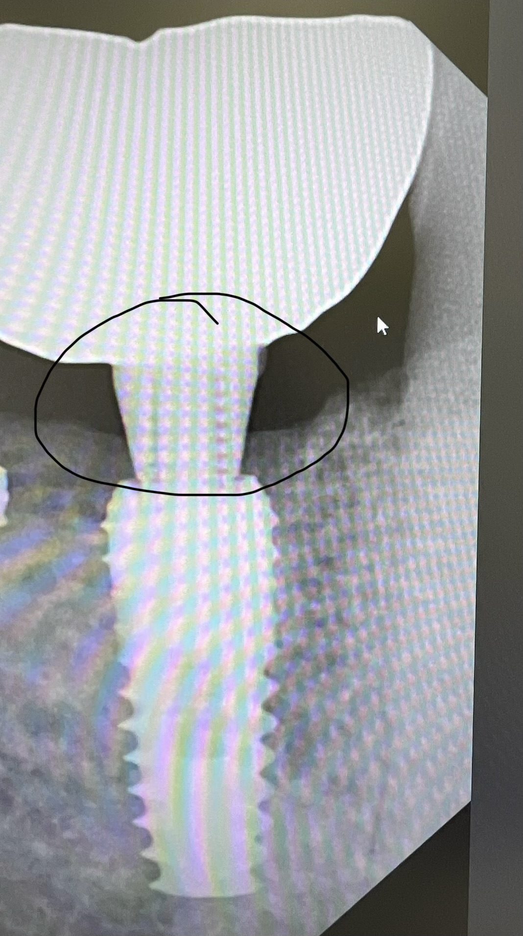
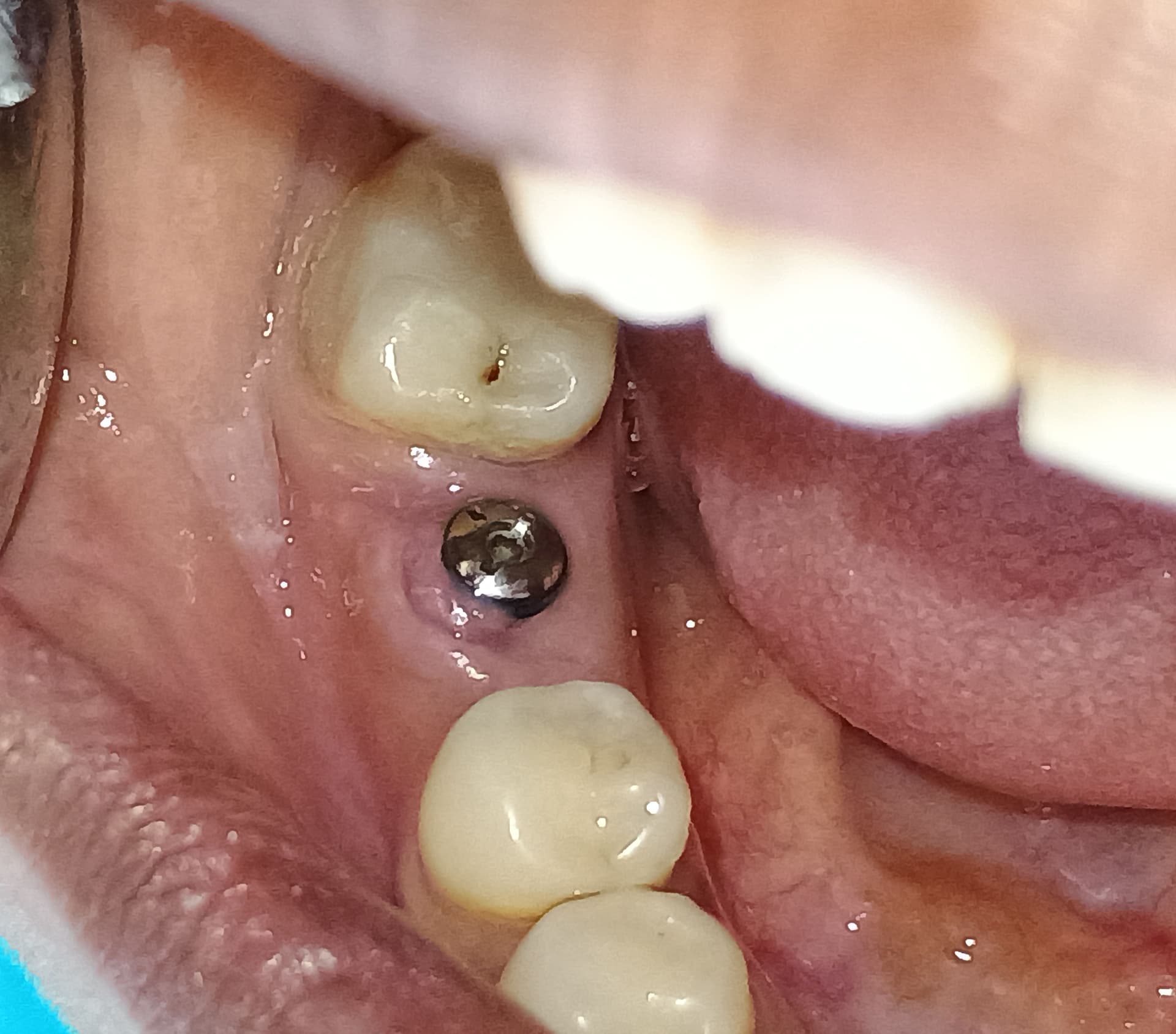
Yesterday, while preparing #18 [mandibular left second molar; 37] for a CEREC crown, I noticed a very small fistula between # 18 and #19. A periapical radiograph with gutta-perch point revealed a potentially large apical lesion #19. A follow-up CBCT revealed 10mm radiolucent lesion extending out to the mesial root of #18 and apically to within 1mm of the mandibular canal. While #19’s mesial root also had a periapical radiolucent lesion, #18’s mesial root seemed unaffected.

I can refer the patient to an endodontist for a consult. But given the degree of severe bone loss, what has your experience with the endodontic healing situation of a “virgin” lesion this large.

Yes, we all have seen lecturers with “miraculous” treatment where the fill is perfect and all of the bone grows back. But are they the “norm?” And I don’t ever remember seeing a single one of these “miracles documented on a CBCT.

So, given the limited but visually significant amount of clinical findings, would you suggest that your patient get an endodontic consult and possibly throw a ton of cash into a seemingly “herodontic” case, or extract #19 & #18 (#18 has only a very small amount of opposing occlusion) and thoroughly remove the granulation tissue, place a bone graft, then one or two implants. It would seem that the extractions and bone grafts would be the more predictable procedures in this case. Then implants could be installed in the healed sites. What is your opinion?

(click images to enlarge)


PA #19 distal fistula track with gutta-perchaPA #19 distal fistula track with gutta-percha
Full CBCT screen shot with "focus" on #19 large periapical lesions.Full CBCT screen shot with “focus” on #19 large periapical lesions.
CBCT tangential view with "focus" on #19 large periapical lesions.CBCT tangential view with “focus” on #19 large periapical lesions.
CBCT crossection Di root #19 with proximity to mandibular canalCBCT crossection Di root #19 with proximity to mandibular canal
CBCT crossection more posteriorl) Di root #19 with proximity to #18 mesial root and mandibular canal.CBCT crossection (more posteriorl) Di root #19 with proximity to #18 mesial root and mandibular canal.

64 Comments on Refer large apical lesion on #19 to an endodontist or place implant?

New comments are currently closed for this post.
ttmillerjr
6/9/2013
Definitely try rct. I've had many like this over the years heal well. I've also had teeth, like 18, that remained vital even when the lesion from the adjacent tooth has invaded the periapical bone. So eval vitality b4 tx.
Pynadath
6/9/2013
Refer to Endo. On another note taking a full cbct is a bit overkill with the radiation isn't it?
CRS
6/9/2013
Is this a vital tooth? If so send it to an OMS for a biopsy, I had a very similar case which turned out to be an odontogenic keratocyst. Same presentation I thought it was a lateral periodontal cyst. Take it out of occlusion. You need to know the etiology I don't see caries perio or traumatic occlusion. Don't miss the diagnosis which is key. This is not a simple root canal Thanks for reading
Keith Komatsu
6/11/2013
CRS, If the tooth tests vital, I would recommend starting root canal treatment first to confirm pulp vitality diagnosis. Multirooted teeth can give false positive responses. In addition, if an OMFS is going to biopsy, they would probably be as aggressive as they need to enucleating the lesion knowing they do not have to worry about devitalizing the tooth.
naser
6/10/2013
it is a common periapical abscess and the source is the crowned non-vital 18, refer to a periodontist for RCT. treating and preserving a natural root is much wiser than extracting and implant placement. here u have 18 and 19 in a very solid state and they can function better than any implant in the world.its a simple case and could be treated in a simple and straight forward manner unless you have another calculation. thank you
CRS
6/10/2013
Also on your axials the nerve is closer to the inferior border, not sure that superior radioluncency is the canal, I like to trace it from the lingula. I think you migh be picking up the radioluncency just inferior to the roots on the distal root. There is something funky going on here. I would open this up and biopsy this bothers me that the etiology of the radioluncency is unclear, interesting case.
Richard Hughes, DDS, FAAI
6/10/2013
Do the Endo or refer to an endodontist.
Dr. Alex Zavyalov
6/10/2013
To my mind it’s a typical burned pulp complication after tooth aggressive preparation. Both roots are involved and the bone lesion is vast. Try to save the tooth, but I’m not sure that this endo treatment will be effective for the bone rehabilitation.
CRS
6/10/2013
It would be helpful to send a biopsy of the lesion the buccal plate is perforated. This seem to be an unusual spread pattern. Are the teeth vital?
Dr Don
6/10/2013
Thanks everyone for your continued help with this case! Here is some additional information. The tooth with the large distal periapical lesion (#19) is non-vital. A close inspection of the tangential CBCT “screen shot” shows a separate and distinct mesial periapical radiolucency. There is a long standing crown on #19. While I don’t have records as to the tooth’s preoperative condition, there is a high probability the crown was done in response to caries and/or a fracture. I too am very uncomfortable with the nature of the lesion and the amount of bone that has been lost. As such, I agree with those that have suggested a biopsy. To perform this biopsy and the a complete enucleation of the “cyst”, without destroying the buccal plate, it has become clear that an extraction of #19 is in order. As I implied in my original post, this patient is not looking for “hero-dontics.” The two endodontists I have consulted on this case give #19 only a fair prognosis. I feel that with the significant “internal” bone loss, the unknown nature of the lesion(s?) and its causative factors, that an extraction, biopsy and bone graft followed by an implant may offer far more potential for success than a “finger crossed” endodontic attempt. Especially since the potential implant site will have its buccal plate intact and would probably present with decent post graft bone height. While I feel that endodontics is now currently considered a first choice for teeth with good to excellent prognosis, I don’t agree that its success rate is consistently better than implants. In fact, looking at multiple CBCTs of my patient’s supposedly “successful”, root canals (most teeth were specialist treated with 2d imaged “proof”), I wonder how well, their now visible residual “cyst issues” will be accepted in a CBCT “standards” future. With thousands of hours of CEs, I am a MAGD / FICOI who is now even more perplexed as to the decision tree process for these situations. And…I am open to any suggestions regarding courses and reading materials. Sure, I can make it the endodontist’s “call.” However, I afraid it will be “endo” almost every time…until, like perio and oral surgery, the discipline is “Limited to Endodontics and Implants!!!”
CRS
6/11/2013
Dear colleagues, whenever I see something unusual I always like to ask qualifying questions vs treating a case in the only way one knows how. If this case was referred to me now knowing it is non-vital and treatment planned for extraction I would biopsy this unusual radiolucency, It is the only way to be sure. That's what I feel a prudent surgeon would do. If it is a simple cyst great but if it is another type of pathology then it has been diagnosed. It has nothing to do with endodontists or RCT but wise council from an experienced surgeon sharing a viewpoint. It is not about implants or root canals but diagnosis and pathology. If you guess you could be wrong. That said an excellent pick up on the cone beam by the poster and asking more questions very astute. Now reading up on this is fine but I feel referral is indicated to someone with oral pathology experience that's my wheelhouse! And yes each practictioner has their bias I've biopsied bone after the RCT or implant take the help that's what my training is for to help. Again thanks for reading!
ttmillerjr
6/10/2013
If the patient understands RBA's and you both feel more comfortable with removing and going the implant route, then no problem, move forward.
Richard Hughes, DDS, FAAI
6/11/2013
This is a classic "paralysis due to analysis". Perform endodontic treatment, if the tooth is not mobile. If it does not work it can always be extracted etc. How would you want to be treated in this situation?
Dr L
6/11/2013
I totally agree with you Richard. Common things happen commonly guys. All this talk about keratocysts, biopsies etc etc is valid but really, just open & drain the tooth first & see how its resolves.
CRS
6/12/2013
I would want the pathology definitively treated and removed from my body. I would also like to be reassured about the diagnosis rather than watching and hoping that the root canal will solve the problem. I hear this on a regular basis from patients.
endodontist
6/11/2013
The diagnosis for this tooth is necrotic pulp and chronic periapical periodontitis. If the RCT is done properly, the prognosis is good for the tooth. It would be unfair to the patient to not present the RCT option for the patient and encourage implant and ext. Success of initial therapy endodontics is very high. If this case was a retreatment with adequate looking root canal therapy, then there would be a better argument of extracting the tooth vs. saving the tooth. Dentists have been doing endodontics for years. Extracting a tooth should be the last option on the treatment plan.
Zaki Kanaan
6/11/2013
Pynadath, many endodontists now have CBCT's in their office and use it fairly routinely for endo. I've been surprised by many largish lesions that have healed completely. General bone levels in the area are too good to 'bin' the tooth IMHO without trying endodontics first.
mrc
6/11/2013
the rct prognosis is good. In this case dentin is better than metal. Refer to your local endodontist and make two friends, the patient and your endodontist.
dentist dave
6/11/2013
All great suggestions from different points of view. Regardless of what the statistics and clinical research says, it is ultimately up to the patient to make the decision with the help of the dentist to explain the RBA's. Money and time is important to most patients so we must give them a chance to consider all options before making the decisions. Sometimes these are tough calls but if we educate our patients on the time, cost and prognosis, its easier with their help to diagnose and treat..
CRS
6/12/2013
Patients don 't decide treatment in non-elective treatment of pathology which could be harmful to them. They rely on the experience of their treating doctor and consulting doctor. Keratocysts and other pathology develop in long standing periapical lesions although more commonly seen in the more posterior mandible. I do want to apologize for sounding harsh many of these posters just don't know what they don't know I think they mean well but have one type of treatment and don't rely on their OMS. Perhaps they do not have a good working relationship with their OMS colleague so I don't want to place any blame but state what I would do to help all involved. I don't set much store in CE training and specialty background but I listen to the content of what is said. There are a lot of very wise dds regardless of credentials. If I hear a CV stated I am usually suspect if the advice I feel doesn't coincide with the appropriate treatment.
Keith Komatsu
6/11/2013
As an endodontist who has had formal implant training, I would like to chime in. An endodontic consultation is warranted in this situation or at least offer this option to your patient. Your endodontist should be communicating to you and the patient the diagnosis, treatment recommendations, and prognosis. It sounds like you do not have a good relationship with your endodontic colleagues by your comment "get an endodontic consult and possibly throw a ton of cash into a seemingly “herodontic” case". We as endodontists have to bear some of the blame as to how our referring doctors feel about the outcomes of endodontic treatment. There has been such an emphasis in doing one visit endodontic treatment. So when that "virgin" root canal comes back from the one shot endodontist and the sinus tract is still present, you will make that assumption endodontic treatment does not work. The point is case selection is important. In this particular situation with the limited amount of information and if the pulp testing indicates there is pulp necrosis, then endodontic treatment could be initiated with placement of intracanal medication placement. If followup finds the sinus tract to resolve and reduction of the periradicular lesion size, then complete treatment.
Steve
6/11/2013
I am an endodontist, and I also have been doing implants for 15 years. This is absolutely not an extraction case at this point. This is one of the very predictable 90+% success cases with proper endodontic treatment. The crestal bone appears to be nicely in-tact. It would be blatant malpractice to remove this tooth at this time. It doesn't matter how large a periapical lesion is -- as long as it is all periapical, and there is no probing to an apex, the prognosis for full bone healing after proper endo treatment is excellent. 18 is not involved at all. Stop looking for zebras on a horse farm.
Brian
6/12/2013
Agree with everything you said 100%.
CRS
6/12/2013
It no longer a periapical lesion with this presentation , perforated buccal wall and this size. It would be difficult to come up with diagnosis based on a root canal treatment. This will continue to fester and drain after root canal treatment. I feel it is a more serious maltreatment not to biopsy this lesion and curette it out around the extracted or not extracted tooth using a buccal access with the perforation as a guide. It is straightforward and easy if you have he experience. I have removed many much smaller lesions on RCT teeth and extracted teeth by other colleagues. Sometimes I am surprised by the path report. I just don 't get the watching and resolution thing that was taught in endo. I feel that philosophy is a disservice to both the training if an endodontist, generalist and patient. Pathology is pathology.Good Luck
Pynadath
6/12/2013
Crs I really don't understand your point. You are saying because there is buccal wall perforation and bone loss it's something more than a Endo lesion? I'm not sure if you are aware but chronic Endo lesions have buccal wall perforations and large due to them being chronic and leaving them untreated. What's so special about this case???
Endodontist
6/11/2013
Well that is embarrassing. I gave the wrong diagnosis. It's necrotic pulp and chronic periapical abscess because there is a sinus tract.
Pynadath
6/12/2013
Zaki I'm fully aware of endobods and a whole bunch of other specialists having cbct at their disposal. But does that mean you should zap every tooth with a paeriapical lesion with a cbct instead of just a paxray? Is it going to change the prognosis? I don't believe so. I'd understand if they had xla the tooth and were about to place implant. But that's not the case here. Or not yet anyway....
PKC
6/12/2013
If it was my tooth, I would follow endodontist comments above.
Peter Fairbairn
6/12/2013
We have an in-house endondontist and every referral is given a second re-appraisal if at all possible to save with a good long term prognosis. I just treat paients as I would like done to myself or my family . Saving teeth is our initial priority . Peter
Zaki Kanaan
6/12/2013
Pynadath, As you can see by the number of opinions and treatment options given on this thread having the CBCT has proved very useful. The general consensus is that this tooth is amenable to endodontics and saving trying to save the tooth rather than extract were also my initial thoughts. I have 2 endodontists who work for me and both have said they would take a CBCT if this was available. Even if you were referring to someone else, it helps you with your initial differential diagnosis and the following discussions with the patient. If it was my tooth and the dentist wasn't sure about the most appropriate option going forward, I would be happy to have a small volume scan to help come to a decision. Doses are coming down day by day and this is becoming more routine IMO.
Pynadath
6/12/2013
Zaki As you know I'm not a endodontist. but I can't see how a endodontist would need a cbct before Endo in this case. I would've thought a pa X-ray is sufficient in this case. There is no doubt a cbct shows more info, especially now it's been taken. But was it needed for Endo? I don't think so. Th Endo guys who have answered haven't specifically referred to the cbct being especially useful and justified over the paxray. Although now its been taken obviously it has a use. All the Endo guys I've worked with haven't generally needed a preop cbct before Endo.
Zaki Kanaan
6/12/2013
Times are changing Pynadath. As Dr Don and others have said I don't refer to an endodontist who doesn't have a CBCT or have easy access to one. Now of course I'm not suggesting every tooth destined for RCT requires a scan, far from it, but what I am saying is that when something approaches cystic proportions, like this lesion, and when you, the treating dentist, are unsure of the best way forward, either xla or RCT, then yes it is indicated. The scan can reveal lateral/interradicular canals, the canal system/morphology and sometimes even cracks not visible on a regular PA. I could go into a comparison of doses for you and it really isn't a big deal with newer CBCT's that can be honed down to 2-3 teeth in a quadrant and the images now use even less radiation than before due to superior image enhancements.
pynadath
6/12/2013
I cant see any special reason to provide a cbct in this case. You mention because of the associated radiolucency of the lesion. what size does it have to be in your opinion when its justified for cbct and not? I wouldnt class this size lesion totally out of norm for a chronic lesion. The endodontist certainly hasnt requested for the cbct from what the op said. From studies the reasons and Justifications for cbct in endodontics:- Differentiation of pathosis from normal anatomy Relationships with important anatomical structures Aiding management of dens invaginatus and aberrant pulpal anatomy External resorption Internal resorption Lateral root perforation by a post Accessory canal identification Surgical management of fractured instrument Aiding surgical endodontic planning references: Cotton et al, 2007 John, 2008 Siraci et al, 2006 Maini et al, 2008 Cohenca et al, 2007 Walter et al, 2008 Patel et al, 2007 Patel & Dawood, 2007 Cotton et al, 2007 Young 2007 Cotton et al, 2007 Nair et al, 2007 Patel & Dawood, 2007 Tsurumachi et al, 2007 Patel et al, 2007 Patel & Dawood, 2007
Zaki Kanaan
6/12/2013
Pynadath, I studied with Shanon Patel and work closely with Andrew Dawood on many cases and refer to them regularly. I consider them good friends and they account for 5 papers you reference above. Both are great advocates of CBCT as you may be aware and they often include snapshots of a CBCT scan on writing back to the referring dentist on far lesser lesions than this. I also work with Michael Sultan and Richard Kahan, all advocates of CBCT. However you seem to be missing the point with my post for you to spend your evening looking up the papers above. I think the original poster did the right thing by taking a CBCT scan as he was unsure of the way forward. Taking a CBCT scan may have aided in his decision making process. I did not say that the endodontist requested a CBCT. If your question to me is 'would I have taken a CBCT?'no I wouldn't because as you can see from my original post, this is a clearcut endo case and therefore my decision making process was clear...but that is me. I don't think anyone here would take a CBCT scan for every case and likewise I don't think anyone would suggest taking a CBCT scan for this case by the OP is wrong as you seem to be implying. The two endobods I asked actually said there is nothing wrong with taking a CBCT for this case also. It all helps. If it makes you feel better I can ask Drs Patel, Dawood, Sultan and Kahan their viewpoint.
Baker Vinci
6/17/2013
I have found way to many missed canals, even from the specialist; it is my opinion that every root canal should get a scan. Bv
Dr Don
6/12/2013
All great suggestions! My patient will get another endodontic consult. However, if this tooth had an “ideal” endo fill, and still presented with a similar lucency on a CBCT, an implant would have a better prognosis. This begs the question: Do you really have a successful endodontic case if the patient develops a post-op, asymptomatic, lesion not visible on a PA that “screams” at you on a CBCT? I don’t! Frankly, I stopped referring to endodontists that don’t have a CBCT or the capability of interpreting my scans.
pynadath
6/12/2013
The xray and cbct reveals 2 apical radiolucencies in both root apices. The distal being larger than the mesial. Likely due to the necrotic nature of the pulp and tooth being non vital. Leading to periapical periodontitis and associated buccal sinus from the distal apical infection. The buccal sinus, like all chronic endodontic fistulas has lead to buccal bone perforation for the pus to drain out from. Why are some posters saying this is unusual? This is normal for a chronic endodontic infection. Listen to what the endodontists who are posting on here are saying. refer to the endo guy!
Baker Vinci
6/12/2013
The scan needs to be done first. Root canal therapy, just delays the inevitable, because two teeth are involved. It is likely that the tooth or teeth need to go and once the Infection resolves, start from square one. You can send the inflamed peri apical Cyst to a pathologist, I'd would like. Bv
CRS
6/12/2013
I would still recommend sending the pathology for a definitive diagnosis. Why wouldn't you? It does not make sense not to and any studies or endo experience is not relevant so what is the big deal in sending the specimen? Do you not know how to interpret a path report?
Pynadath
6/13/2013
Zaki You must think I'm crazy if I looked at papers just for this! That was just a copy and paste from a power point presentation. I think your missing my point. let's go back to the points you made. I have no doubt cbct is useful and used by many endodonists and it is useful info.i think this is common knowledge to most dentists and I never denied this.I totally agree and am not sure why you needed to state this as I didn't say anything to disagree with this in the first place. I didn't even say it was wrong the do the cbct in this case. HOWEVER I can't see why it's needed (yet). What's the justification? You said yourself you wouldn't have taken it. Neither would I. If the op was unsure of he way forward due to prognosis for the Endo, don't you thik it wouldn't have been better to let the endodontist decide on doing the cbct after referring to him? Like the other endodontists who have commented, it looks like a straight forward Endo case here just from the paxray. So what's the special reason for the cbct. It can't be just to see size of radiolucency as I don't thik it's to the size or character to be something more than a paeriapical infection. Remember this isn't a sectional cbct here. It looks as though the whole upper and lower jaws were zapped. Now you could ask Patel et al, but unless they can say from the paxray there's a special need for cbct I can't see how it's needed.
Zaki Kanaan
6/13/2013
I couldn't agree more Pynadath. I am also unsure why a full CBCT was taken to investigate the LLQ. That is a question you will need to ask the OP. All my references were to small FOV and that if taking one helps both the dentist and patient come to a decision, then so be it. Codiagnosis. No harm done. Justification "I was unsure how to proceed with this case and had initially thought the LL6 may require extraction due to the size of the lesion visible on the PA. I decided to take a CBCT to determine the extend of this lesion and to help make my decision as to whether RCT was a viable option for the patient."
Pynadath
6/13/2013
Zaki, You do realise my comments were in reference to why a full mouth upper and lower cbct was taken for a localised area. It wasn't a small section cbct of just the lower molar teeth. Hence my first post "taking a full cbct is a bit of overkill" What's the justification for the full mouth cbct? Why were you referring to a small fov cbct when that's not the case in this? I was presuming you were talking about the full cbct was ok??
Zaki Kanaan
6/13/2013
Pynadath, I was aware you're initial comment was about a full CBCT. My response to that was that "... many endodontists now have CBCT’s in their office and use it fairly routinely for endo". I was just making a general satement of fact that they are becoming more commonplace. An endodontist would never have taken a full CBCT, so I wasn't referring to the specific use of a full CBCT in this case but just in general. However your subsequent post refers to single teeth with lesions and reads "But does that mean you should zap every 'tooth' with a periapical lesion with a cbct instead of just a paxray?" so hence it was you who initiated the debate about the use of small FOV CBCT's for single teeth. For this specific case, I think we agree in broad terms...we both agree that this is an endodontic case and we both would have initiated or referred for endo rather than extract and implant. Where we differ is that you would have let the endodontist decide on whether a CBCT was required (and actually so would I, as I have mentioned previously), BUT as I have also said and probably where we differ is that " I think the original poster did the right thing by taking a CBCT scan as he was unsure of the way forward. Taking a CBCT scan may have aided in his decision making process". Admittedly a full CBCT was probably not needed but a small FOV one would IMO have been justified. As a side note, I had a chat with my endodontist about this case and his response about CBCT's in general was "Life is changing and in a few years every endodontist will have a ct machine-low dose and volume...buy shares" :-)
Pynadath
6/13/2013
I'm saving up for the nursery fees let alone for shares! :)
CLK
6/13/2013
Hi, why don't we open drain the tooth after clearing the necrotic debris and review after a week or two. If there is improvement in the signs and symptoms treatment planning will be easier. Thanks.
Richard Hughes, DDS, FAAI
6/13/2013
CBCT will not be the standard of care, until they are in the majority of dental offices. This will only happen when they are sold at a much more reasonable price! I have seen cases that looked bad resolve with endodontic treatment and remain in service for many, many years. If #19 is not mobile, give endo a chance. You can always take it out later.
Baker Vinci
6/13/2013
Richard is correct, IMO, but it should be the standard of care. CRS, I still have a microscope and can see cholesterol with my "naked eye". You might as well get a chem 20 and CBC with diff.. The odds of this being anything other than reactive disease is hugely unlikely. I don't think it is worth a 75-100$ path fee. Bvinci
CRS
6/13/2013
Standard of care is what a reasonable dentist would do in an identical situation with care and concern for a patient, it is determined by a jury. Experts opine on treatment choices, literature is only helpful as a guide since anything can be refuted. Biopsy will tell you if there is any significant pathology. And by the way with all this talk about oral systemic inflammation and infection why not remove the periapical pathology of a significant size instead of waiting for the endo to clear it up, makes sense doesn't it? We all know implants are not a panacea for basic dentistry .
Baker Vinci
6/13/2013
Standard of care is not determined by a jury. " It " determined by what is done by the majority of doctors in your geographic area. I have yet to be sued( it will happen ) , but I have sat on several peer review panels and jurors have no affect on the "standard of care". CRS, have you ever seen an ameloblatoma, myxoma, keratocystic tumor, or scca develop at the apices of a Perio/endo involved tooth. Do you send the follicle of every partially impacted third molar to a pathologist? BV
CRS
6/14/2013
I'm sorry BV the standard of care is determined by a jury in the legal sense, trust me on that. My clinical protocol is that if there is a significant amount of tissue that has an unusual presentation I am always glad I sent the specimen. And yes I have seen unusual path reports in unusual locations and have been surprized. I don't routinely send follicles either sounds to me you may want to send more specimens and I hope you never get sued for missing pathology. The chem 20 and cbc is just an arrogant comment it has no bearing on sending a specimen. Not sure why you stated that. The patient deserves good care and the excuses I hear in this entire thread of posts confound me. It is just I would do Thanks for reading.
Baker Vinci
6/14/2013
Not trying to be arrogant, but we have some surgeons that send every follicle to the pathologist and charge for the removal . Bv
CRS
6/15/2013
I hear you no worries!
Dr Don
6/13/2013
CRS is on the right track regarding “standard of care.” Sure a CBCT is a very expensive purchase for myself and others. So was the decision, at age 60, to take a hundred and fifty hours of implant CE’s, and start performing procedures. Interestingly, every one of my instructors stated (in 2010) that you never place an implant without a CBCT. With hospital access to CBCTs, X-ray technicians driving them around in vans, local endodontists, orthodontists and oral surgeons having machines, the availability and/or cost of this device, in my region, is completely irrelevant in a court of law. If you don’t take a pre-op CBCT and you “screw-up” an implant case, you might as well give your full consent to a malpractice award. Like it or not, in the United States that is how “standard of care” is determined. Lastly, I took a full CBCT, (using a new machine that takes them with the purported dosage equivalency of my old film based pan) because the patient was having additional, symptomatic issues in UR areas of previous endodontic treatment. Two of these areas proved to have significant radiolucencies at their root apices (also not visible with 2d imaging) and are also being referred to the endodontist.
Edward Dergosits
6/13/2013
I am quite surprised that many think this case is complicated. When I see stripes I usually think of a Zebra not a horse. It is almost certainly a chronic apical lesion of endodontic origin. The tooth is solid. I personally would not even discuss implant treatment until an experienced endodontist treated this tooth and the lesion did not heal. In one year there will be complete bone fill and the patient will be healthy. Biopsies are not needed. What this patient needs is a competent endodontist and another new well fitting and properly cemented crown. If it were my tooth I would insist on it. Ed
Peter Fairbairn
6/14/2013
Like all diagnostic tools you use them when you you need them and not as a source of revenue . The issue with buying is the tendancy to scan too much to pay the machine off . Hence you see before and after scans etc ( I am Guilty as well of this but rarely ). Fortunately in all big cities there are companies that provide a great sevice with the added benefit of a qualified radiologist to help in the analysis . The idea of all cases needing scaning may only have value for the lawyers but limited diagnostic value in most cases. The other issue with buying is that todays latest technology is scrap in 3 years , so let the hospitals and scanning services take the hit and work with them. Regards Peter
CRS
6/14/2013
Peter again very helpful feedback, I'm waiting for the prices to come down like VCRs and microwaves (what's a vcr?) Or I'll get one from a hospital! Regards!
Richard Hughes, DDS, FAAI
6/14/2013
CLK flapping, curettage, detoxification, synthetic particulate graft (OsteoGen) with PRP is a distinct option after successful endodontic treatment. One does not have to do the above grafting but is would sure help!
Baker Vinci
6/16/2013
Personally, I see no need to graft an area that has had appropriate root canal therapy. If the periosteum is intact, bone will regenerate. I do have cbct documentation of a successful case, that is similar to this one. I have always encouraged the patient to remove the Perio/endo involved tooth or teeth. I believe this is the exception. Bvinci
Steve
6/17/2013
Another thing -- The reason why this is NOT a "virgin" tooth is that it has been prepared and has a crown on it. This is the MOST COMMON causative factor for endodontic problems (for those of you in Rio Linda). Again, have an endodontist do the endo and be done with this. 90%+ success rate. Not even a question.
Baker Vinci
6/17/2013
Yuup, Steve I was concerned that maybe I didn't understand what virgin meant. Yeh the tooth has been " violated ". Bvinci
CRS
6/18/2013
Somebody missed a pulp exposure or a traumatic occlusion which devitalized the tooth. Now there is large lesion which no one seems to want to remove. Now cbct Is great however a microscope is used during the root canal to see the canals during treatment and to correlate the X-ray findings. Perhaps this treatment path is why there is the hesitancy to remove the tooth. So do the root canal under a microscope and remove the lesion to be sure. The benign neglect over the last two years has not helped the patient, fix it and move on! Thanks for reading sorry to be so harsh!
M. Friedman, DDS
6/18/2013
I believe the OP meant "virgin" as in no history of endodontic treatment. He stated "endodontically virgin" which was confusing to me as well.
Bgr
6/18/2013
I have seen and treated teeth with bigger lesions endodontically and it healed - and I am not an endodontist (Periodontist/general dentist). In my opinion it would be malpractice to not even try or suggest it to the patient. I hope you tried to keep the tooth. Don't you read the literature that periimplantitis also becomes more and more of a problem.
Dr Don
6/19/2013
Steve, this case was one of two #19 cases I simultaneously submitted to Osseonews. (The other case, now "top listed", had endodontics.) Both #19s had crowns. However, to distinguish this case from the other, I posted this one as: "This endodontically “virgin” #19. I hope, given the tooth had a crown, no one else thought I was referring to a “virgin” tooth. Also, I don’t feel that "broad" statements indicating that crowns are the "MOST COMMON causative factor for endodontic problems" are helpful to the profession. Rather than assume that full coverage preparations cause irreversible pulpitis, I tend to believe the real causative factors are most often vital teeth with deep caries, trauma, undermined/fractured structure that don’t fully express the extent of their viability until long after the crown was placed. Obviously, that doesn’t apply to truly “virgin” teeth that were “cut down” for FPDs or cosmetics. As for this #19, your advise and the majority of others has been taken to heart. My patient is being treated by an microscope utilizing endodontist with CBCT interpretation experience. Due to the long standing chronic nature of the lesion, and the amount of bone loss, she was advised of a guarded prognosis. I will keep everyone posted!

Featured Products

OsteoGen Bone Grafting Plug
Combines bone graft with a collagen plug to yield the easiest and most affordable way to clinically deliver bone graft for socket preservation.
CevOss Bovine Bone Graft
Make the switch to a better xenograft! High volume of interconnected pores promotes new bone. Substantially equivalent to BioOss and NuOss.