Splitting the Ridge Instead of Block Graft: Details on Procedure?
Dr. A. asks:
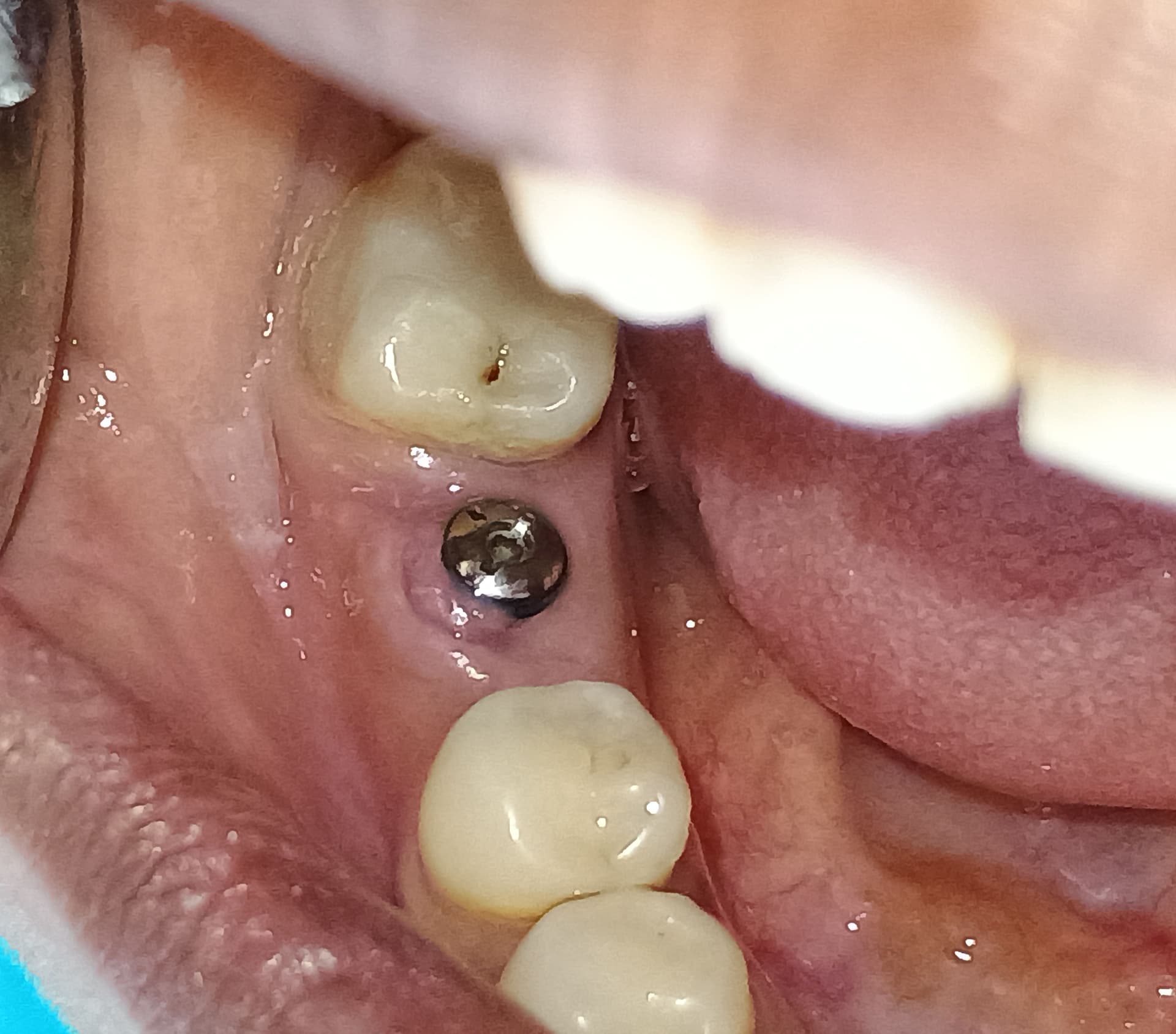
I have a patient who requires an extraction of a non-restorable maxillary lateral incisor and immediate placement of an implant. There is a slight buccal concavity that complicates the placement because of the lack of bone volume at that part of the site. I have read about splitting the ridge to increase bone volume at the recipient site instead of doing a block graft on the buccal of the cortical bone. What instrument sequence should I use to split the ridge? What narrow diameter implant would you recommend for this procedure? What bone graft material should I place around the implant? Do I need to cover the site with a membrane?
20 Comments on Splitting the Ridge Instead of Block Graft: Details on Procedure?
New comments are currently closed for this post.
IP
8/3/2009
This is a very complex scenario involving pre-existing tissue deficiency, extraction of non-restorable tooth (endo? fx?), immediate placement?, and in the esthetic zone. IMHO, if not 100% comfortable with all types of complications, this isn't the case to try out...
Anyways, where is the concavity? Is this a case of a shortened root and apical bony deficiency? In my experience, the most predictable method to get this right is to extract and preserve the ridge without attempting primary closure, using mFDBA and a long term collagen membrane. Then after healing for ~6 weeks I go back and do a block graft. I've tried doing ridge splits in this situation and haven't felt that the result are as predictable. A couple mm short in results in the esthetic zone could be harsh.
I've also attempted to tunnel particulate graft to the apical deficiency from a vertical incision and have gotten mixed results. In my hands, a block graft gives the best and most esthetic results.
Dr Dwayne Karateew
8/4/2009
A few questions:
-Have you completed a CBCT to assess the bone volume and presence/absence of buccal plate of bone?
-Are esthetics an issue for the patient?
-Your level of experience?
In my opinion this is not the situation for a ridge split technique as it seems to be an extraction case with either 1. immediate implant placement or 2. delayed with socket preservation grafting. Ridge splitting tends to be done in long term edentulous areas where there is a thin knife edged residual alveolar ridge (with adequate height) and not usually associated with recent extractions.
I would recommend extracting the tooth (after a CBCT) and either immediate implant placement with concomitant bone allograft+resorbable collagen membrane or a staged approach with bone allograft and membrane and then placing the implant later. This clinical decision will be based on a number of factors the most important would be perhaps your comfort level and surgical experience.
Just my $0.02
Dwayne Karateew
DDS, Dip Perio, Dip Prosth
Gerald Brown
8/4/2009
I have done a number of anteriors, laterals, centrals and canines with both immediate and delayed placement.
I would consider doing the following: A CT will not show how thin the buccal plate is. It is a good idea to have a CT radiograph. I will tell you it is less than 1.0mm thick. As far as the apical concavity, if you can place the implant to the lingual of the extraction site you may miss the concavity.
The key is to remove the tooth without any damage what so ever to the buccal plate. If you want a failure then fracture the buccal plate and put in the implant. I use the Easy X Trac system to remove the tooth. Basically you remove the crown and post if possilbe using ultrasonics or whatever you prefer. The system involves a large post placed into the canal space and a ratchet is used to pull the tooth straight out of the socket. There is a learning curve with the Easy X Trac but it is very helpful in removing anterior teeth without damaging the socket.
Clean out the extraction site to make sure you remove 200% of any granulation tissue. You can use a twist bur at slow speed if needed but do not damage the buccal plate. I would place a 3.5mm diameter tapered implant and leave about 2.0mm between the buccal plate and the implant. You will want to do the osteotomy to the lingual. If you did not flap, check the osteotomy with an explorer to make sure you have not penetrated the buccal plate at the apical area. If you want you could flap the area to make sure that you do not have a communication thru the buccal plate at the apical.If you do a flap there is a greater chance of tissue shrinkage.
If there is a communication thru the buccal plate all you need to do is graft the area after you have placed the implant. Some prefer to graft inside the osteotomy. I have done grafts of this nature in prior extraction sites by elevating a flap.
The implant is placed about 1/2 way into the osteotomy. You then fill in the space between the implant and the buccal plate with Puros or your graft material of choice. Continue placing the implant so it is 3mm below the adjacent CEJ. You want the implant to the lingual.
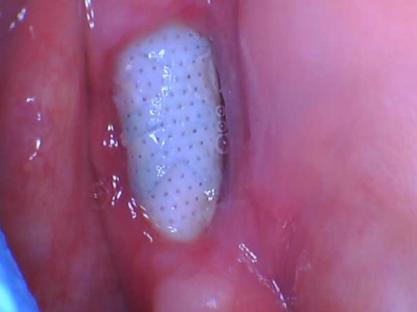
The next step is going to be a crescent tissue graft. A piece of tissue from the palate by the premolar is taken. It is about 3mm wide and about 3 mm thick. It is a crescent shape, like a half moon. A healing cap is placed on the implant, the cresent tissue is sutured over the buccal gap that is filled in with the bone graft. The tissue is used to hold the graft material between the implant and the buccal plate. It is sutured on the mesial and distal. The tissue graft holds the graft in place but more importantly it prevents tissue shrinkage due to the extraction. In days of old we would super errupt the tooth and let the bone fill in and then do the implant. Dr. Tom Han show's the Crescent tissue graft on DentalXP.com.
Generally all of this can be done at one appointment. This is much more convenient for the patient and the dentist.
Good Luck
Jerry Brown
Caesar Wong
8/4/2009
My advice to all dentist doing implants is to think simple and do simple. Don't complicate your work and life with fancy procedure. Especially if you are in private practice, you want a sure win in your treatment. Patients coming to you to do implants because they trust you and can wait for you. Why not graft the socket and the alveolar ridge first and do the implant later?
DYNAGRAFT demineralized bone matrix is the appropriate bone grafting material to use. There are two compositio - Dynagraft-D which is pure matrix and Dynablast which contains cancellous chips in addition. They are good stuffs.
ED
8/4/2009
You sound like you are new to this field. Keep it simple and predictable. Do one step at a time and be patient. As Carl Misch said "One miracle at a time". Especially if you are new to this don't try to package everything into one surgery.
First of all extract atraumatically. There probably is no reason to graft at this time especially if you think subsequent ridge augmentation will be necessary any way. If you need a temporary dont use a flimsy "flipper" they are very traumatic to the tissue. Make a temporary "Maryland"type bridge. If this is not an option make a cast partial with cingulum rests on the adjacent teeth.
Go back in 4 months and open up the site. If you have atleast 3mm thickness you can place the implant using "Ridge expansion" techniques either with osteotomes or mechanical expanders (BTI or MIS or Meisinger). I prefer expanders they are gentler. If buccal plate is too thin or perforated after placement then you can augment with particulate allograft or autograft covered by long lasting resorbable collagen membrane.
"Ridge split" will not work in a short span but ridge expansion will.
If you have less than 3mm thickness then you must consider a block graft or particulate graft and delayed placement of implant.
good luck
Dr. P
8/5/2009
Dr.A,
Ridge splitting is appropriate for the posterior or in contiguous extraction sites. I recommend a 10mm a-p distance. The maxillary anterior facial bone is very thin and almost exclusively cortical (inflexible).
Block grafts are a pain in the... Ridge splitting is very predictable. Particulate bone w/a membrane and bone pins(MTF)can further augment the deficient ridge. You may enter the same site a second time, w/o laying a flap, to further widen the bone (12 weeks healing each time). If the bone is resistant, the implant can be placed at the same time (USE A GUIDE- DePlaque, otherwise the implant will fall into the weakest area).
Patient psychology (you have lost bone over decades, we can't make it overnight) and proper pharmacology are important. Breaking the jaw hurts like hell (especially the lower posterior where the mm of mastication warp and flex the jaw).
PLACE THE IMPLANT, USE A SEMILUNAR INCISION, FILL W/GRAFT, AND COVER. OCCLUSION IS PARAMOUNT- CROSSOVER SHOULD BE DELICATE.
Thanks for the question and opportunity for great dialog.
MJP
Richard Hughes DDS, FAAID
8/6/2009
Ridge expansion is a needed tool for the implantologist. I have had success and failure. Each failure was due to the lack of understanding on my part. I strongly recommend that any serious doc learn the expansion techniques fron Dr. Hilt Tatum or from Tatum Surgical 1-888-360-5550. Tatum Surgical also sells the instrumentation which is well designed. You also need to train your surgical assistants and be open minded.
IP
8/7/2009
ED, now Misch is stealing Tarnow's sayings? ;)
Nailesh Gandhi
8/10/2009
I fully agree with Caesar Wong's opinion.Always think simple.Block graft will be much better in post extraction healed ridge case.
Peter Fairbairn
8/10/2009
There are many ways of achieving consistent sucessful long term outcomes here but after nearly 20 years placement would I use a block graft on myself or family , NO way.
Richard Hughes DDS, FAAID
8/10/2009
I agree with Dr. Fairbairn. Also one may want to learn Tatum' concepts of NIRISAB.
Dr. T
8/11/2009
If the buccal plate is too thin, you may end up with buccal plate resorption after splitting the ridge and placing the implant at the same time. The matter will be worse if you cannot get non-tension primary closure or the site get exposed during the healing. The defect now becomes more costly and harder to fix in this aesthetic zone. Otherwise, it's pretty reliable to split the ridge and place the implant at the same time.
Dr. Dennis Nimchuk
8/11/2009
The connotation of the term "ridge splitting" is to create a fracture and the best tool to do this with are chisles. More desirable is to expand or dilate the ridge with progressive dilating osteotomes (tap-in designs or twist-in designs). The best protocol is that this be done without lifting the periostium so as not to lose the blood supply from this area. The worst scenario is to lift the periostium, lose the periostial blood supply and then fracture the ridge. Edentulous sites can be dilated a surprising amount a.k.a. Arun Garg and Hilt Tatum and there is a very good success rate if proper case selection and good technique is utilized. The case selection should optimumly involve a single missing tooth site and preferrably be in the soft bone of the maxilla. Contiguous multiple missing teeth as recommended by Dr. P. above are much riskier to do and the likelihood is higher that the ridge will split rather than dilate. In these instances onlay grafting may be the preferred method of preparing an adequate surgery site for implants.
Robert J. Miller
8/11/2009
The decision to use particulate or a block graft should not be made solely on the basis of clinical expertise. For the greatest success you must also look at the quality of the remaining bone. Angiogensis and osteoblast migration requires medullary bone. In a B-w and C-w ridge, the top 5-10 mm og bone can often be almost entirely cortical in nature. If you attempt a ridge split in these cases, be prepared for complete failure as there is no cellular component to these plates. These are cases that require block grafts with an inlay approach (converts a one-wall into a four-walled defect). If there is a good medullary component, you can split the ridge provided, as Dr. Nimchuk stated, you leave the periosteal supply to the distracted segments intact. The only way to determine the quality of bone in these ridges is a cross-sectional tomograph using a CBCT scanner.
RJM
Peter Fairbairn
8/12/2009
I totally agree, but the origional question stated that there was only a slight buccal concavity.In extreme case such as congenital odontia ( I have had cases ) there is only thin cortical bone plate as there had never been any teeth to dilate it. Here you cannot split the ridge ( I tried once ) and a block is maybe the choice.
Currenly there is a synthetic block ( Beta TcP) undergoing final animal testing which will be interesting.
When we ridge split I agree too, always keep the periosteum attached until the implant is placed then raise it and graft , here we use BTcp in hydroxyl sulphate matrix
Richard Hughes DDS, FAAID
8/12/2009
Dennis Nimchuk, You hit the nail on the head. Also if you can tap deep enough you may obtain an internal blood supply in addition to that from the periosteum.
Dr. C
8/15/2009
I have a similar case in which I would like to either block graft or use a Ti-reinforced membrane. I have done both procedures before with good results. But, this is my first case on a completely edentulous patient, If you block graft or graft with a Ti-reinforced membrane what do you have the patient due in the heaing phase with the denture? Do you relieve it and place a soft-liner? Do you completely shell it out and have them "deal with it" during the healing phase? I dont want pressure from the denture to compromise my graft. Any adivce would be greatly appreciated.
Richard Hughes DDS, FAAID
8/16/2009
Dr. C, Sounds like a neat case. You can relieve the areas where you graft, reduce the flanges and secure the Max CD with an orthopaedic screw. This will also protect the graft. Also a prolonged soft diet. Remember patient compliance is a major issue that will sink the case.
Milo Garcia Tempone
11/22/2009
The way I would treat this case is to first extract the tooth carefully preserving the buccal plate and graft the area with mixture of dFDBA mix with CaSo4 cover by CaSo4 fast setting and wouldnt try to cover it with the tissues. Let the tissue grow over it to increase queratinized gingiva. Meanwhile I would fabricate the patient a Maryland bridge without pressure in the area. Three months latter I would enter the area with a split thicness flap and do ridge splitting with a piezo surgery unit and work the area with microdent hand expansors and place a conical implant with a conective tissue graft from the palate to improve the concavity it had in the buccal.
Jeevan Aiyappa Consultant
11/23/2009
Very interesting blog! Considering the varied responses and approaches of all the names on the blog... it just goes to show that there are definitely more means to the end than one can count!!!
However, some of the best teachers I have had - (including my guru and Maxillofacial Surgery mentor Dr Ranganath Nayak, the skillful Head & Neck Oncosurgeon Dr Deo whom I shared many years of my life with and two highly respected Implantologists Dr Ady Palti and Dr Hom Lay Wang who shaped much of my Implant career) taught me to respect and follow EVIDENCE-BASED medicine.
I would therefore be wary of surgical tenets that are based on "works in my hands" ... I recall Dr Arun Garg repeatedly mentioning during the course of his lectures to our group, that anyone who has "golden hands" who assures that a particular procedure works best in his hands,is best left complimented and not followed!!
The discussions regarding Block graft vs. Ridge Split are probably not completely justified (pardon the transgression of sensibilities here guys!).....
what else can you expect when
1)We don't know how old the pt is ?
2)We have no idea of the soft tissue Biotype?
3 We have no clue about the pt's systemic background?
4)What is the clinical situation that makes the lateral Incisor "non-restorable"according to Dr A ?
5)Is there a pre-existing pathology in the vicinity?
6)Sinus tract ? Fistula ?
7) Need CT evidence of labial bone (or the lack of it), as some colleagues mentioned above
8) The bite / Occlusion ? Is there something contributory there which is making the tooth fragile and caused its non-salvageable status ?
9) Do we have only the cortical plates remaining residually, or is there some Cancellous component left that will enable the Split to be carried out successfully?
.............
The blogs are a great forum for expression, Treatment Planning however, requires more than just a few relevant peices of information !!??
I daresay (at the cost of upsetting a few colleagues) our really enjoyable posts on this forum may turn into an arena for amusement if we are not guarded in our expression , which will then cause a confusion in the ranks of those that do take these seriously.
NO HARD FEELINGS FOLKS! ITS ONLY MY OPINION AFTER ALL, AND SHOULDN'T MATTER TO ALL!
Cheers
Jeevan