Thoughts on bone loss/appearance for 4-unit bridge case?

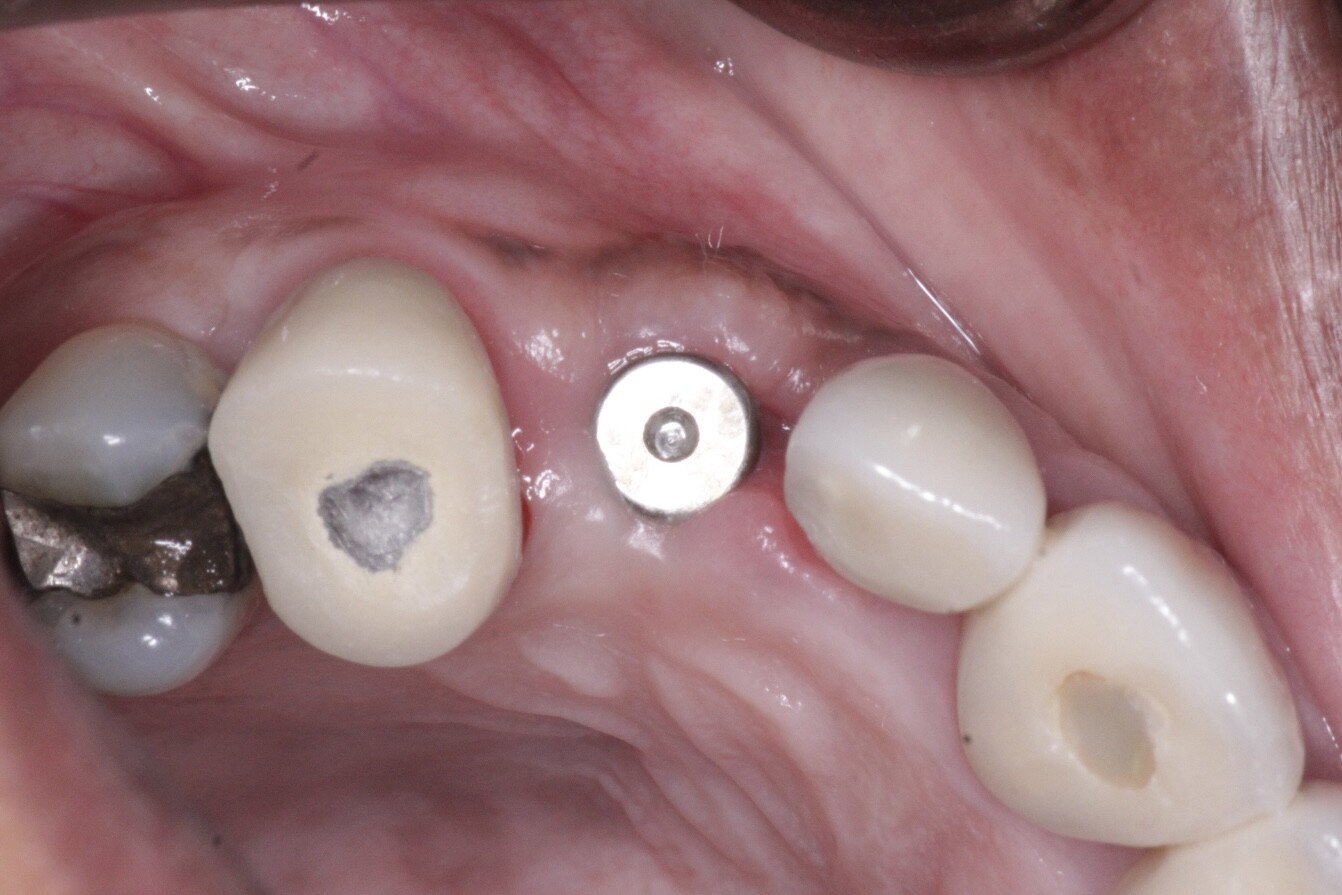
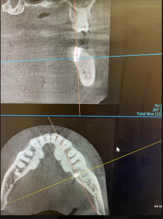
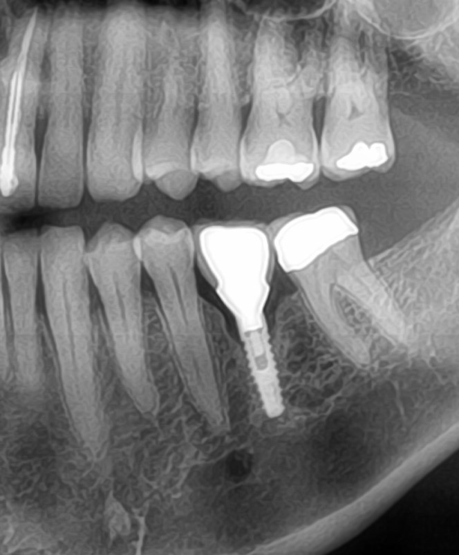
In July of 2014. we removed #26, 25, 24, and 23 [mandibular right lateral and central incisors and left central and lateral incisors; 42,41,31,32]] and placed immediate implants in #26 and 23 extraction sockets]. The treatment plan is for a 4-unit bridge on the implants in the lateral incisor sites. After 3-months, we removed the cover screws and replaced them with transmucosal healing abutments. The periapical radiographs revealed significant bone loss on the mesial of #23.

My thoughts are resorption of the thin alveolar crest following removal of centrals. The centrals did have significant bone loss around them prior to removal. The implants feel solid and integrated, during both removal of cover screw and fixating the healing abutments, however the density of the bone mesial to the 42 implant prompted me to repeat the PA. I’m not sure if this is a sign of failure or delayed healing of the socket where 41 was. Nothing is significant clinically and the patient notes no pain or swelling. Advice on how to proceed would be appreciated. Thoughts very much appreciated.


1008996-X-20140722-145138-XCZTWYQDERZQ-4


1008996-X-20140722-161251-XPN4MIXKDI6H-4


1008996-X-20141024-094816-XJOD0R9ZH6P7-4


1008996-X-20141024-095239-XE2WX7ECLCE3-4


1008996-X-20141024-094541-XVQ61C0U2WSF-4

24 Comments on Thoughts on bone loss/appearance for 4-unit bridge case?

New comments are currently closed for this post.
Don Callan
11/18/2014
If the truth be known, the success of implants into sockets at the time of extraction is about 50%. The problems are infections, blood supply to the area, bone quality, loading early and possible lack of closure. WHY RISK IT? Prepare the site first, then place the implant, The success rates are better. There are no short cuts.
DrT
11/18/2014
Not enough information. Did you do any hard tissue grafting when you placed the implants? Also, did you have 2 mm of bone at the crest of the fixtures. The PAX just show what may be happening mesially and distally to the implants...this is obviously only one aspect of this case....I am more concerned about the facial, especially given what we know about the anatomy of this area. Thank you
Robert J. Miller
11/18/2014
Don; Don't know why you are getting such a low success rate in extraction/immediate implant placement. Just need to address the different biologic response in these sites. But there is not a single parameter that needs to be changed; each sequence of implant preparation needs to be tweaked. After extraction, how do you debride the osteotomy? If you are simply curretting the site, be prepared for post-insertion infection. We use an ablative erbium based laser to decontaminate. Next, how much bleeding are you seeing from the osteotomy walls. If you are removing a failed endodontically treated tooth, the bone will be more sclerotic in nature; it needs to be aggressively decorticated. Finally, how are you treating the gap in the osteotomy defect. We are using L-PRF alone as a graft. In this way, we speed up early vasculogenesis and PRF has an anti-infectious effect. Our success rates are identical to those in healed sites using these parameters. RJM
Ez
11/18/2014
Robert as many more people own diodes, are there any studies showing what effect diodes have on the socket??? Thx
Robert J. Miller
11/19/2014
Diodes are good for decontamination and photobiomodulation. There are not used for debridement of granulation tissue. In extraction/immediate placement cases, teeth are removed for periodontal or endodontic reasons. The pathogen load will be relatively high and are usually associated with formation of pathologic granulation tissue. Diodes have a very limited depth of field (<1 mm) and will not penetrate larger lesions. This is why we use ablative lasers prior to implant placement. Also, there is a high thermal coefficient with diode lasers and you have a good chance of thermal damage to bone if not used correctly. RJM
Dr L
11/18/2014
I don't think the success of immediate implants is as low 50%. It really depends on case selection and practitioner. As for this case, I'd say the most probable reason for the bone loss is that the implants have appeared to been placed directly into the sockets, without any bone/ridge reduction. The bundle bone will resorb once a tooth is taken out, and what I think youre seeing is just that. If it could be done again- you would reduce the ridge by a bit, removing all the unsupported buccal wall, and probably down to the apical socket. This will also help you align the placement of the osteotomy. It could also be other issues of infection etc, but I'd say is due to the above
Sb oms
11/18/2014
The truth be known? I do about 3-4 immediate implant cases a week. Sites are infected, teeth are cracked, there is bone loss. Each case is handled with intense care to clean the socket, maintain and maximize blood supply, and maximize bone implant contact. My success rate is not 100%, but way more then 50% as DC states. In the last five years, which is as long as I've been keeping track, 98.7 % meet criteria of success and patients are happy. This case shows infection. That's the only kind of process that could lead to bone loss like that in such a short time period. Lower anteriors have the worst perio bugs of anywhere in mouth. Do not load this case. I agree with many good points made above
Michel Raad
11/18/2014
Not a good idea to load the case and fabricate a bridge because you will be throwing good time/money after bad. For some reason you got some infection going on around one of the implants ( You did not mention what is your prior experience with immediate placement ). Not all implants with excessive bone loss will fail or appear loose in the mouth immediately. Having said that you don't want the patient back in your chair in 1.5/2 years with two implants failing and a new bridge to be made with all the lab expenses to incur all over again. Because of the extent of the bridge the failing implant will need to be removed, the location grafted and left to heal. Go back after the bone has healed and place a new implant. If it was a single tooth I would have suggested you try and open a flap and see if you can debride the area and graft it before you move on with restoring. Hope this helps and good luck
Mike Heads
11/19/2014
Sorry Don, but I think you need a lot more training if you are only getting 50% success. The immediate dental implant in my hands actually gives the BEST results. The only thing I would say is that I use Bio-Oss round every immediate implant, no matter what the gap size is, just to be sure and as I said the results are fantastic and the soft tissues hardly ever move. For anyone worried about immediate dental implants please do not give up on them just get some more advice and training and reap the rewards of great cases and very satisfied patients
Tuss
11/19/2014
Immediate placement plus bio-oss - I have seen several perio review articles that are starting to show increased rates of peri-implantitis around sites grafted with material of bovine origin compared to fully synthetic graft materials. Agree sucess rates on immediate placement (esp full arch load cases) is higher, I would ask if the gap between the implant and socket was grafted at placement or if nature was left to her own devices - on the placement film a wide gap can be seen at the head of the implant so its probably lack of grafting that causing the 50% failure rate combined with the other points made
Peter Fairbairn
11/19/2014
Yes Tuss , I have seen a few vey compelling research cases on that another factor is that although it can make a nicer looking x-ray , it is bone ? probably not. With time the bones ability to turn over naturally may be impeded due to foreign material resulting in reduction of Osteocytes present which again could have longer term implications. As per Immediate placement I do a few but generally all cases are a delayed immediate at 3 weeks post extraction. But as with all these cases site preparation is the key to success . Everyone has their own preferred materials and protocols and they all probably work well if the core surgical protocols are adhered to . Peter
Tuss
11/19/2014
Thanks Peter, I do the same, prefer to let infected sites heal for a few weeks prio to implant placement and tend to go immediate in selected cases (good buccal bone width, no pre-existing infection, no perio issues and good keratinised tissue). With immediate implants combined with grafting I have seen issues arise later post restoration rather than at an early stage so my feeling are if the sites are infected or require significant bone augmentation then stage the placement - heal, graft then implant. Patients may not want to wait and may shop around which is fine but atleast in this day and age you will have less leagl issues to deal with
CRS
11/19/2014
Some really helpful advice, many good rationales. I think the mandibular anterior is tricky due to the anatomy, less cancellous bone.On immediates I like to see bleeding bone after the extraction sites have been properly prepared. These implant infections are similar to a localized osteomyelitis. I like to treat the extraction site with ablation of granulation tissue using an Nd Yag to target the pigmented bacteria. I think that many methods such as curettage with some form of chemical disenfect ion and oral antibiotics all work. I don't know much about the synthetic bone grafts but I use human bone. There is also the rationale that the implant itself maintains the space nicely. I think it boils down to experience and judgement with an understanding of the biology iof the area. I usually extract, graft and wait to see what I get then place the implant. If I have a perfect extraction site then I will place immediately. Very good discussion.
osurg
11/19/2014
50% failure with immediate placement. Where do such figures come from. Please if we make such statements lets make them "evidence based". I have not ever seen any studies that support these figures. If this is personal experience please state as such. Too many things we are taught in school lack scientific support. Lets not add to that.
Dr.Ahamed
11/19/2014
Is there a correlation with high implant insertion torque and crestal bone loss? Or Is it periodontal pathogens of adjacent teeth causing Peru implant crestal bone loss?
Robert J. Miller
11/19/2014
There is an absolute correlation between insertion torque and crestal bone loss. Above physiologic deformation (about 50 Ncm) you get pronounced microfracture of the crestal region and compression remodeling. Higher insertion torques (70-120 Ncm) may be one of the prime causes of crestal bone loss after implant placement. With regard to periodontal pathogens, any reservoir of pathogen load can contribute to this type of bone loss. Most often, it is really just a failure to completely debride and decontaminate the site prior to implant placement. RJM
Dr.Ahamed
11/19/2014
Thanks indeed Dr. Robert J. Miller for the detailed discussion.
Dr John H.
11/19/2014
The OP commented that although the radiographic evidence of bone loss the implant at present is immobile and symptom free. Should the discussion move around what protocols to carry out or is removal the only way forward. Recently with peri-implantitis, some debridement regimes have been suggesting full flap, debridement, non metal curettes/ ibrush /tibrush, citric acid & chx rinse combined with tetracycline mixed in with graft material and close. Do Peter & others have any thoughts on Btcp / fortoss as a membrane free option here. My experience has been with bovine materials though Vital etc may have more of a bacteriostatic use here? Good discussion
peter Fairbairn
11/20/2014
John H , as you probably know I have not used a traditional membrane once in the last 13 years all graft materials I have used "set " to be stable throughout initially. The elements that ensure they are stable CaSo4 and in the other case Polylactide coating are bacteriostatic ( Petri Dishes studies show the Bioloinker for the poly-lactide to be bacterioscidal ). I have used them in cases to treat crestal bone loss with many great results , but feel predictability can be an issue . But again strict surgical protocol is very important . But again all graft materials must fully fully bio-absorbed to have only true host bone and allow for correct long term post op radiographic monitoring . Merely having some HA in the area taking an x-ray and saying it is bone is fanciful. Regards Peter
Alex Zavyalov
11/20/2014
As the dentist mentioned, the implants are clinically asymptomatic. I do not see a real problem on the current X rays proceeding with temporary bridge.
DrT
11/20/2014
If you have inadequate labial bone thickness, or are losing bone interproximally at this early stage, I think the first priority is managing these problems before proceeding with any restorative therapy. I cannnot see settling on a compromise situation at this stage of treatment....if this were 5 or 10 or more years out, then, yes I might be looking for a way to maintain a compromised case.
Tuss
11/20/2014
Most implants don't present with pain until they are about to fail. The radiographs show that bone is being lost so delaying any remedial treatment is just creating a problem for late on (kicking the can down the road). You have to inform the patient of what you can see on the radiographs and give a prognosis. The longer you wait the more bone you could lose and the more complicated the future treatment. If the patient says "go ahead and lets see what happens" then fair enough but you need to make sure they fully understand the issues and potential problems. At this moment in time (unles you ISQ or reverse torque test" you can't say if there is good degree of osseointegration . Plaque control in this situation may be an issue and if there is not a high degree of control you will get framework/ implant show through. The provisional bridge is an option as long as the patient fully understands the prognosis.
Dr. C
11/20/2014
I am always curious how doctors handle these cases. If YOU were the surgeon that placed the implants and you felt additional treatment was necessary to correct the situation do you charge the patient? If YOU placed the implants and feel they need to removed and replaced would you charge the patient? I don't charge for replacement of a failed implant, if it has failed prior to restoration placement. But, there are some doctors in my area that do. I am just curious how other doctors handle these situations in their own offices. Dr. C
Tuss
11/21/2014
If this happens in the first 5 years then I personally do not charge the patient, if its +5 - 10 years then a reduced fee is charged. If you can see progressive bone loss happening from day 1 and the ptient regularly comes in for check up/ hyg appts then look after them. If this patient had treatemnt then never returned for 5 years then I have no ides if it was down to poor OH, lack of follow up etc so as part of my treatment plan I make sure they know recall/ review/ hyg post restoration are a must otherwise charges may apply

Featured Products

OsteoGen Bone Grafting Plug
Combines bone graft with a collagen plug to yield the easiest and most affordable way to clinically deliver bone graft for socket preservation.
CevOss Bovine Bone Graft
Make the switch to a better xenograft! High volume of interconnected pores promotes new bone. Substantially equivalent to BioOss and NuOss.