Very Enlarged Incisive Canal: Experience with this Situation?

Dr. D. asks:
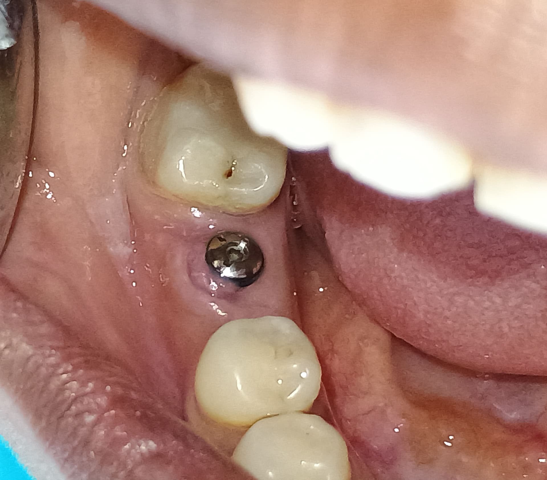
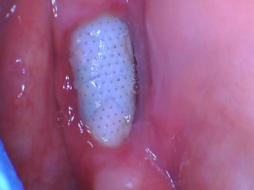
I have a 21 year old male patient who presented for dental implant placement in #8 area [maxillary right central incisor]. The buccal cortical plate in this area has a concavity and bone resorption at this site was clinically evident in the consultation exam. The CBVT scan reveals a very enlarged incisive canal which runs parallel to and extends well beyond the adjacent roots. The width of this canal is approximately 3-4mm.

I have bone grafted the area of the incisive canal in the past on other patients without any complications. This is even suggested by Misch and he does not believe it causes any complications. However, the width and length of this incisive canal for the present patient concerns me. He will need buccal bone grafting to augment the site for sure, but the prosthetically acceptable positioning of this tooth, will place the implant right into the canal towards the apical one third. Is there anyone who has had an experience with this kind of situation? Any complications from bone grafting into the canal of that length and width? Any suggestions?

16 Comments on Very Enlarged Incisive Canal: Experience with this Situation?

New comments are currently closed for this post.
Dr SDJ
1/19/2009
I have 2 such cases waiting for me to do and even I am waiting for an answer from osseonews.
GC
1/20/2009
dear colleague, grafting the incisive canal is not a major issue but if your case includes placing of the implant straight into the canal, I'd rather use autogenous bone to be fully sure of the quality of bone for primary stability of the implant,quality of integration ,long term success and precise placement in apico coronal direction. like many others, I use a large roundbur handpiece to obtain very good cleaning of the canal before the grafting.one important point is not to leave tissue remnants in large canals and make a good packing of the bone with some bone pluggers to condense bone ;in my previous cases, I used block of bone from ramus to complete the buccal side of the graft and provide bulk for soft tissue support and esthetic enhancement. Good coverage with a resorbable membrane of your choice and flap release to get excellent primary closure are keypoints to this procedure. Hope this point of view might help you in some way. friendly yours, GC
dr.e
1/20/2009
reading the original post, I realized that I questioned about incisive canal. The anatomical structure that I am actually concerned about is the Nasopalatine foramen and canal, It runs the entire lenght and parallel to the the root of tooth #9, even on the CT it seems like it never ends!, I don't think I could completely remove soft tissue from this structure except for the more coronal parts and obliterate with bone graft.... please comment.... thank you
R. Hughes
1/20/2009
No big deal. Graft and do it.
GC
1/21/2009
dear dr e , as stated above, graft and do it...this pedicle is the constituted by the junction of the two ending sphenopalatine vessels and nerves and it has a secondary role in innervation and blood supply for the anterior palatine , so no big deal,you cant go too far and have problems in this location.
GC
1/21/2009
please read anterior palate , bad typing...
dr.e
1/25/2009
what is the chance of nerve tissue growing back and encapsulating the implant? implant failure?
Robert J. Miller
1/25/2009
Have you ruled out a nasopalatine cyst? On a CBCT tomographic slice, the nasopalatine canal is well defined and relatively parallel. If the walls show a widening at the apical third of the adjacent centrals, you must rule out pathology before you graft the area. RJM
Amar Katranji
1/26/2009
I agree with previous posts, this shouldn't be an issue. I would graft the area with autogenous if possible,allograft is fine, too. Loss of either central almost always makes the incisive canal larger and can create issues during implant placement. Just drill it out, graft, and position implant properly. If you need help, refer it and ask to watch the implant surgeon/specialist (whoever you send it to) and you'll see it's not too difficult. If it's a pathology case then obviously you need to deal with it differently. Hope this helps.
JM
1/27/2009
There is absolutely no need for harvesting autogenous bone to graft the incisive canal since it is a small self-containing defect that usually has a high success rate if the neurovascular bundle has been completely removed.
Dr HArold Bergman
1/28/2009
It appeasrs that you are discussing the naso-palatine canal which is located between the maxillary central incisors not the anterior palatine canal which is located further back opposite the molar teeth. One should consider pathology on any radiograph exibiting an area larger than 1 cm in this area, the differential diagnosis being firstly a nasopalatine cyst. If grafting the area, any possible damage to the contents of te canal are usully minimal and of little cosnsquence to the graft or the contents. First do no harm.
Rz. Radwan
2/3/2009
Dear Dr.D, U said that ur implant is going to be placed in the incisive canal only in the apical 1/3 , so why not placing a tapered implants using only one dril to make an undersized osteotomy , it going to save u a lot of bone , u r going to find such implants in the market such as OSTEOCARE , it's a british implant company. they r offering this new concept (not just tapered implants) it actully conical in shape. I think it's going to help u avoiding the canal from the first place. Good Luck.
satish joshi
2/6/2009
Dr.D Last week We had case for anterior ridge aug.from #6 to #11 and grafting of canal.If you are interested to look at photos of of entire procedure.Let me know.I will try to e-mail.I am on vacation right now,so it may be possible after two weeks. Satish Joshi
Dr. Tassos Irinakis
2/24/2009
Here are my two bits. 1. although i'm a big fan of "bottled" bone i always use autogenous in the canals (with membrane) and have had great success 2. you indeed need a radiologist's report regarding the possibility of pathology and if it's presence is of any consequence 3. Good luck. Smart of you to ask for advice on such a case.
dr.e
3/2/2009
Dr Joshi, would you be able to post those pics? On the radiologist's report of the scan, there is no pathology. I am certain that the structure is nasoplatine canal, with parallel walls (3-4 mm wide). No cyst is present.
Dr. T
3/5/2009
I placed bone graft into the nasopalatine canal (only the coronal 1/3th), did not remove the Apical portion of the soft tissue content of the canal. the patient developed late Swelling on the buccal aspect of Anterior teeth (from #7 t0 #10). I also had a titanium reinforced membrane for the buccal bone augmentation. This membrane never got exposed. But I removed it after the swelling in the site happened. No exudate was present at the time of membrane removal. I am not sure why this late swelling appeared. I have the patient on Antibiotic for 7 days. Patient does not have any pain in the site. Could this be because of the bone graft placed in the canal? On the peri-apical radiograph, a radiolucency is evident in the apical 1/3 of the canal. This could be because the coronal third which now has bone graft content, so the apical third looks more noticeable? I read on this site that the content of the canal will need to be completely removed. Is infection a consequence of not removing this soft tissue content entirely?

Featured Products

OsteoGen Bone Grafting Plug
Combines bone graft with a collagen plug to yield the easiest and most affordable way to clinically deliver bone graft for socket preservation.
CevOss Bovine Bone Graft
Make the switch to a better xenograft! High volume of interconnected pores promotes new bone. Substantially equivalent to BioOss and NuOss.