This healthy, non-smoking patient had his 46 (rct, apicectomy) removed by a maxillofacial surgeon. The tooth came out piecemeal, but healing was uneventful and the implant was placed 5 months later. It was torqued in at just under 30 Ncm, follow up at 1, 3 and 10 days had no pain, gingiva adapted and healed well at the 10 day follow up. This was a most straight forwardly executed case.
3 weeks after placement he presents with mild swelling and pain. The implant is not loose, there is no drainage or gingival recession.
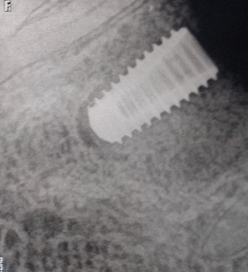
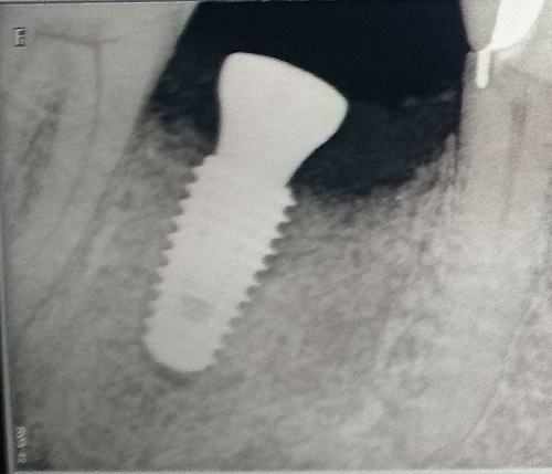
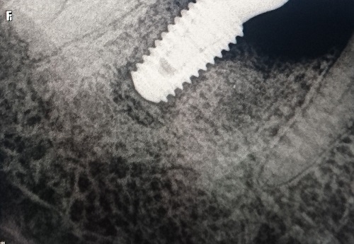
The xray shows an apical area.
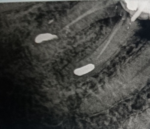
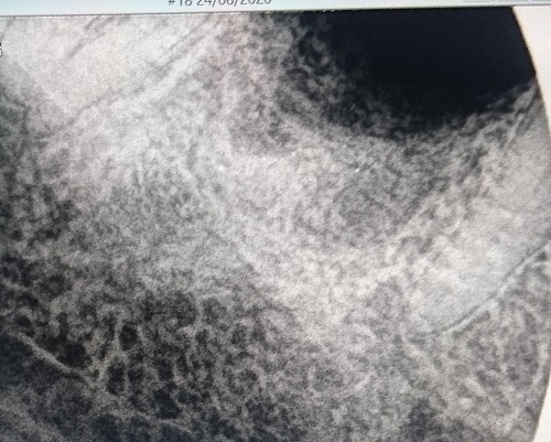
Upon looking back, I can see a denser area than the surrounding bone where the root apex used to be, as well as scattered white spots which I assume to be spread out retrograde filling. If the denser area is Condensing osteitis, could this be the cause? or is the scattered retrograde filling material the cause?
Or more importantly, can this be treated with antibiotics? And if not, when this implant is removed, how to avoid failure in the future: is proper curettage enough?