Foreign Object in Implant Site: Best Option?

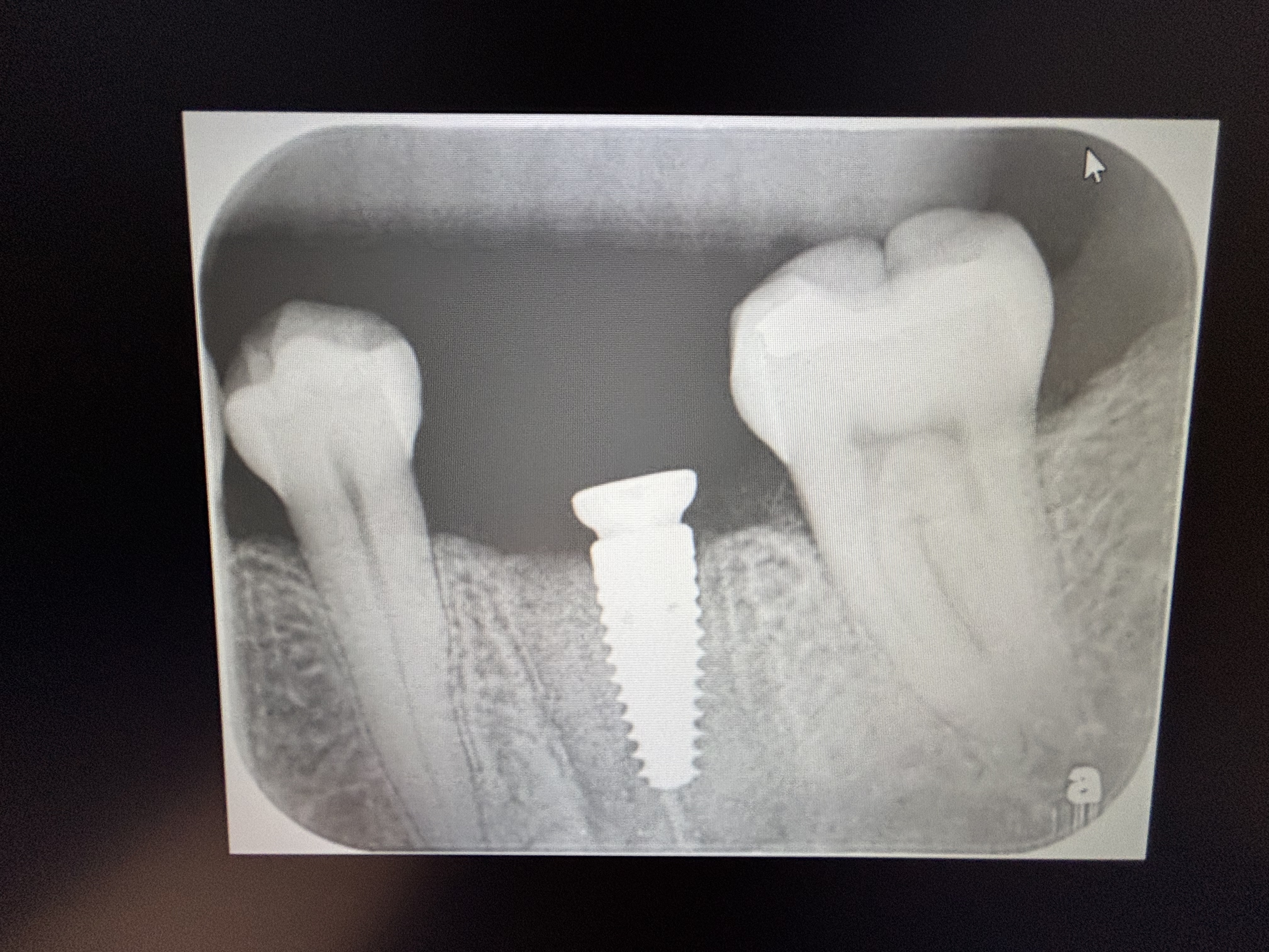
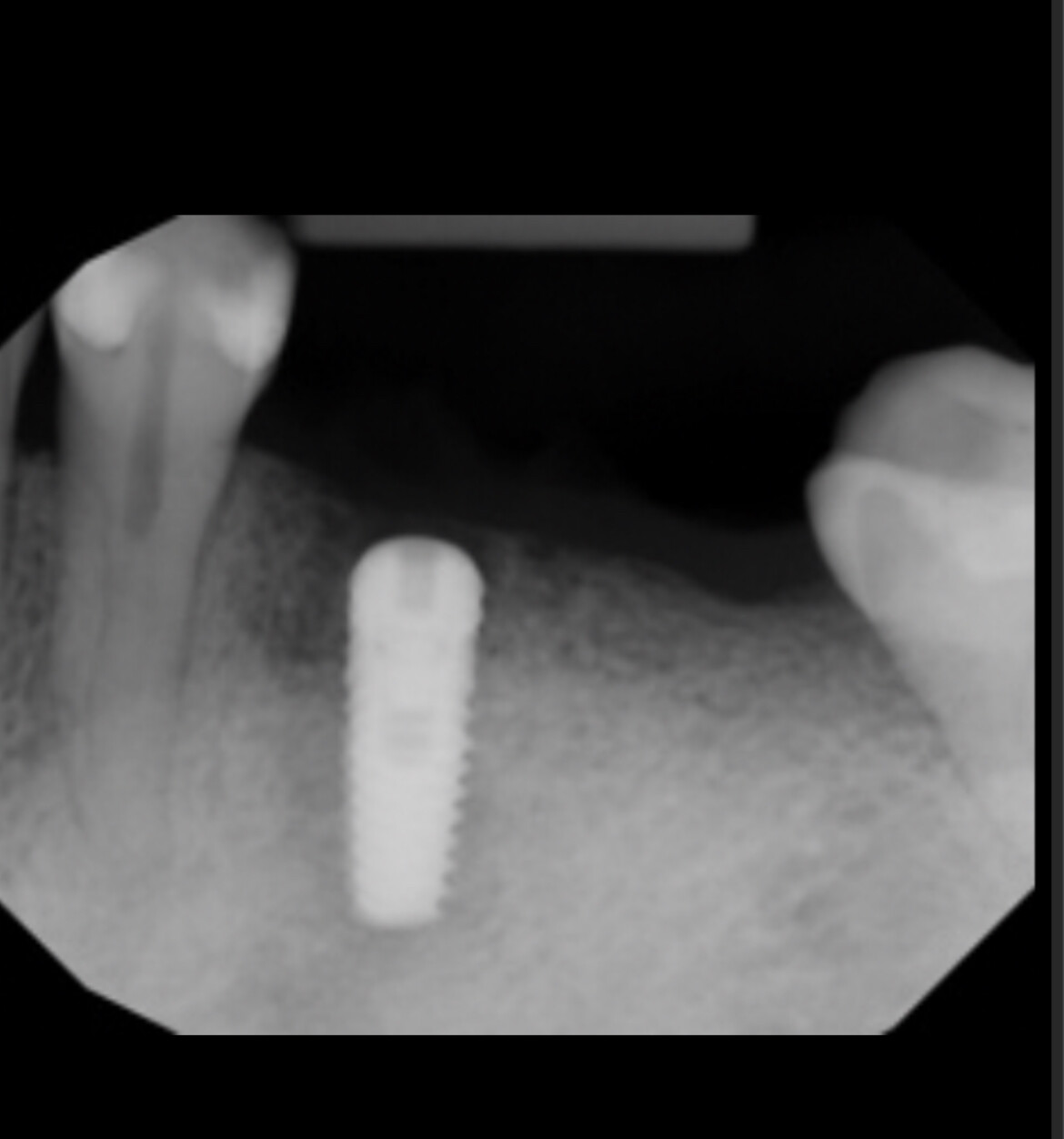
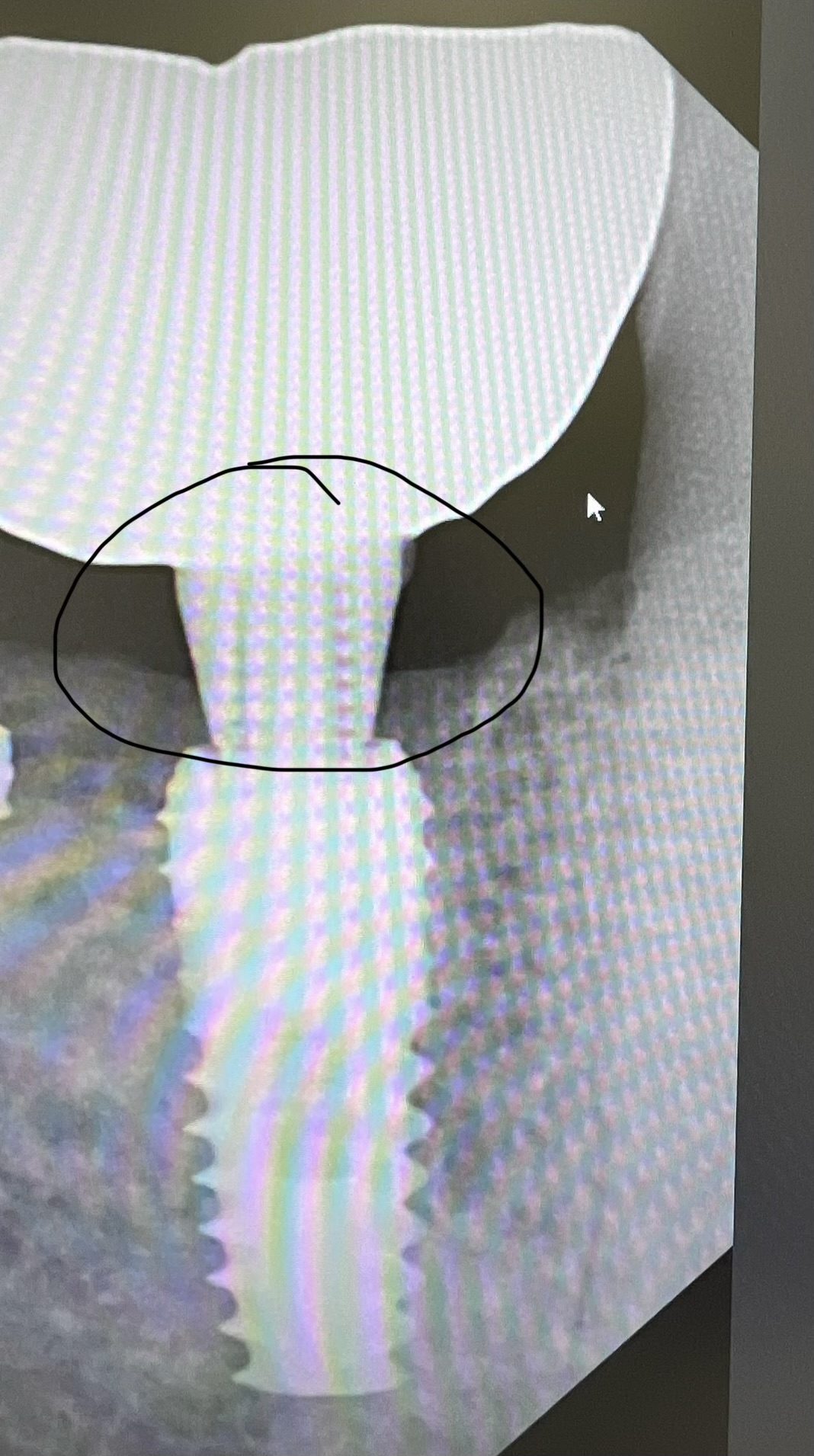
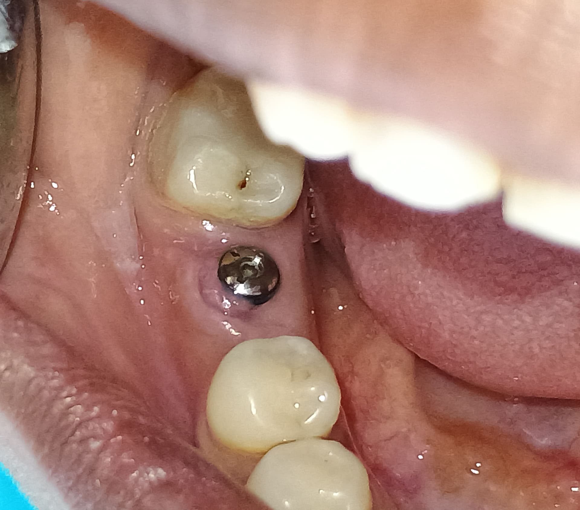
I have a 63-year old female patient requesting implants to replace first and second upper right premolars ( 14, 15). 14 and 15 were extracted about 6 months ago and
periapical x-rays prior to the extractions reveal a radiopaque mass most probably sealer or extruded gutta-percha in the apical area of 14, with no obvious radiolucent lesion or pathosis in the immediate vicinity of the radiopaque mass. The radiopaque mass had not been removed with the extraction of 14. The location of the mass lies potentially in the path of the osteotomy preparation and implant insertion. Will this cause any problem for the implant insertion in the 14 area? Which of these is the best option: to surgically remove the mass prior to the implant surgery? Or, include the mass in the bone osteotomy site with the hope that it will be removed? Or, choose an osteotomy path that avoids the mass all together?


9 Comments on Foreign Object in Implant Site: Best Option?

New comments are currently closed for this post.
John T
8/20/2018
Good grief. Can you really justify a full arch CBCT scan for such a teeny weeny fragment of root filling material?! Ignore it.
Adibo
8/20/2018
It is very unfortunate to see unnecessary comments to a valid question. The foreign body seems to be very small and superficial, easy to remove during the osteotomy. However, it is less likely to cause any problems if not removed.
Sean
8/20/2018
I agree completely with Adibo. Obnoxious comment John to a very reasonable and clearly stated question. I would also add two other thoughts, really excellent healing of this site and should it be "standard of care" to obtain a CBCT scan prior to any implant placement?
Dr. Gerald Rudick
8/20/2018
I think the question of the poster is valid, and shows that he/she is very concerned about doing a good job......as stated above, most probably the small root fragment with some sealer and gutta percha in it, will come out while preparing the osteotomy....however... you never know...if left, it may come back to haunt the implant surgeon...so the question is valid, and do try to get it out, in a minimally invasive way
DrG
8/20/2018
1. It’s sealer or residual gutta percha. It will come out while you are preparing the osteotomy. No worries. 2. In this day and age implants are treatment planned in 3D prior to surgery. That way ideal dentistry can happen. Seeing a full arch CBCT is more common. I assume the angry poster probably still uses a PA and eyeballs the placement. After placing over 10,000 implants I can tell you one of the most difficult eyeball placements is two adjacent implants.
Wesley Haddix
8/21/2018
Outstanding images. A surgeon can never know “too much” about the tissues he/she is planning to modify. CBCTs frequently disclose improved information, and are of firther assistance in a case such as this. Given the patient’s history and appearance of the foreign object, I concur with other opinions that this mass is likely extruded endodontic sealer or filling material. It does not appear pathologic on the images and I would feel comfortable making note of it, informing the patient, and proceeding with the implant. I might spend a few moments after the final osteotomy looking for the particle, but would not worry if I did not find it. A final notion with regards to avoiding the object by modifying the path of the implant: put the implant in its best prosthodontically guided position; with luck, the osteotomy drills will remove the particle. Best to you and your patient.
Matt Helm DDS
8/21/2018
This is definitely the remnant of a root canal sealer overfill that was larger at the time of the original RCT and has been gradually been resorbed over time. Note the proximity to the mesial apical accessory canal (where in fact it seems to originate) and the fact that it has been resorbed from that accessory, although very slight traces of it do remain in the accessory canal. Evidently considerable time has passed since the original RCT. Definnitely not gutta percha over fill (gutta percha never resorbs). This will deffinitely come out during the osteotomy. Although it sounds like a bit of a novice's question -- a dentist with a lot of experience would know almost instantly this is a filler overfill from the pre-extraction pa -- the poster is definitely to be admired and commended for their attention to detail and their desire to do the best they can for the patient. As they say, there are no stupid questions, only stupid answers. Agree with the other posters: good call on the CBCT, use guided surgery if you think you need it, and don't worry about this spec of sealer. It will come out with the osteotomy, and no need to redirect the implant and possibly cause yourself problems with the restorations because of an implant missangulation. You're doing fine! Good luck with your case!
Jalil Sadr DMD, MSD
8/21/2018
Sorry and excuse me, first and second upper right premolars are not # 14, 15. They are #4, 5 as in PANO and PA show. Also to me radiopaque area is in # 5 apex. even 10mm implant may not get there or may come out during the osteotomy. Beside this regard, how many implant do you want to insert? did you measure space between tooth #3 and # 6 as we see in radiography (J1). It seems the space collapsed and is less than at least 14-15 mm for two narrow implant. what is your plan? thank you and for everybody response and good luck.
Greg Kammeyer, DDS, MS
8/22/2018
CBCT to evaluate the bone and confirm the object doesn't have a root tip with it.

Featured Products

OsteoGen Bone Grafting Plug
Combines bone graft with a collagen plug to yield the easiest and most affordable way to clinically deliver bone graft for socket preservation.
CevOss Bovine Bone Graft
Make the switch to a better xenograft! High volume of interconnected pores promotes new bone. Substantially equivalent to BioOss and NuOss.