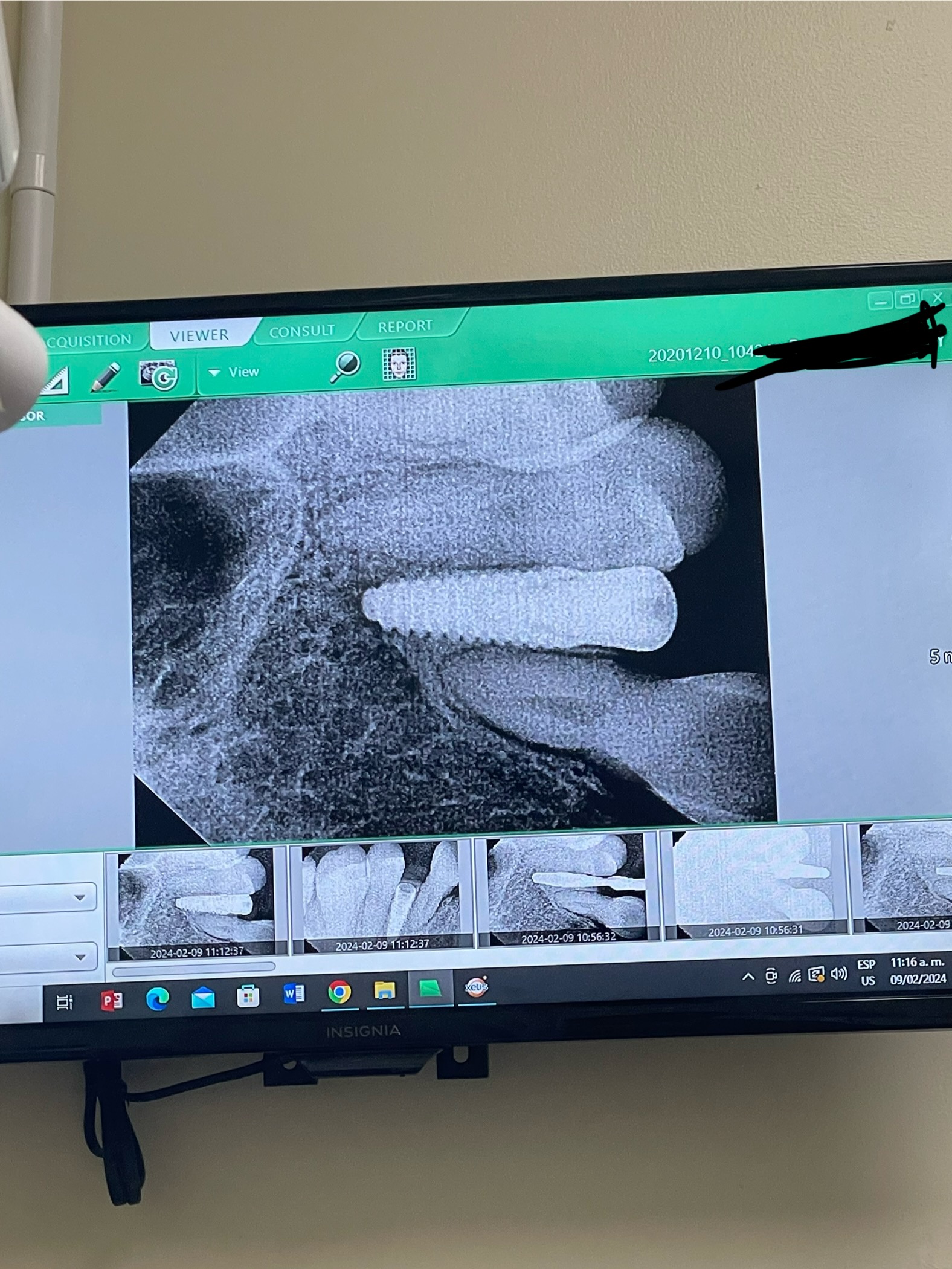
Best way to rescue this implant case?

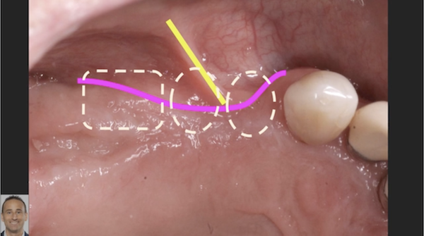
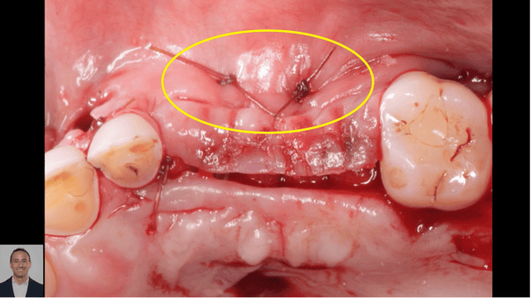
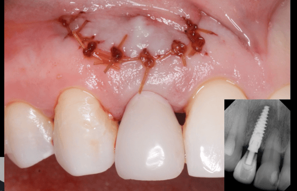
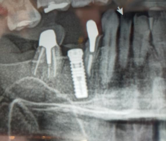
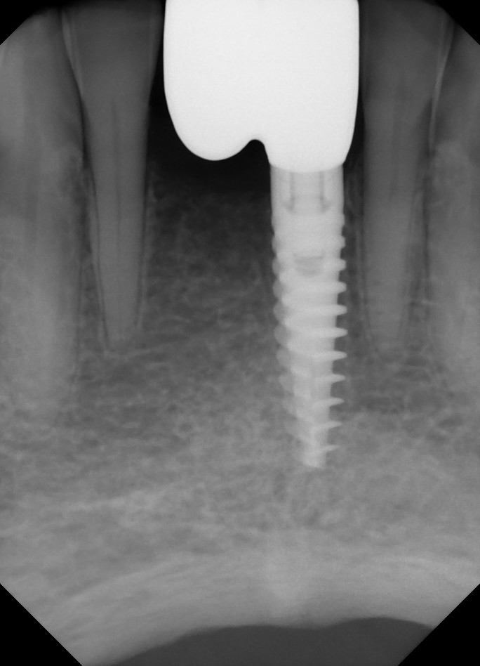
To replace a lost failed post-crown, a Straumann BLT 3.3 x 12mm Implant was placed early at 5-6 weeks after the tooth was removed. The implant was placed 1mm subcrestal with a thin intact buccal plate. I was anticipating a little bone loss, but I got a bit more than I had bargained for, and we lost the papillae. For the past few months I have been altering the contact points on the temporary crown to promote papillae formation and the papillae have now filled in a little. I have now managed to reform the mesial papilla and greatly improved the distal papilla. This may be at it’s limit of improvement or there could possibly be a little scope to improve it further. Unfortunately I cannot find the latest picture showing this and so have sketched the latest appearance. The papillae are now of acceptable appearance for the patient, but there is still approximately 1 1/2 -2mm Soft tissue deficiency. What are the options for improving this further?


17 Comments on Best way to rescue this implant case?

New comments are currently closed for this post.
Kevin Calongne
3/27/2018
The only way that you can fix that is to remove the implant and place a bone graft, then orthodontically erupt the cuspid. It would take a year to 18 months if the patient is willing.
Philip Christie
3/27/2018
Why remove the fixture ? Could you flap and graft and coronally reposition the flap using the graft or graft/membrane to support the soft tissue.
Kevin Calongne
3/27/2018
Because the bone is the blood supply for that soft tissue, and you won't get any with that fixture in place. I'm telling you this from 25 years of experience as a periodontist. You won't get that soft tissue back until the bone level is improved. Studies show that you can't get soft tissue more than 4mm past the bone, you are at least 3mm short in that papilla now, so you need bone to about 1mm below the current gingival margin on the distal. I'm guessing that you need about 3mm more bone to accomplish anything. Erupting 11 will help, but you need more at 10 as well. A soft tissue graft will get you little or nothing.
Matt Helm DDS
3/27/2018
And where exactly would you erupt the #11 to??? Have you carefully looked at the photos? Have you not noticed that the #11 is in its proper position from all points of view? Also, have you not noticed the wear facet (read "occlusal interference", or "bruxism", whichever you prefer) in the distal incisal of #22? #11 has nowhere further to go! Clearly! Not only is the bone level adequate on the x-ray, but so is the labial gingival margin on the #10, if we're to nitpick the aesthetics. Besides, the later photos show a greatly improved distal papilla. With a proper interproximal emergence profile in the final crown that papilla will be just fine! My 30 years says so! :)
Kevin Calongne
3/27/2018
The cuspid would have to be shaved down as it is erupted. I'm guessing you've never been to one of Maurice Salama's lectures on eruption. I realize that there are multiple ways to address an issue, mine was aimed at getting the best aesthetics possible. Notice the bone loss on the mesial of 11. I'm guessing that there is at least 3mm of bone loss there. I'm also guessing that there is a deep probing depth there. I'm looking at this from a long term perspective.
Matt Helm DDS
3/28/2018
While I do agree that the mesial bone loss on #11 is about 3mm (which, by the way, could have been corrected prior to implant placement, had the implant not been placed so early) it is highly doubtful that the full 3mm of mesial bone can be regained by erupting the canine. Furthermore, since the enamel is only about 1-1.5mm thick, shaving the tooth to accommodate for its further eruption would put us squarely into dentin, both lingually and incisally, thereby creating yet another set of problems. I am also looking long term (I always do), and in that context there are many other factors which can, and likely will, cause natural age-related bone loss as the patient ages. There is such a thing as over-treating and, considering that the papilla has regenerated nicely (and will continue to do so with proper subgingival contouring of the #11 crown), erupting the #11 would be a good example of over-treatment.
Kevin
3/28/2018
I respect your opinion, but, having done several eruption cases, I disagree that this is overtreatment. It really comes down to biology and the patient's expectations.
DrDave
3/27/2018
The patient finds the result acceptable... Why then subject the patient to an additional surgery and ortho? While our aim is to do perfect dentistry. Often our ideal isn’t met but if the patients function and esthetics are acceptable to them then stop. Certainly give them future sequelae and tx options to “make perfect” . To me I’ve seen enough of these cases in immediate placements whereby the esthetic outcomes are unpredictable to make me pause and plan the site a little longer. Sometimes pts need site development, ie acrylic partial(flipper) bone and ct grafts to get them where we’d want them. Just my 2 cents. Been doing this 30 years and a preceptor and mentor told me once “when your have a good result stop. Trying to make it perfect will often result in a substandard outcome “
Neil Bryson
3/27/2018
My first thought is that maybe a graft and membrane would help this case but I also believe that Dr Dave is on to something here. You have done a very acceptable job of papillae development and the patient is satisfied. It is possible that you could maintain this contour for years but you could also get additional recession and bone loss 8-10 years down the road and the patient should be made aware of this possibility. From your photos, I find your temporary is still somewhat bulky close to the margins and on the buccal aspect. Why rush forward? Give the patient the information and see if they would be open to adjusting the temp and waiting several more months. You need to have more space between the margins and the bone. If you will under-contour the interproximals, you will probably find that the gingival will fill the space even better over a few months. This tissue may not be as supported by bone as you would like but the esthetic effect should be better and sustainable with superior hygiene. Flatten the buccal more and taper the interproximals AND make sure your margin is not too far subgingival. After a few more months , if you and the patient are in agreement, you might even try a porcelain or zirconia crown placed with temp cement. The margins will be much smoother and hopefully the contours will be more perfect which will aid in a better result. Just don’t over treat to the point of making things worse.
Dr Ep
3/27/2018
try to inject i-PRF into the papilla
Matt Helm DDS
3/27/2018
Perfection does not exist, nor should it be strived for in such cases. The present outcome may not be the epitome of ideal, but it is certainly good enough. Subjecting the patient to further interventions is unnecessary and counter productive, for it will not produce any truly appreciable difference in the end. However, having said that, the one thing that I feel will produce an appreciable difference long-term (and in fact would have probably produced a better, faster result in the distal papilla), in the interest of encouraging further natural improvement (growth) of the papillae (specially in the distal one), is a shallower subgingival emergence profile in the final crown in the interproximal areas. The subgingival emergence profile of the present temporary crown, from its cervical margin toward the incisal should be more shallow, or straighter, if you will, allowing more room. The temporary crown is presently grossly overcontoured subgingivally in the interproximal distally, at least according to the x-ray. Note the almost blocky contour in the .5-1mm immediately adjacent to the crown's subgingival cervical margin on the distal. It may sound like I'm nitpicking, but that emergence profile contour is essential. A shallower (i.e. somewhat flatter) distal contour should be adopted in the final crown, so as not to impinge on the papillary space. That distal contour should match more the present mesial contour, which is much better -- point in fact: the mesial papilla has regenerated faster and better. As for Kevin's suggestion, I shouldn't say it's nonsense because it's not polite, but it's total nonsense. Sorry Kevin, no disrespect meant. Not only is it nonsensical to remove a perfectly integrated implant and subject the patient to unnecessary additional surgery but, there is absolutely no indication, nor space, for extruding the cuspid any further. Not only is the cuspid properly aligned in occlusion as well as within the anterior incisal curve but, if anything, the cuspid next to the implant is erupted even a little more than the other cuspid. The wear facet on the distal incisal of the opposing lower cuspid is a clear giveaway of this. It is also a clear indication that the upper cuspid has nowhere to go and, it is actually a contraindication to extruding this cuspid any further. Doing so will only exacerbate the already existing inter-cuspid occlusal interference, and will also accelerate the attrition of the distal incisal of the lower cuspid.
HR
3/27/2018
Thank you all for your comments and suggestions. To clarify the pt is now happy with the position of the papilla, but not the zenith which needs to be 1 to 2mm lower for patient satisfaction
Matt Helm DDS
3/28/2018
HR, the zenith on the implant looks fine, according to the photos. To correct the zenith on the canine, which is indeed lacking, perhaps you might want to consider a reverse tunneling technique like the Chao pinhole surgical technique.
Bill M
3/27/2018
It may be that you need to remove the implant but the bone level next to the teeth is high and you can get a high degree of success if you graft to that level then keep your next implant more lingual. I think you are too far facially Not sure about erupting the cuspid and what that would accomplish when the bone level on the cuspid appears at th right level
Dr TC
3/27/2018
I agree with undercontouring the provisional at the gingival margin and giving it more time. After that, measure the gingival zenith of both 7 and 10. You may find that some cosmetic gingivectomies using a laser may work well. I would not want to remove the implant in this case. It seems workable both functionally and aesthetically with some manipulation
Jlperio
3/29/2018
I agree that the distal contour of #10 crown may have impinged the papilla and if that could be recontoured a fuller papillae may result utilizing the idea of working with the proximal contact height.
Wes Haddix
5/16/2018
FWIW at this late date, the outcome of this case was cast when the implant was placed in a site not optimal in a vertical dimension. Failed anterior post/crowns almost always have vertical/facial bone loss owing to the mechanics of the failure. An implant placed more than 3-4mm below adjacent CEJs and 5-7 mm below proximal contacts stands a good chance of being in violation of physiological conditions necessary for optimal hard and soft tissue contours. In our eagerness to place the implant , we sometimes fail to consider if the placement site is optimal for the titanium root upon which we will build our porcelain house. If obtaining the optimal esthetic and periodontal conditions is your goal, the best option IMHO is removal of the implant with concomitant initial grafting. Soft tossue mangement and tension-free primary closure are mandatory. A second graft may be necessary depending on the result of the first, as well as keratinized tissue grafting. This is a chalenging procedure in a challenging area, and experienced assistance will prove invaluable. Only after such optimization has been accomplished along with provisional development of the implant site should conservative, possibly guided placement take place. I am guilty of this very sin, subjecting patients to less than optimal outcomes and extra procedures. so when I say it is best to slow down and make a receptor site the best it can be regardless of the desire of you and/or your patient to jump in with an implant, it is a suggestion made from experience and the state of the art at the time. Currently available respurces make management of such a case straightforward to the practitioner who seeks to be well-versed in dental implants. It is a difficult learning experience that will not be the last despite your skill level, but failures in and of themselves can teach us more if we learn from them, correct our treatment approach, make things right by the patient, and most importantly recognize how not to end up here the next time a similar case presents. Prayers and all the best to your patient and your practice, and don’t let this stop you from continuing to expand your skills and knowledge.

Featured Products

OsteoGen Bone Grafting Plug
Combines bone graft with a collagen plug to yield the easiest and most affordable way to clinically deliver bone graft for socket preservation.
CevOss Bovine Bone Graft
Make the switch to a better xenograft! High volume of interconnected pores promotes new bone. Substantially equivalent to BioOss and NuOss.