Bone density less than expected: what complications may one encounter?

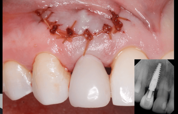
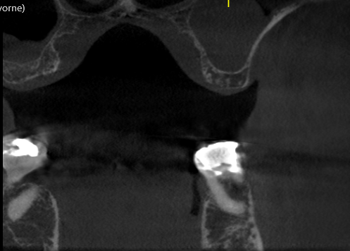
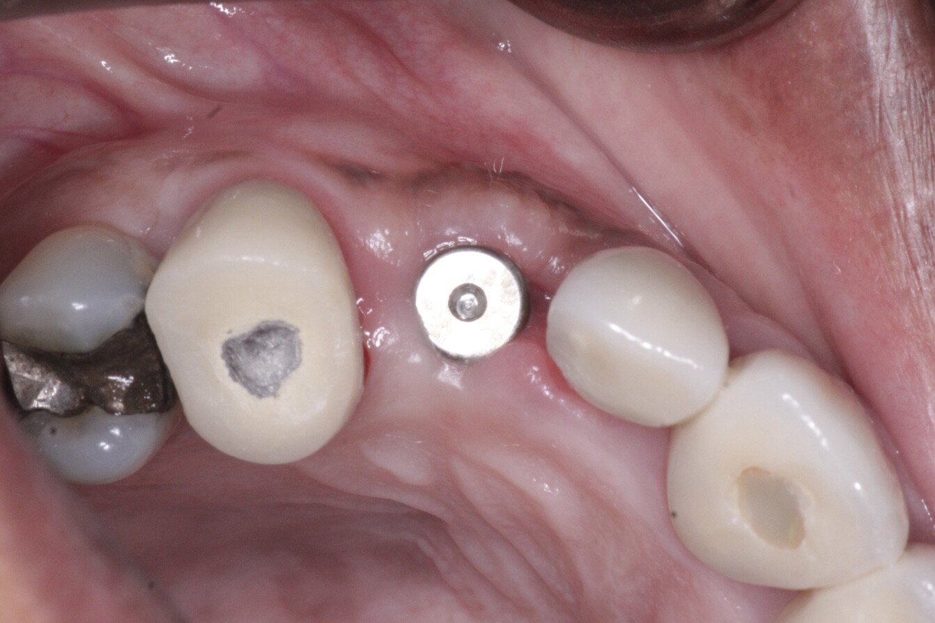
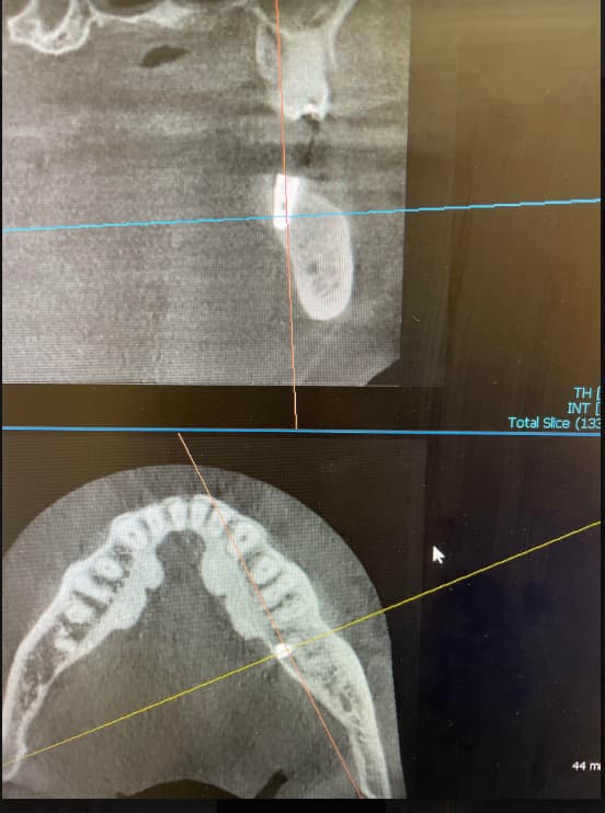
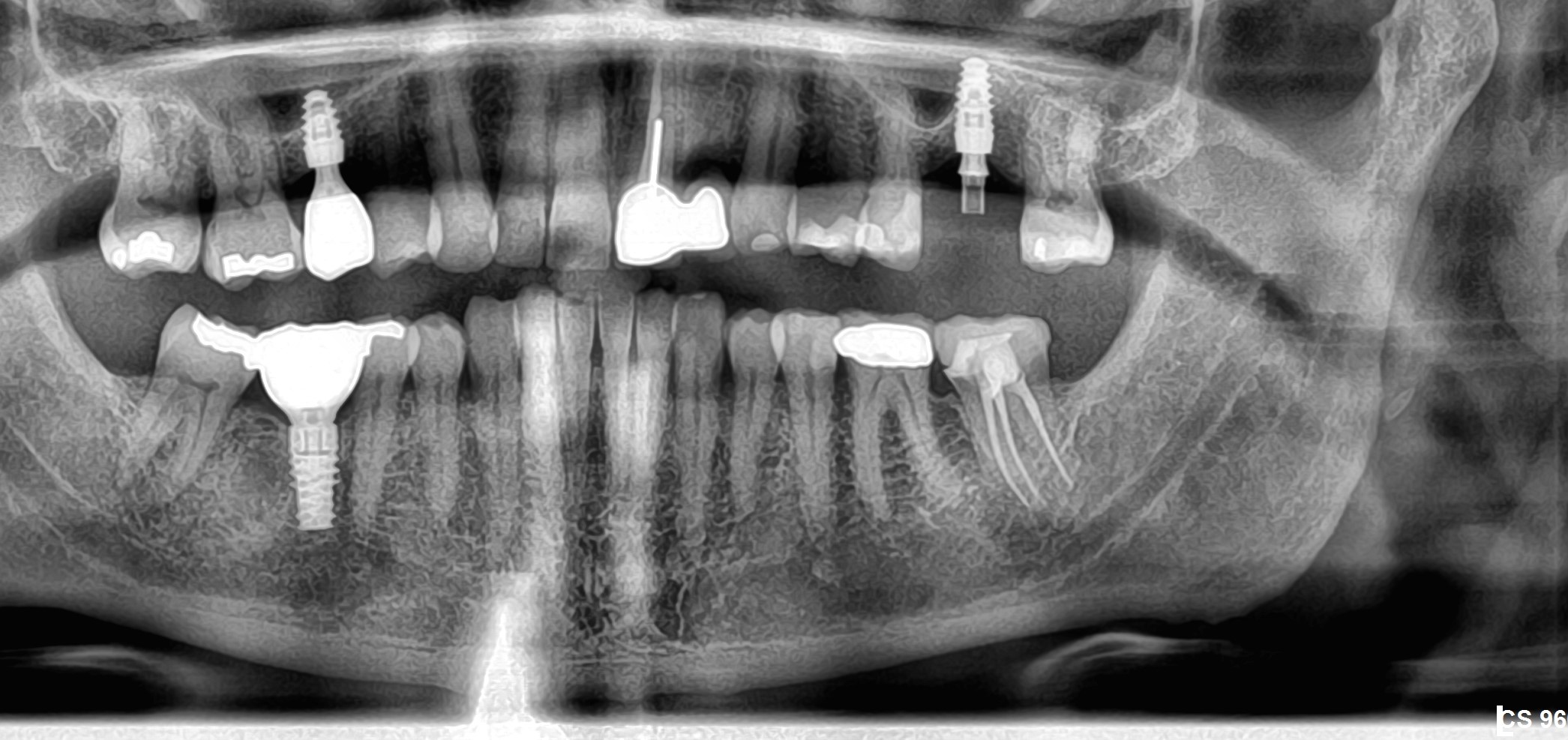
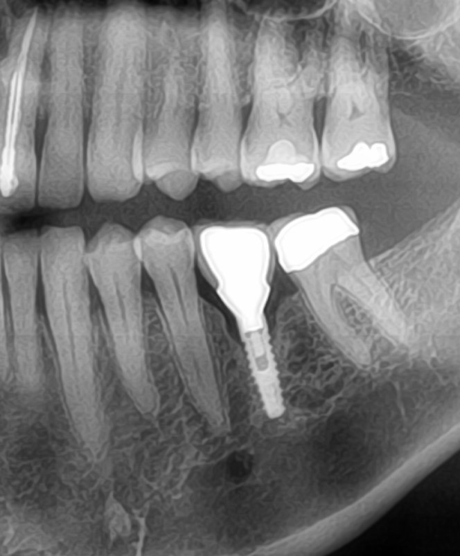
I have a 40 year old patient in excellent health who lost his #20 [mandibular left second premolar; 35]. I extracted the tooth which had endodontic therapy 15 years prior and fractured. I removed the roots and grafted the socket with the allograft, Dynablast.  Six months later I installed the implant. One unexpected complication was that the bone density was less than I expected and actually somewhat soft. The radiograph posted is taken in October 2012.  What are the complications I may encounter when I restore it? What are your recommendations?


![]RFsystem](https://osseonews.nyc3.cdn.digitaloceanspaces.com/wp-content/uploads/2012/12/20121031182925.jpg)


![]RFsystem](https://osseonews.nyc3.cdn.digitaloceanspaces.com/wp-content/uploads/2012/12/20121031182816.jpg)


![]RFsystem](https://osseonews.nyc3.cdn.digitaloceanspaces.com/wp-content/uploads/2012/12/20121031182726.jpg)

35 Comments on Bone density less than expected: what complications may one encounter?

New comments are currently closed for this post.
CRS
12/6/2012
Were you able to get primary stability and why is the implant buried so deep? Is there no lingual plate? I 'm suspicious that there may be retrograde peri-implantitis based on the endodontic history is there a preop film? Personally I find that dynagraft does not give a great result I like cortico-cancellous bone with a Teflon membrane over the top to regenerate the lingual plate. A lot of factors here in six months should have been plenty of time. I think it would be wise to remove the implant and regraft the socket as above into decent bone I've had this happen and always felt better correcting it early on vs rolling the dice and hoping for osteointegration I just don' t like the look of the bone on the radiograph not much density.
Joe Fanti
12/8/2012
Will the crown to implant length also be an issue?
R
12/9/2012
Is 50/50 crown root ratio acceptable?
Guy Carnazza
12/6/2012
The implant seems to be very deep below the crest of bone. If possible try to remove ,place closer to the crest and pack some graft apically because trying to restore will be very difficult in current position.
Ali
12/6/2012
It is deep, but it looks like an ankylos, and this looks normal subcrestal placement for ankylos.
Peter Fairbairn
12/6/2012
CRS This is the new fashion 8mm implants placed 5 mm subcrestally We will see .
CRS
12/7/2012
Interesting I don't have any experience with ankylos implants. My background is Zimmer,Nobel,BioHorizons and Straumann. This placement would be difficult to restore with those systems. Hope it integrates, in my hands I would not trust it but I would have grafted differently. Thanks for the post.
R
12/7/2012
This was an ankylos implant placed one mm subcrestal according to the buccal bone height and as a result it was two mm sebcrestal lingually. My question is that in nine months when I want to expose the implant as a second stage of surgery and initiate restorative stage, what challenges will I be facing both surgically and prosthetically.
CRS
12/8/2012
Dear R, I can only vouch for the surgical make sure it is integrated.The soft bone is concerning but if you had good primary stability, torque, it should integrate. I see an undercut on the molar which may have to be adjusted. I would assume that the Ankylos kit would have the proper countersinks to allow the prosthetic healing heads and abutments. I would respectfully defer to my more experienced restorative colleagues. I just think that this is a tougher system to be subcrestally but I am willing to check into it. What are the advantages to going subcrestally?
R
12/9/2012
By placing it subcrestally the biological width and the platform switching are both addressed and make a more naturally resembling assembly.
greg steiner
12/6/2012
The most common reason for socket graft failure is retained foreign bodies such as root tips and gutta percha but in those cases you get granulation tissue rather than poor mineralization. You have placed number of foreign materials in this socket in form of demineralized freeze dried bone and mineralized freeze dried bone in a medium to make it into a putty. I suspect that your patient produced an immune response to one or more of these potential l antigenic agents which interfered with mineralization. Graft failure aside, no graft material is perfect and you will experience failures and in the case the best course of action would to have increased the length and width of the implant so that the majority of the grafted area would be removed during the osteotomy and you would then be placing your implant in normal bone. When you are ready to expose this implant if it is not integrated be ready with that bigger implant and place it at the time this implant is removed. Greg Steiner Steiner Laboratories
CRS
12/7/2012
Good post I like the rationale. My experience with dynablast has been softer and less bone. Could it be the filler? Zimmer had a similar product with a similar result.
Dr. Alex Zavyalov
12/7/2012
If I were you, I would proceed with a temporary crown fabrication at your own expense and monitor the case. I think it’s immoral to ask the patient to pay when the outcome of the treatment is uncertain.
Bill Schaeffer
12/7/2012
This will be fine. You had less dense bone than you were expecting and you've buried this implant as a two-stage procedure. A nice safe approach. It will be fine. As for too deep - this isn't an implant that has to be placed at the crest, nor is it an implant that will lose bone if placed below the bone. Peter thinks it's been placed 5mm below the level of the bone - but his eyes clearly aren't what they used to be ;-) This implant appears to have been placed 2-3mm below the level of the bone AND FOR THIS SYSTEM that is a perfectly restorable position and one in which you would not expect to lose crestal bone. It's a nice placement - though for clinicians not familiar with the system it may look strange. Kind Regards, Bill Schaeffer
R
12/7/2012
Stability was checked and it was good at the time of the placement.
R
12/9/2012
Dear Dr, schaeffer This implant was placed one mm subcrestally in the buccal aspect of the bone and about 2-3 mm subcrestally at the level of lingual bone. Hence the pictures looks terrifying. Please advise if bone will grow at the apex of this implant in nine months time.
Bill Schaeffer
12/10/2012
Dear R, THIS IMPLANT WILL BE FINE. Yes, bone will grow at the apex where you have over-drilled the length of the osteotomy - it does every other time we do this so why should your case be any different? The appearance of the PA is only "terrifying" to clinicians not used to implants that are designed to be used like this. It looks perfectly normal to me. Kind Regards, Bill Schaeffer
Bill Schaeffer
12/10/2012
Sorry R, I just saw you said 9 months. Uncover this implant in 3 months (4 if you really have to!). It will not need longer than that. Kind Regards, Bill Schaeffer
R
12/10/2012
Thank you Dr. Schaeffer. Your opinion means a lot.
CRS
12/7/2012
Thanks for the clarification it did look strange to me!
naswe
12/8/2012
a longer and wider implant should have been placed engaging the normal apical and axial bone , here your implant is floating in the graft material only .
CRS
12/8/2012
Greg Steiner will be all over this!
greg steiner
12/18/2012
CRS I like implants in grafts as long as they produce vital functional bone. Most all of my implants are in grafted sites. I don't like implants in the artificial tissue created by allografts of xenografts which are low in vitality and have no ability to adapt. It all just depends on the material. Greg Steiner Steiner Laboratories
Richard Hughes! DDS, FAAI
12/8/2012
For this situation, I would perform the typical socket graft procedure (degranulate, detoxify and decorticate then place a slurry of OsteoGen or whatever you want to use). Then place the implant. This will increase the BIC at this site. In the maxilla I would do the same with a socket. A virgin site in the maxilla, where the bone is soft D3 or D4, Use osteotomes to improve bone density and OsteoGen. Under prepping the osteotomy by width is an option. These are basic procedures that have proved to be most reliable.
Yassen Dimitrov
12/9/2012
I think Greg Steiner is right! Soft bone in a mandible socket 6 months after extraction? It must be related to the bone remodelling of the demineralized bone.I checked Keystone Dental, and the indications they give for the use of Dynablast. I don`t think you should use similar materials for bone grafting at all .Here is why: First-the demineralized part of the material will trigger fast resorbtion (predominantly osteoclastic activity),resulting in soft bone. Second-each graft material should have porosities, to alow fast vascularisation of the grafted site (within 3-4 days), otherwise oxigen tissue level in the grafted site will be reduced, resulting (again) in osteoblastic activity.In a material with a putty shape, or gel-no porosities! Remember- osteoblasts (the "good guys",around our implants, which produce new mineralized bone) come at the end, when there is enough oxygen in the grafted area. Otherwise only soft tissue and soft bone will result. Excellent planning and perfect implant placement for a bone level implant (Ankylos). My advice-after 5 months uncover the implant, measure its stability (with Osstell, Periotest, whatever), if values are acceptable, place a healing abutment, then-provisional crown with moderate to light occlusal contacts (train the bone), after 1-2 months re-measure the stability and complete the case. Keep us informed, Yassen Dimitrov
Dr. Alex Zavyalov
12/9/2012
It’s a molar mastication area, and the apex-implant-bone void will cause the implant sagging after loading.
Mike Heads
12/12/2012
Every one is talking about grafting the socket for six months. I can never see the point of this because if you leave the socket alone then after 6 months you will have the most beautiful natural bone to place an implant into, what could be better. It also gives the body time to get rid of all the problems that were associated with the old apical area, that has been described in the comments above as having a detrimental affect on any grafting material. When you come to place the implant, if you have any deficiencies around the implant you can then simply graft these regions, which will be on the outside of the implant where as the main body of the implant, where we want all the strength and stability to be, will be into natural bone, which surely is what we want. Having said all of this I would actually be back into a socket, radiographic evidence permitting, in three months so as to reduce the amount of crestal buccal bone loss and I can guarantee you at 3 months there is normally good bone to work with.
Baker k. Vinci
12/12/2012
I have to agree with mike. Either you place the implant at the time of extraction or, if you are going to wait six months, do nothing. I'm not suggesting that you are intentionally over treating, but what is the rationale? Bvinci
rsdds
12/12/2012
i would remove this implant asap 3 to 4 mm subcrestally is unacceptable follow dr. hughes advice.. good luck
Baker k. Vinci
12/12/2012
I have had a few cases like this . One was on a friend that I caused a paresthesia on and she would not let me place a smaller fixture back in it's place. 13 years later she has severe osteoporosis. That is my only permanent paresthesia case, with implants. Other cases, I have filled the osteotomy with a mixture of dfdb and autogenous bone, or autogenous only and half had no failures, that I can recall, with this type of case. Today, I would use mineralized and autogenous. Bvinci
Baker k. Vinci
12/13/2012
Addendum : I place the fixture on top of the grafted bone, in the soft site. Sometimes with less than 35ncm. Certain cases I have placed membrane, even though the grafted bone was not exposed. Bvinci
Peter Fairbairn
12/13/2012
Bill has years of amazing cases with this protocol , which is correct for this system so as to the depth of placement , there is no issue . Anyone who has been to a Ankylos meeting will understand . And yes Bill the "5mm " was a figurative statement , and yes I do place sub-crestally in cases where it warrants it The surrounding graft material may have some issues , but best to leave the body will heal and this will work. Peter
Bill Schaeffer
12/14/2012
Thanks Peter. I have to say that my favourite comment on this thread was from the guy who wrote this one; "It’s a molar mastication area, and the apex-implant-bone void will cause the implant sagging after loading." Kind Regards, Bill Schaeffer
Baker k. Vinci
12/14/2012
You have got to image your pilot drill and final position,even on the "simplest" cases. Bvinci
K. F. Chow BDS., FDSRCS
12/15/2012
I have used Ankylos implants before. They are supposed to be inserted until flush with the crestal bone or a little below. Initially, they left the collar polished where it is next to the cortical plate. Logic was a rough surface is susceptible to germs attaching and causing an infection. Thus they left the collar polished just in case part of it ended up above bone. Later, they gave all the collars a rough surface. My experience is even if the implant is slightly above bone, the bone rises to the same level and sometimes even cover up the whole implant in which case, I had to remove bone to uncover and use the implant. All things considered, we should respect the body's natural tendency to want to heal and recover itself with minimal help from humans. Please study this link: http://smalldentalimplants.blogspot.com/2012/12/bone-climbing-up-mini-dental-implant.html. In this case, it would be better to have inserted the implant until it is flush with the crestal level though part of the implant may be exposed. However, this particular implant can still be completed satisfactorily and no attempt should be made to improve things.

Featured Products

OsteoGen Bone Grafting Plug
Combines bone graft with a collagen plug to yield the easiest and most affordable way to clinically deliver bone graft for socket preservation.
CevOss Bovine Bone Graft
Make the switch to a better xenograft! High volume of interconnected pores promotes new bone. Substantially equivalent to BioOss and NuOss.