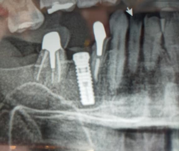
Bone resorption because of bruxism?

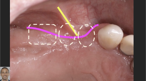
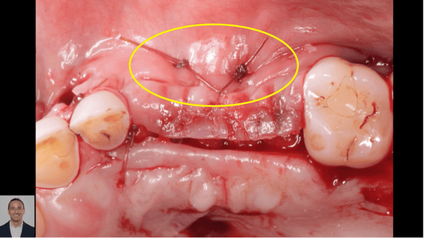
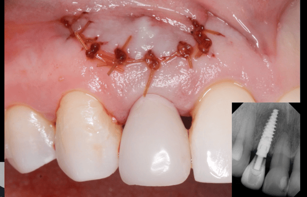
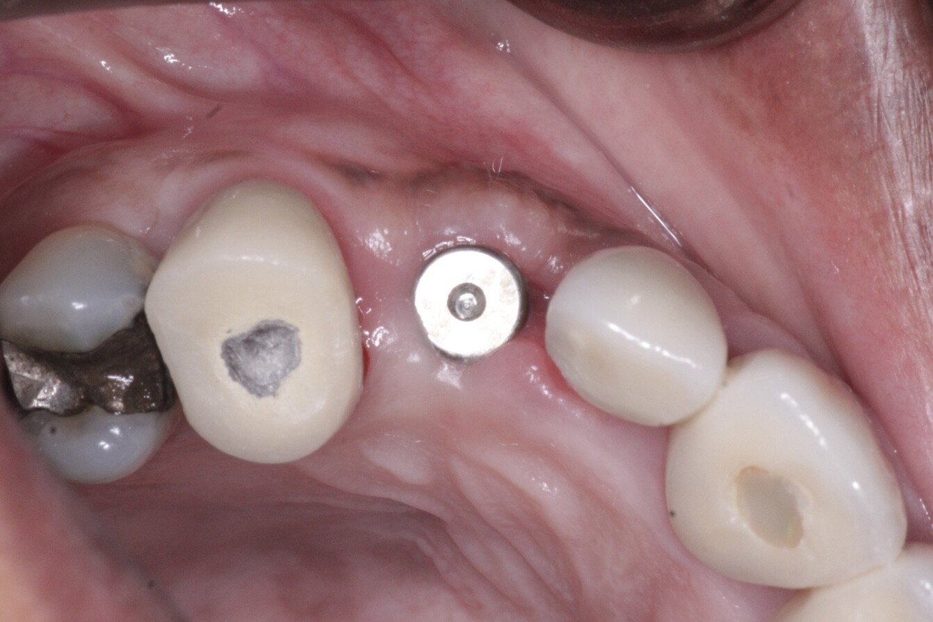
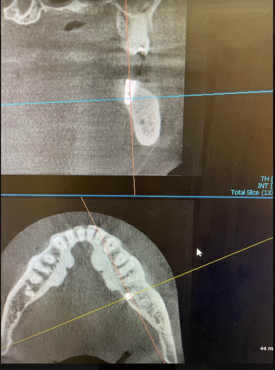
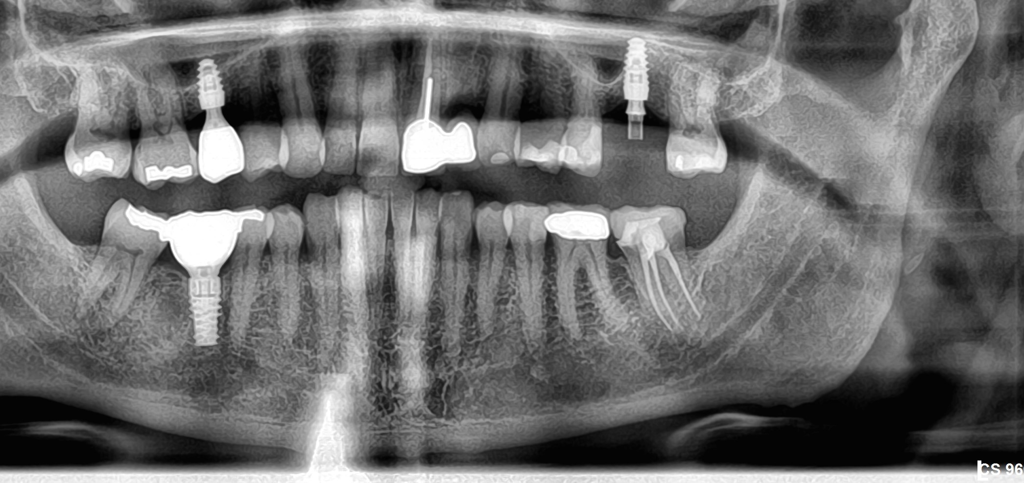
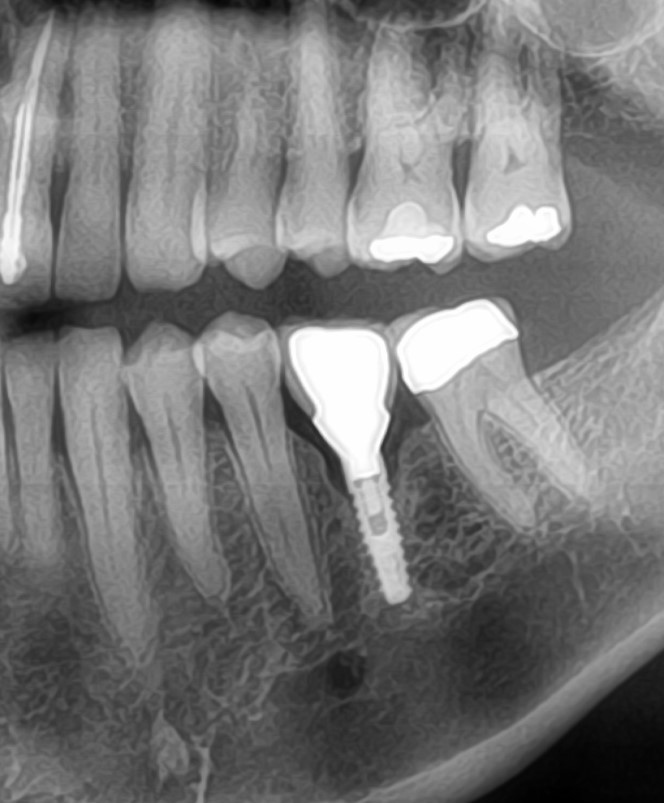
An implant was placed 3 months after extraction of a lower first molar, bone quality was good, implant was placed 1mm below bone and a cover screw was placed (first image). After 3 months fixed, screw- retained crown was placed. 6 months after functional loading patient comes in and complains about suppuration (second image). Clinically suppuration was seen on the buccal gingiva about 3 mm from gingival margin. During cleaning of the area with CHX, I rinsed out something similar to a small fish-bone (?). Afterwards antibiotics were prescribed. The crown was obviously receiving lateral forces and patient admitted that he is a bruxer. I made a night guard and reduced the height of the crown. Patient came in for check-up after 2 months and has no complaints but the bone in the X-ray seems to be resorbed (third image). There was no bleeding on probing and no suppuration. What should I do next? Leave it for further follow-up?


11 Comments on Bone resorption because of bruxism?

New comments are currently closed for this post.
Dr. Gerald Rudick
2/6/2019
I think the #36 implant tooth is doing well, but possibly the suppuration you mentioned is coming from #37.... in the 2nd and 3rd xrays, there appears to be a hairline fracture in the furcation area, with possible pathology at the apex of the mesial root....check it out, do a vitality test for the #37.
Wadhwani c
2/6/2019
You cannot determine the level of the bone form the radiographs shown, they are not at all standardized. Remember the image you show is of a 3 d object in a bone environment, flattened into a 2D image. You have parallax and magnification errors. I suggest you consider improving radiographic technique and read Malloy: IJOMI 2018 on errors in radiographic technique relative to marginal bone loss. I do not see a fracture- just superimposition of the root form, though bone radiolucency at the apex of the adjacent molar is evident. I also note image manipulation likely unsharp masking effect which could induce an artifact at the apex- see Schweitzer J prosthet dentistry 2012, and Clark J prosthetic dentistry 2018 articles that explain this very nicely.
MWeizman
2/6/2019
It all looks good to me and you should keep it shy of the occlusion and get him to wear a nightGard
Greg Kammeyer, DDS, MS, D
2/6/2019
How did you set the occlusion? no contact in light biting and light contact in full force biting. I have seen numerous implants that had problems because of parafunction.
Timothy C Carter
2/6/2019
Leave it alone. It will be fine. It is not at all uncommon to get a "gingival abcess" secondary to food debris etc.. around an implant. The supuration was probably more of a serous exudate rather than the thick mucous type from a chronic infection. Also even with new connections/platform switching/medialized abutment interface there is still some potential for micromovement at the implant/abutment interface and the classic bone loss to the first thread on a 2 piece system is not uncommon. This appears to be a variation of normal and I would not complicate the situation.
Timothy C Carter
2/6/2019
Recall also on the classic 2 piece/bone level systems the bone loss never occured until the transmucosal abutment was placed (healing abutment or final abutment). At that time the microgap begins to do its thing with the crestal bone.
Dok
2/6/2019
Leave it. Check it again every few months for changes or check it sooner if the patient complains of developing symptoms. Good that you reduce the occlusion and made a guard.
oralsurgeryjj
2/6/2019
My theory on bone resorption are.... 1. routine marginal bone loss due to machined surface of fixture. - I dunno what this implant is but some internal type looking fixture offers machined surface on top level and I sometimes inserts to deep level and it never stops bone resorption once exposed transmucosally. 2. Too thin marginal crestal bone perimplantly+ A bit flared emergence profile. -Check out CT for bone thickness arount the implant -I personally prefer keeping abutment's profile as 'slander' as possible keeping cuff higher than 3mm, divergence no more than 30 degree to axis. -a bit 'fat' emergence profile leads to immediate bone loss for several months once it is loaded 3. Cement infiltration -RMGI sometimes leads to cement infiltration to perimplant soft tissue when you set the prosthetics. -The cement never gets out when the emergence profile is 'fat' My first Tx on immediate bone loss after loading is unscrew the prosthetic part and clean up transmucosally with alcohol sponge. I have found some infiltrated resin cement on the spot or around the abutment on my early years of practice. After then, I always unscrew the abutments and clean up around the abutments and fixture immediately after resin cement setting. Also I try to avoid using cement type as much as possible.
R Gangji , DDS, AFAAID, F
2/6/2019
I Totally agree with Dr Timothy Carter , and this is not an uncommon phenomena , to get a gingival ,localized infection , that resolves once you debride and clean the site . Also crestal bone loss can occur even w/ platform switch fixtures from micro movements of an abutment or even a loose healing cap , even a loose cover screw ! We have a two week post op apt, after cementation cases ( especially ) or delivery of screw retained crowns and then even a one month post op check . In my experience, if not monitored periodically at early stages , a few cases every year end up like this . Always wise even for non bruxurs to create shallow cuspal anatomy as long term lateral forces may cause issues especially with posterior teeth where most of your load bearing forces are. Agree with Dr Kammeyer, light contacts where you can pull the shimstock, or no contacts if you’re worried! I would just monitor, this case looks like it is resolving ., monitor. Thank you for posting case.
ltd.healthy
2/11/2019
Thank you for your comment. What exactly I should be looking for during the check up after 2 weeks (1month) ? Should I try to move the crown and see if it is moving? But if it is only micro-movement how am I supposed to diagnose it? Since this is a screw retained prosthesis, my hypothesis is that patient received lateral forces on the crown because of bruxism and poor crown design. I was not the one doing the crown, however during the check up I noticed quite a lot of reduction on buccal cusps made by prosthodontist. I am guessing that the crown was poorly made and needed a lot of in-chair correction.
Jason
2/7/2019
Nature is re-establishing biological width. Crown bottom design should be more concave to allow gingival cuff height. Should have at least 2mm buccal bone at placement. Watch radiolucent cement. Just my 2 cents.

Featured Products

OsteoGen Bone Grafting Plug
Combines bone graft with a collagen plug to yield the easiest and most affordable way to clinically deliver bone graft for socket preservation.
CevOss Bovine Bone Graft
Make the switch to a better xenograft! High volume of interconnected pores promotes new bone. Substantially equivalent to BioOss and NuOss.