Bone Ridge Reconstruction after Failed Implants Removal
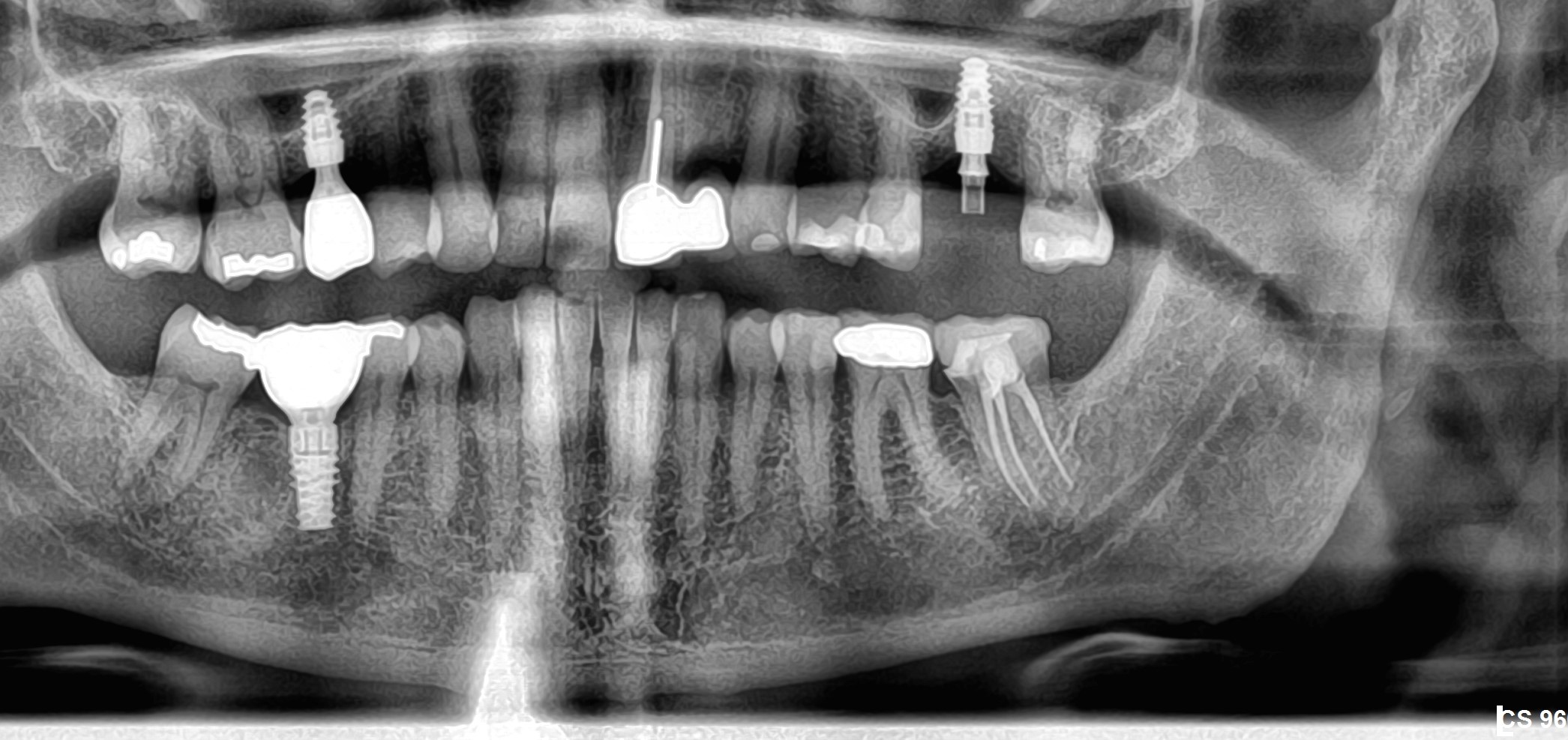
A 67-year old female came to my office with the chief complaint of being dissatisfied with the aesthetic appearance of her lower anterior prosthetic bridge due to the exposed implant threads. The implants were placed 8 years earlier. During the clinical and radiographic examination, all implants were stable, with large buccal bone deficiencies and exposed threads and loss of the attached keratinized gingiva.
The treatment plan was to remove all involved implants, augment ( with Bond Apatite – Bone Graft Cement by Augma Biomaterials) and reconstruct the deficient bone ridge and soft tissue, and to place a new implants.
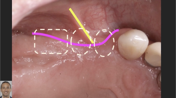
The flap was reflected according to recommended Bone Cement Envelope Technique, by performing mid-crestal incision continued with intra sulcular incisions in the teeth nearby and then reflecting a full thickness flap to create a pouch. It is important to emphasize that during flap reflection the periosteal elevator should not go past the mucogingival junction by more than 2-3 mm. In that way, we prevent the engagement of the muscles of insertion. This takes the influence of muscle movements out of the equation. At this stage, implants were removed and complete debridement was performed.
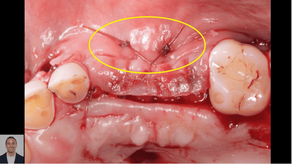
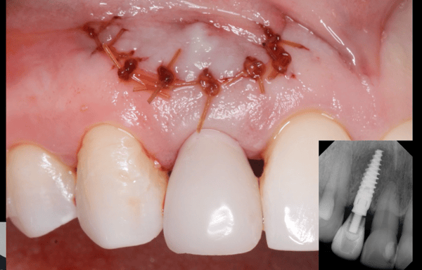
At this stage before Bond Apatite placement, a stretching was performed by grasping the mesial corner of the flap with a needle holder and stretching. Then the distal, than the middle. If we want to release it a bit more, we insert the periosteal elevator into the mesial apical corner of the pouch with a 45 degree angle and then distally. Then Bond Apatite cement was injected into the site, a dry sterile gauze pad was placed and pressed above the material for 3 seconds, and the flap was closed by stretching the mesial corner of the flap and suturing it and then the distal, then the middle. After 3 points of suturing, a predictability test was performed by placing a finger in the vestibule and vibrating it. If the sutures do not move at all it means that the muscles movements will not influence the stability of the graft during the healing phase, which can indicate that a high success rate will be guaranteed.
Healing went uneventfully and reentry and new implant placement took place 3 months post-op.


