Can bone be regenerated in this case?

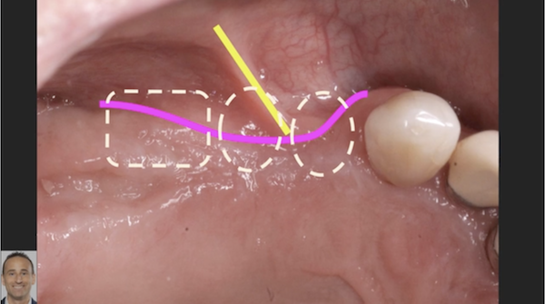
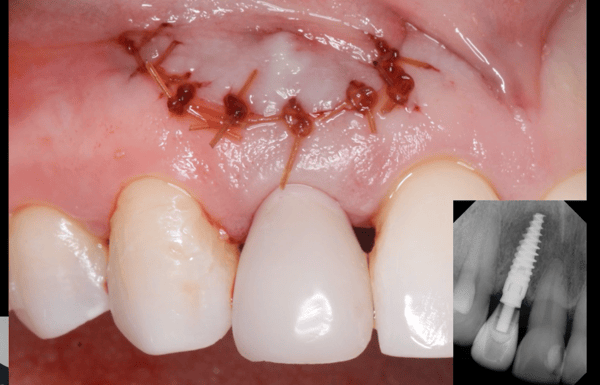
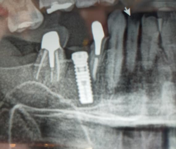
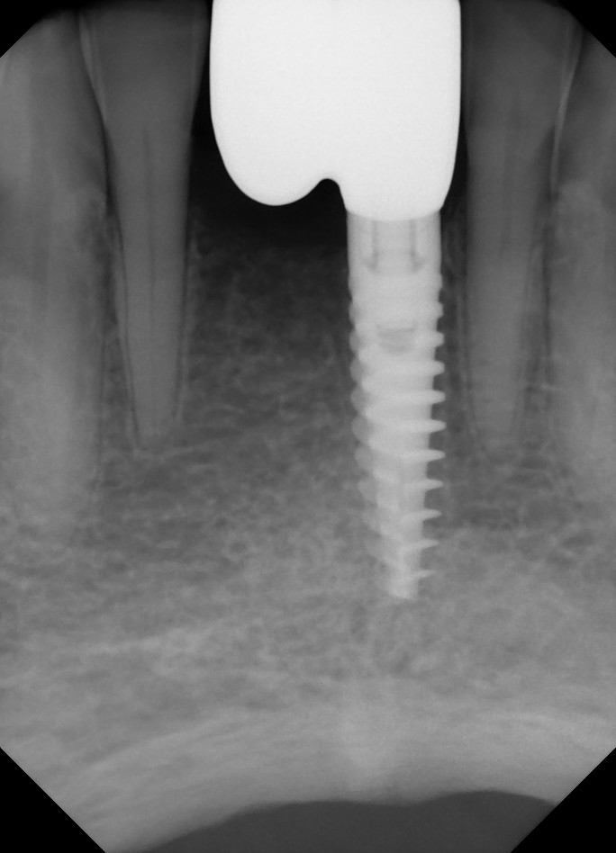
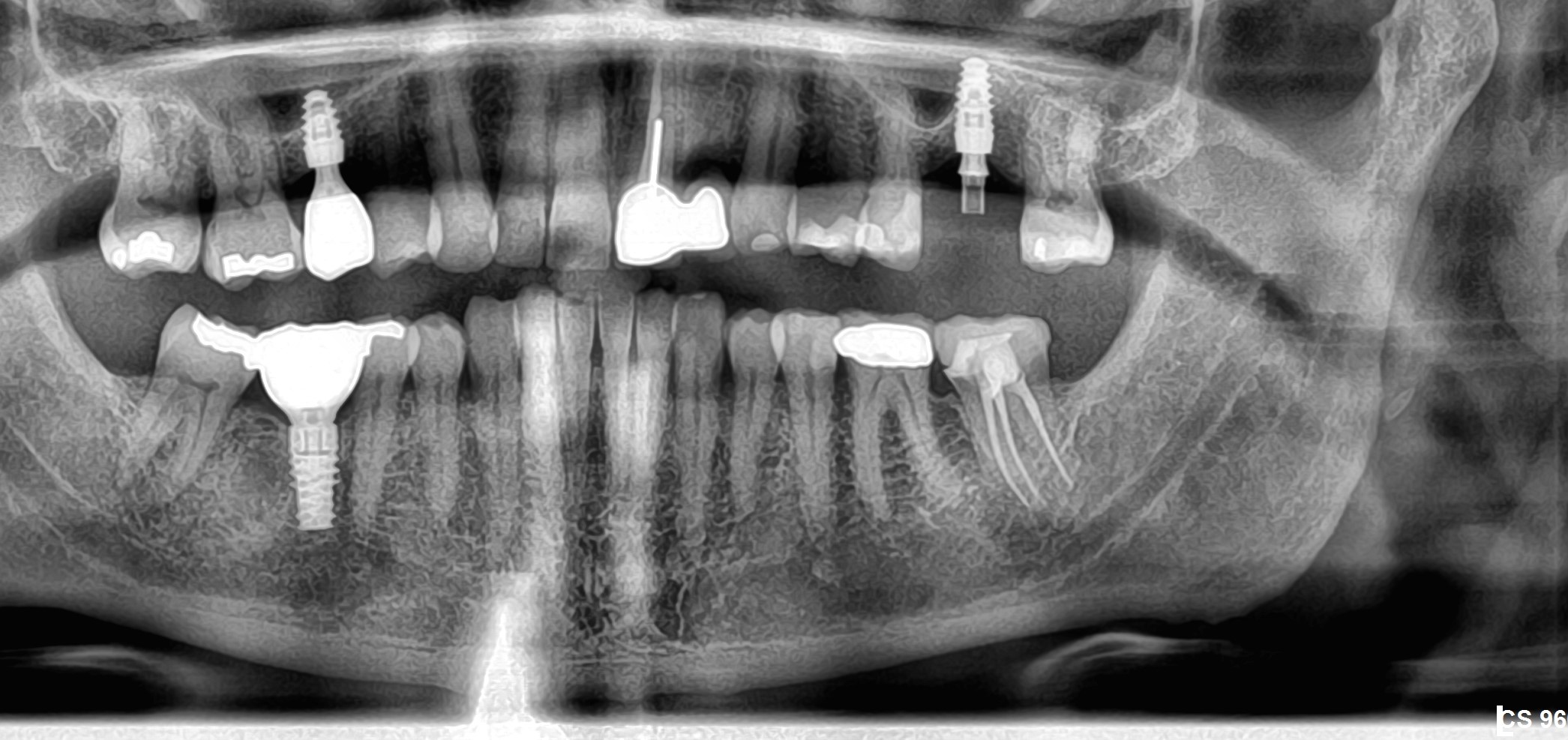
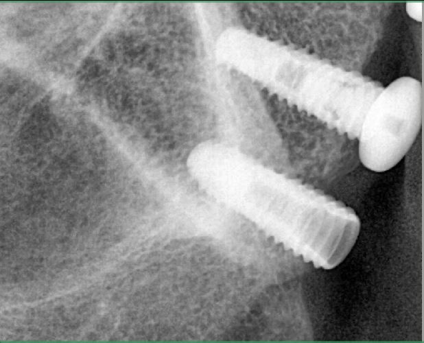
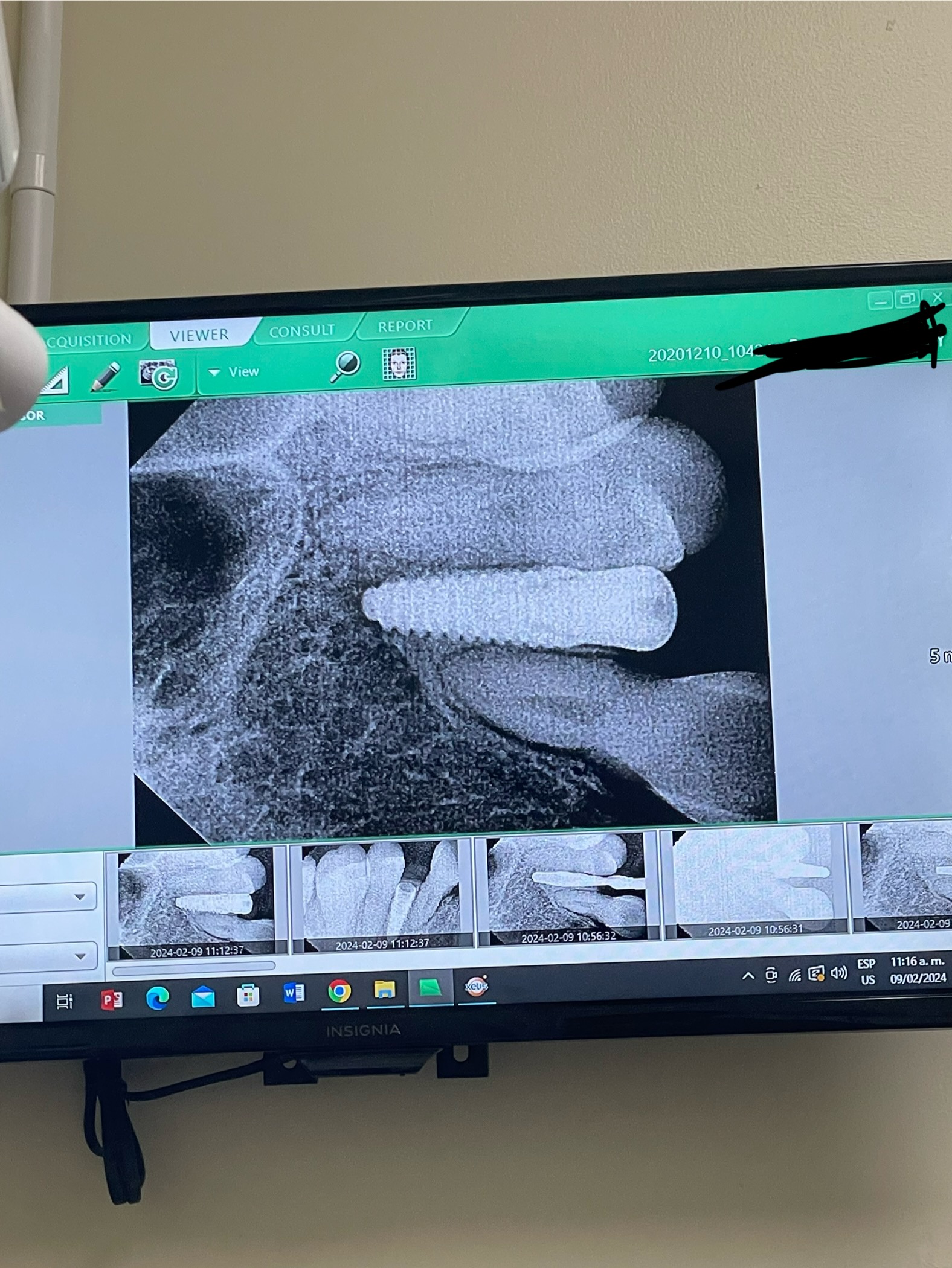
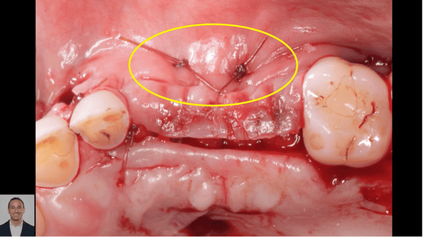
This 47-year old female patient presented with Xive [DENTSPLY] 15mm long implants installed in #2, 4 and 5 sites [maxillary right first molar, second and first premolars] 5-years prior. The implants were placed following an autogenous block bone graft in another clinic. None of the implants were mobile and #5 resisted torquing. #5 had a purulent exudate on palpation. I laid a full thickness flap and determined that there had been significant bone loss. I curetted the area and irrigated with chlorhexidine and replaced the flap and sutured, in order to reassess the case and get some advice from the more experienced doctors. What do you think can and should be done here?


![]20150709_185732-1-1](https://osseonews.nyc3.cdn.digitaloceanspaces.com/wp-content/uploads/2015/07/20150709_185732-1-1-e1436708158435.jpg)


![]20150709_1857532](https://osseonews.nyc3.cdn.digitaloceanspaces.com/wp-content/uploads/2015/07/20150709_1857532-e1436708172483.jpg)


![]20150709_114353_HDR-1](https://osseonews.nyc3.cdn.digitaloceanspaces.com/wp-content/uploads/2015/07/20150709_114353_HDR-1-e1436708190800.jpg)

7 Comments on Can bone be regenerated in this case?

New comments are currently closed for this post.
CRS
7/12/2015
I think what is holding this together is the fact everything is splinted. With the pus around #4 you may want to consider section into this and removing. If #5 is mobile the try and save #3. The patient would most likely benefit with an implant supported partial, tough to regenerate vertical and horizontal bone and a block was performed before. Sinus lifts and implant supported partial prosthetic is next step.
mike shulman
7/14/2015
hi, if you would remove the anterior implant (4), clean and detox second, performed GBR. or augment with non resorbable or even synthetic HTR. You would have this construction serving for many more moons. Likely, i would do that. Thanks for posting. Good luck mike
Geoff
7/15/2015
I would recommend removing the anterior implant. If you don't have experience pulling connective tissue from the palate to help with closure (this could be a tough one in this narrow space if your facial flap is not wide enough or you have no facial keratinized tissue), then place a free connective graft under the facial flap at extraction time and let it heal for 5-6 weeks (no longer than that or the "struts" on the edges of the socket will blunt and you won't have them to help support the membrane at the borders). At 5-6 weeks (or concurrent with extraction if you are confident in your skills to close without risk of the site opening later), reflect a facial and palatal flap with vertical incisions at least one full tooth/implant on either side of the failed implant site, bone graft (GBR) using at tenting screw, a good bone putty such as Optecure, and tack a longer term collagen membrane (of which there aren't many) such as Osseoguard on the facio-apical area and pull to place with a suture on the palatal aspect with a suture started on the palatal aspect of the palatal flap- through the membrane-back through the palatal flap and tie. Now you have your bone graft contained and you can release the periosteum for tension free flap closure without the ensuing bleeding washing your graft away (using a horizontal mattress suture about 7-8 mm from the flap margin on the facial to help "pull" the flap coronally till the wound edges lay in approximation to each other, then one horizontal mattress suture about 4 mm from the flap edge to evert and approximate the flap connective tissue edges and then interrupted as needed to assure the flap is secure. You don't want this site to open during healing or the risk of failure is high. So be sure to release the flap and suture well. Then come back in 6 months with another implant. Hope that helps, Geoff
Dr. Gerald Rudick
7/19/2015
I would agree with the opinions above in removing the first implant #4 or #14, and let the soft tissue develop over the defect and wait about five weeks before returning to the site. At five weeks, the soft tissue is keritanized, and a full thickness flap can be opened, with viertical release incision mesial to #6 or #13, and a distal vertical incision 4 mm distal to the #2 implant. Open the flap and release the tissue on the buccal with blunt dissection to free it so it can be pulled over the area to be repaired. Carefully examine the remaining two implants, make sure there is no granulomatous tissue on the threads, and reduce the bone further if there are suspicious areas harbouring bacteria or granulation tissue that you cannot access directly.Remove the abutments. Small rotary titanium brushes are available to mechanically scrape and clean the threads, followed by soaking with citric acid or peridex....or use a laser if it is available to you for detoxification. Irrigate the cleansed implant surfaces with saline solution. A semi rigid titanium mesh ( Ace Surgical) can be cut to shape and formed as a saddle that will sit on the two implants, and be secured with cover screws or the cleansed abutments . A PRF procedure should be done to obtain growth factors and the liquid that is expressed from the pressing of the fibrin clots ( Fibronectin and Vitreonectin) is used as a wetting agent for the particulate grafting material of your choice. Decorticate the crest of the ridge, and surfaces that the mesh will cover and drill holes into the surrounding bone to promote bleeding that will nourish and feed the particulate graft. Pack the particulate graft around the implants, and place the compressed fibrin membranes over the graft, before folding down the titanium mesh. The titanium mesh is now tented off the remaining implants, with a space containing the particulate graft mixture. Place a piece of PTFE membrane over the titanium mesh, and attempt to approximate the buccal and palatal flaps together in a tension free manner. Suture together in a tension fee manner. Because of the bulk created by the mesh, full approximation of the soft tissue may not be possible and the PTFE may be visible through the opening....... not to worry....... The PTFE should stay in place for about 3-4 weeks before starting to soften and appear like overchewed gum, but during this time an osteoid with immature nonkeritanized soft tissue will be forming under the titanium mesh.......try to leave this 5-6 months.... parts of the titanium mesh will be exposed when no longer covered by the PTFE which has been pulled off or sloughed off by iteself, and the patient instructed to swab with Peridex on a Q-tip twice daily...... if a sharp edge creeps out, fold it or cut off the sharp edge to prevent irritation to the tongue or cheek..... Remember that R.A.P. will develop because of the irritation to the area ...... R.A.P. = Regional Acceleratory Phenomenon that increases the healing rate between 2-10 times) In 5-6 months, open a small flap, remove the cover screws or abutments, and pry out the titanium mesh which should be firmly bound down to the newly created and repaired bone. This is a simple procedure, keep an eye on it, make sure the patient maintains home care, and if you are going to use the abutments to secure the mesh, you can, during the healing period, place a fixed plastic temporary prosthesis for esthetics and minimal function. For further information, please check out my publications on this technique in Implant News & Views, or contact me directly. Gerry Rudick Montreal, Canada
Dr. Gerald Rudick
7/19/2015
Please note that when preparing the titanium mesh as described above, make sure the mesh is extended to cover the defective bone right up to the natural canine #13 or #6..... you want to rebuilt this site, and the damage that was created by the implant #5 ( #14) which has been removed.... as well as improving the periodontal health to the natural canine. Six months after the grafting an implant can be placed into the repaired ridge. Gerry Rudick Montreal, Canada
Gregori Kurtzman, DDS, MA
7/21/2015
With the amount of circumferential bone loss on all 3 implants you will not get bone grafting to be stable to the top of the implants which means you may (and I underline the word may) get some height but likely not much. I would recommend remove all three implants clean the site well, fill a preformed titanium mesh cage with osseous graft mixed with APRF fixate the titanium mesh with screws then cover this with a APRF membrane and get closure allow to heal for 4-5 months open remove the mesh and place implants reclose and allow to heal 5-6 months. I think the implants at 4-5 developed the bone loss due to them being to close together. also lack of prosthetic passive fit and occlusion may have been contributors to the overall bone loss. My question is can the patient remember how long after implant placement was the bridge placed?
George
8/9/2015
First let me thank osseonews for giving all of us the opportunity to share our cases and the esteemed colleagues for the time they took to give their valuable advice.what i have done is what i felt that would work in my hands.i removed the first implant using elevators from the palatal side. And forceps to luxate and extract it.i did an implant plasty removing the threads and polishing the implants for supraosseous part i detoxified #15 surface and i grafted the defect left by the removal of #14 covering the site with a biomend membrane after removing the abutments and placing coverscrews and after getting tension free closure i am now crossing my fingers ...

Featured Products

OsteoGen Bone Grafting Plug
Combines bone graft with a collagen plug to yield the easiest and most affordable way to clinically deliver bone graft for socket preservation.
CevOss Bovine Bone Graft
Make the switch to a better xenograft! High volume of interconnected pores promotes new bone. Substantially equivalent to BioOss and NuOss.