Case Report: Removing Implants with the Neo Fixture Remover Kit
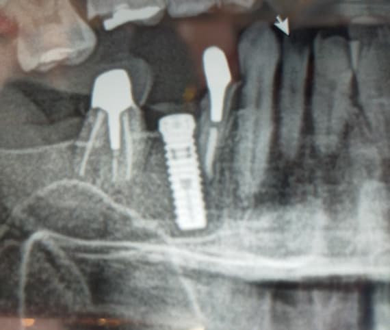
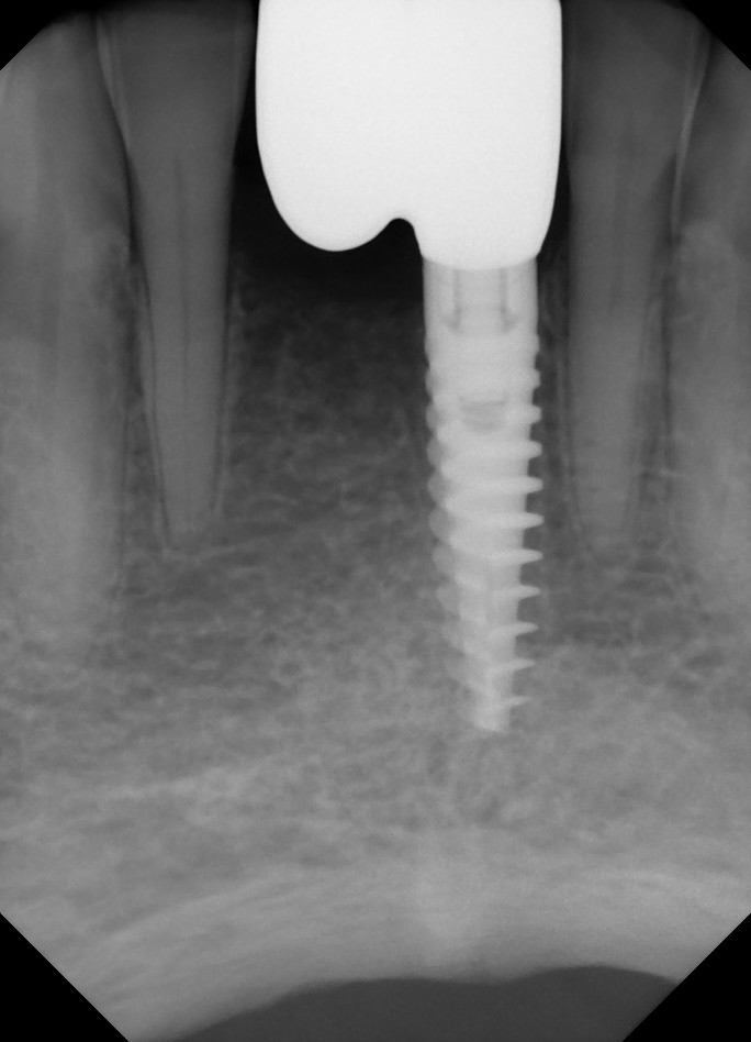
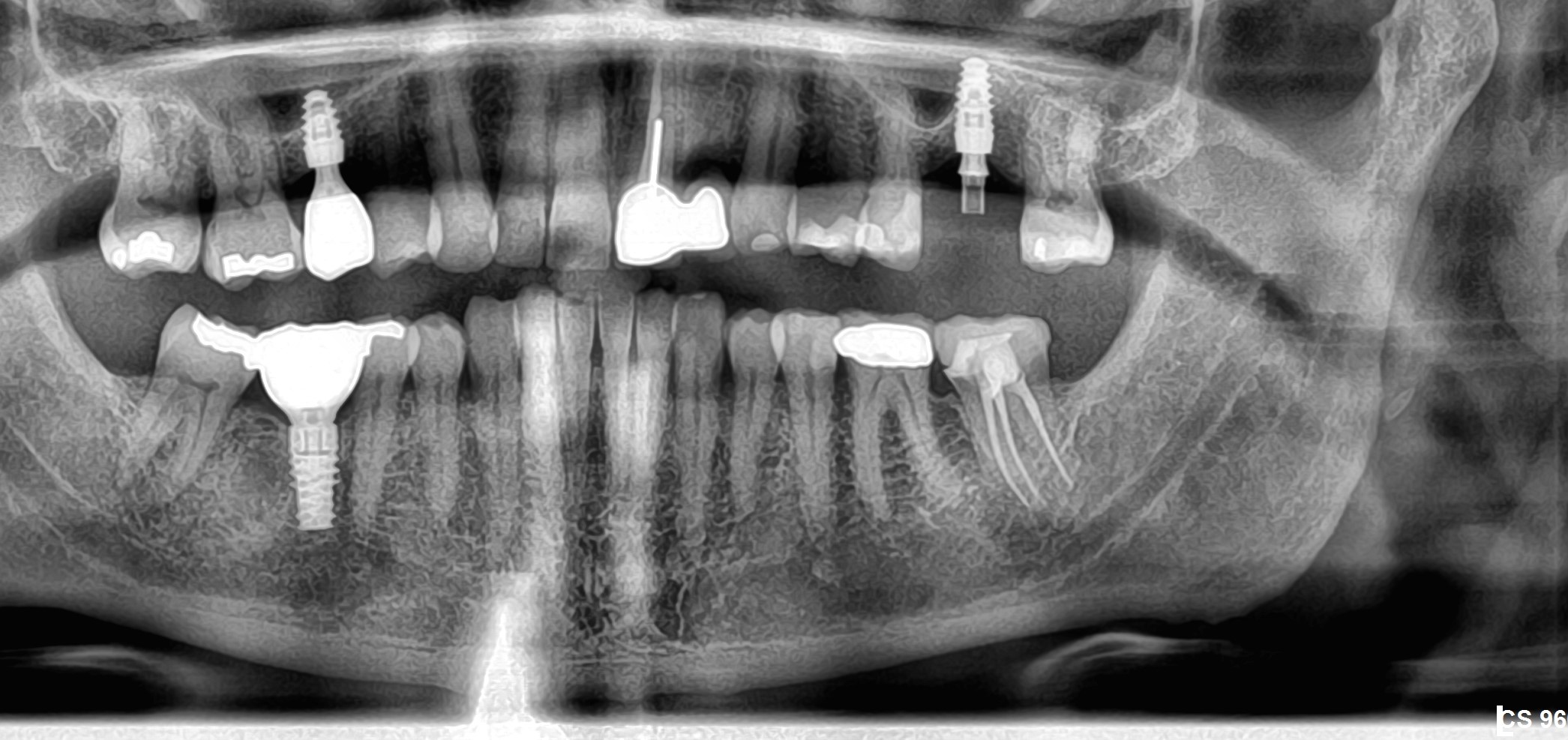
This case demonstrates the use of the NEO FR fixture remover Kit. The Fixture Remover Kit is a specially designed dynamometric key which when used in conjunction with reverse components, effectively removes failed implants. Though Trephines, burs, piezoelectric devices and various forceps have been used by clinicians to remove integrated implants, use of these instruments may compromise the surrounding bone and cause damage to neighboring structures. The NEO FR fixture remover Kit, however, uses a new technique to remove an implant by “unscrewing it†or “breaking†osseo-integration. The technique is predicated on breaking the mechanical bond (resistance to back-up the implant counter clockwise) at the bone-implant interface. Most studies assessing implant torque removal show that fixtures with rough surfaces can be explanted by applying a sufficient amount of force in a counter clockwise direction. The NEO FR fixture remover Kit therefore allows preservation of the surrounding bone obviating the need for major bone reconstruction, making implant removal and replacement more predictable. To learn more about NEO FR fixture remover Kit, and watch a video, please click here.The following two case illustrate how implants have been removed with the Neo Fixture Remover Kit. The case images are at the bottom of the page. Please click any image for a larger view.
A 20-year-old female consulted for the undesirable appearance of the gingiva on the labial aspect of her implant-supported crown at #21 (Figs. 1a-e). Her dental history revealed that both teeth, #11 and #21, avulsed as a teenager following an accident. At the age of 18, reconstructive and regenerative grafting procedures were performed to reconstruct the prospective implant recipient sites. After six months of healing two standard external hex implants of 13 mm in length were placed (Fig. 1b). Two years after implantation, the patient had noticed moderate gingival recession corresponding to the implant replacing tooth # 21 (Fig. 1c). A consultation with the same surgeon resulted in another grafting procedure utilizing particulate xenograft with the goal to provide better support for the soft tissue. A few weeks following the surgery, the gingival recession has increased. Then, another corrective surgery was performed consisting of the removal of the labial frenum. The recession did not reverse but remained at the same level.
![]](https://osseonews.nyc3.cdn.digitaloceanspaces.com/wp-content/uploads/2012/12/neo1a.jpg)1A. Figures 1A-E. Clinical presentation of the patient before removal of the implants. Note pink porcelain (1c) added to the crown of 2.1 to mask the underlying metal component. 1e shows the position of the two abutments from an occlusal view.
![]](https://osseonews.nyc3.cdn.digitaloceanspaces.com/wp-content/uploads/2012/12/neo1b.jpg)1B
![]](https://osseonews.nyc3.cdn.digitaloceanspaces.com/wp-content/uploads/2012/12/neo1c.jpg)1C. Note pink porcelain (1c) added to the crown of 2.1 to mask the underlying metal component.In spite of a favourable smile line the patient expressed a strong desire to address the recession. On clinical examination the recession on the facial aspect of #21 measured 3mm and there was a clear discrepancy between the levels of the marginal gingiva when compared to the adjacent right central incisor. The probing depth was 7mm. After removal of the two cemented and splinted crowns (Fig. 1d), it became evident that the mesio-distal positions of both implants were favourable with adequate inter-dental and inter-implant spaces. However, while the implant at #11 was slightly buccaly positioned the implant at #21 was severely misplaced in the bucco-lingual dimension (Fig. 1e).
![]](https://osseonews.nyc3.cdn.digitaloceanspaces.com/wp-content/uploads/2012/12/neo1d.jpg)1D
![]](https://osseonews.nyc3.cdn.digitaloceanspaces.com/wp-content/uploads/2012/12/neo1e.jpg)1E. shows the position of the two abutments from an occlusal view.After discussing the different esthetic issues and treatment possibilities ranging from maintaining these implants to removing both implants, the patient opted for the removal of both implants in order to proceed with an orthodontic treatment to ideally realign the lateral incisors and canines before an attempt was made to re-treat the sites with two implants. As explained, further bone augmentation may be necessary.
A provisional restoration was fabricated and cover screws were placed on both implants for 2 weeks to maximize soft tissue closure (Fig. 2). To prepare for the eventual bone reconstruction, a preliminary approach was planned to remove the two implants utilizing a novel technique rather than using trephines that results in at least 1.5mm bone loss around the original osteotomy site. The fibrotic and scarred tissue in the anterior area in conjunction with a shallow vestibular fold and a prominent nasal spine posed a significant challenge for bone reconstruction and in achieving tension-free soft tissue closure. It was therefore planned to reconstruct soft tissues at the time of implant removal with a periodontal plastic procedure.
![]](https://osseonews.nyc3.cdn.digitaloceanspaces.com/wp-content/uploads/2012/12/neo2a.jpg)Figure 2: Incomplete soft tissue closure over the cover screw of implant at the 21 site.Following a full thickness flap reflection the implant at #21 site a horizontal bone loss exposing up to the seventh thread on its facial aspect was noted. The implant at the #11 site also showed bone loss exposing up to the third thread (Fig. 3a). Since both implants were integrated on a large percentage of their surfaces, using conventional devices such as trephines or burs would have resulted in substantial bone loss of the already thin alveolar ridge. Instead, a specially designed implant removal kit including a dynamometric key was utilized to remove the implants. The torque wrench allows for an application over 450 Ncm in a antirotational direction, therefore providing sufficient force to brake integration and mobilize most implants regardless of their surface configurations (Figs. 3 d,e). Both implants were successfully “unscrewed†with a reverse torque of 350 Ncm.
![]](https://osseonews.nyc3.cdn.digitaloceanspaces.com/wp-content/uploads/2012/12/neo3a.jpg)Figure 3A. Excessive loss at the buccal alveolar bone following misplacement.
![]](https://osseonews.nyc3.cdn.digitaloceanspaces.com/wp-content/uploads/2012/12/neo3d.jpg)Figure 3D-E. Atraumatic removal of the implant with reverse torque.
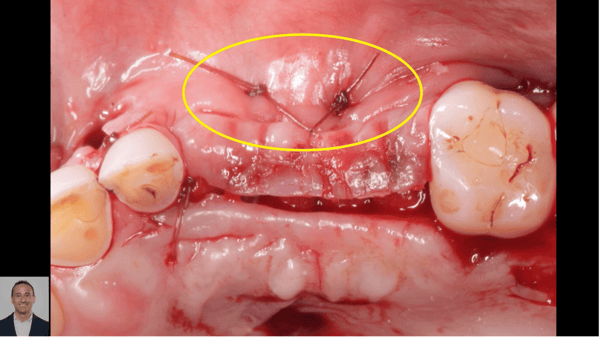
![]](https://osseonews.nyc3.cdn.digitaloceanspaces.com/wp-content/uploads/2012/12/neo3e.jpg)3E.Following implant removal, two autogenous connective tissue grafts were harvested from the hard palate and sutured in layers perpendicularly to each other over the defect covering both sites (Fig. 4). After three months of healing, the bone reconstruction phase was completed (Fig. 5 a-g). To achieve tension-free soft tissue primary closure, a split thickness flap was dissected from the palatal aspect toward the buccal. Evidence of severe bone atrophy was noted at the recipient site an autogenous bone block was harvested from the chin, adapted and screwed in place. Particulated autogenous bone was used to fill in the small voids at the periphery of the block and complete de reconstruction.
Currently the case is in the healing phase and the orthodontic treatment is in progress. The site will be reevaluated for implant placement with tridimensional imaging approximately 4 months post-op. Since the purpose of this paper is to illustrate the use of the “The Kit†the author felt that publication was warranted before final retreatment results were available.
![]](https://osseonews.nyc3.cdn.digitaloceanspaces.com/wp-content/uploads/2012/12/neo4.jpg)Figure 4. Autogenous connective tissue graft used to reconstruct the soft tissue component of the defect in preparation for the future bone reconstruction surgery.
![]](https://osseonews.nyc3.cdn.digitaloceanspaces.com/wp-content/uploads/2012/12/neo5a.jpg)Figures 5A-C. Soft tissue healing 2 months following the soft tissue periodontal plastic procedure.
![]](https://osseonews.nyc3.cdn.digitaloceanspaces.com/wp-content/uploads/2012/12/neo5b.jpg)5B
![]](https://osseonews.nyc3.cdn.digitaloceanspaces.com/wp-content/uploads/2012/12/neo5c.jpg)5C
![]](https://osseonews.nyc3.cdn.digitaloceanspaces.com/wp-content/uploads/2012/12/neo5d.jpg)Figure 5D. Partial thickness flap dissection to allow tension free soft tissue closure.
![]](https://osseonews.nyc3.cdn.digitaloceanspaces.com/wp-content/uploads/2012/12/neo5e.jpg)Figure 5E. Severe bone deficiency.
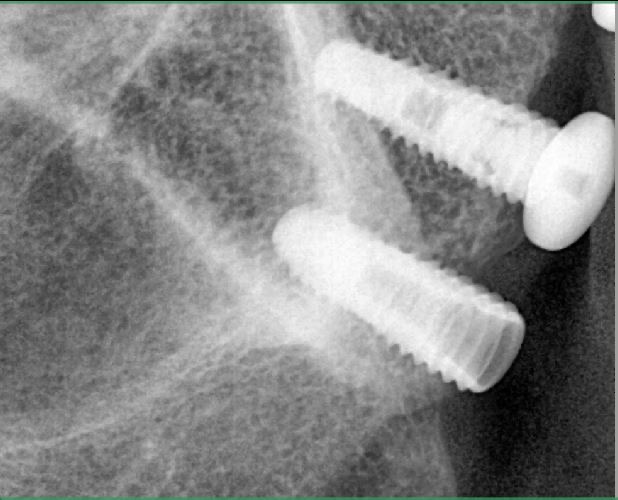
![]](https://osseonews.nyc3.cdn.digitaloceanspaces.com/wp-content/uploads/2012/12/neo5f.jpg)Figure 5F. Autogenous block graft harvested from the chin screwed in place.
![]](https://osseonews.nyc3.cdn.digitaloceanspaces.com/wp-content/uploads/2012/12/neo5g.jpg)Figure 5G. Tension free soft tissue primary closure.Case Author: Mathieu Beaudoin, DMD, MSc. Perio, FRCD(c)
Dr. Mathieu Beaudoin maintains a private practice in Montreal and focuses on periodontal therapy and implantology. He has no affiliation with and received no incentives from companies distributing any of the aforementioned devices.
Special thank to Dr. Peter Birek for his contribution to this article


