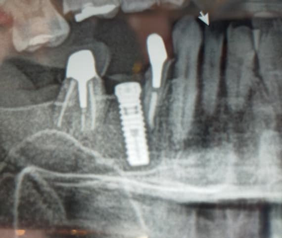
Cone Beam CT for Old Extraction Sites?
Maggie asks:
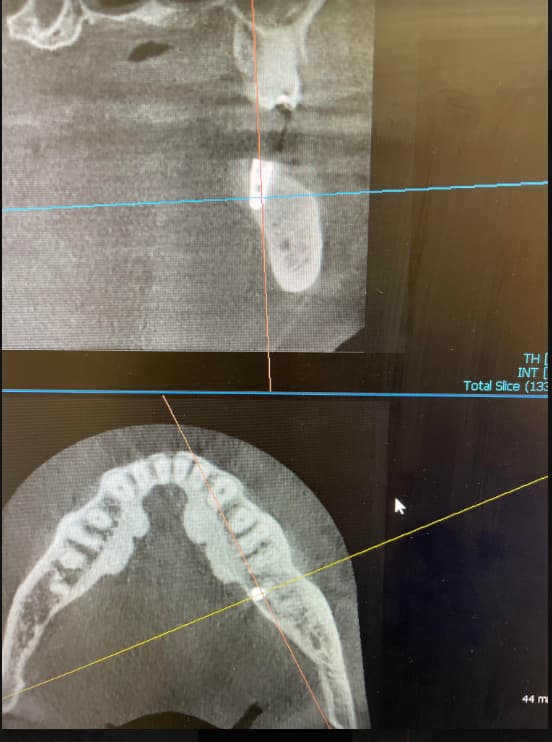
I am a patient who has a question about cone beam CT. I read on here that it can be used to evaluate old extraction sockets, and whether they have healed properly or not.
At this time, I am not looking to have dental implants, but I have concerns about my
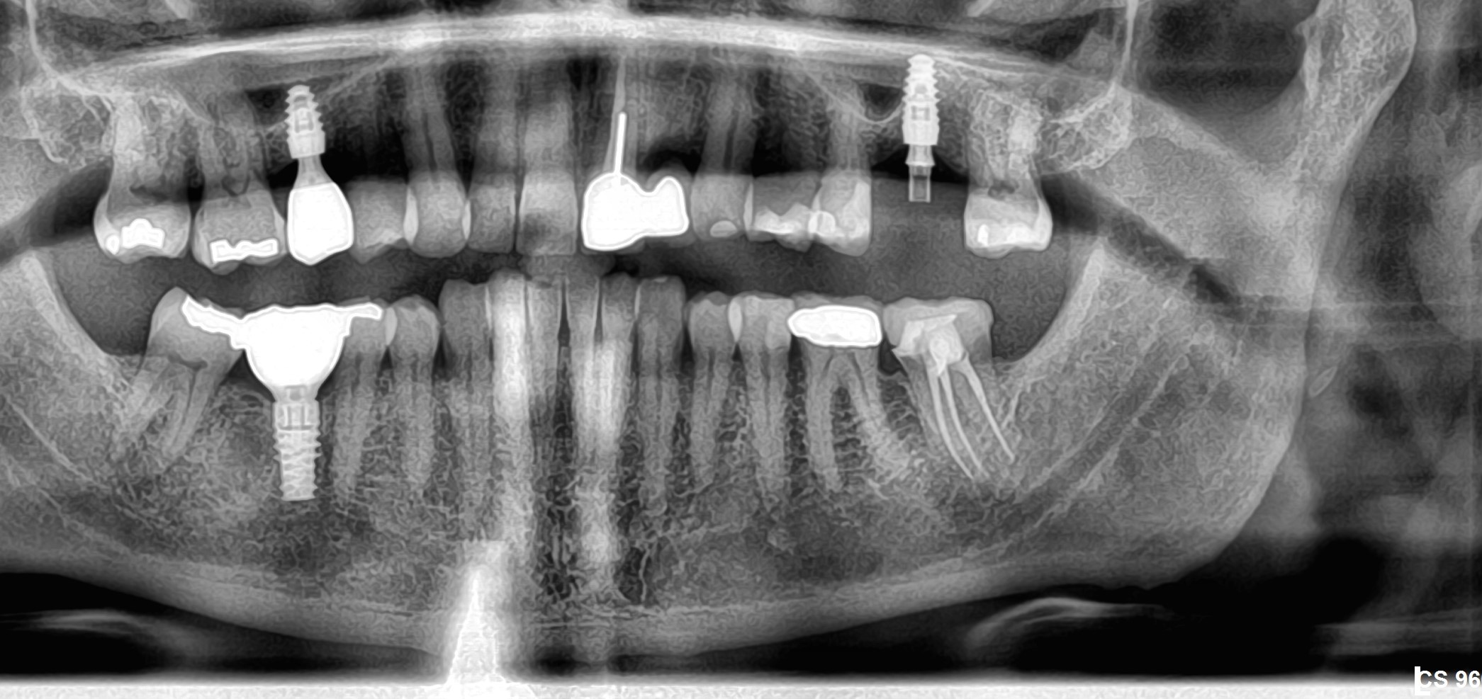
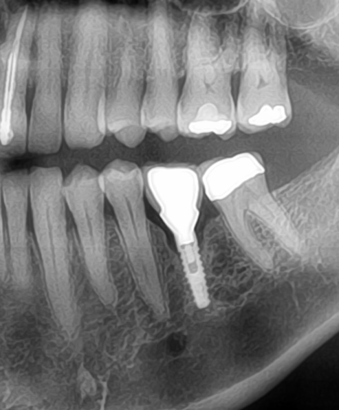
old wisdom teeth extraction sites. My teeth were extracted two years ago, at the age of 26. The lower right extraction site has been causing me discomfort, and on panoramic x-rays, I was told it looks “abnormal.”
Would it be wise to have a cone beam CT scan done to find out what is going on? Would this reveal more information than the panaromic x-rays? If so, who would be qualified to read the scan for this purpose?
Thank you for your help.
4 Comments on Cone Beam CT for Old Extraction Sites?
New comments are currently closed for this post.
Brian
5/1/2007
A cone beam CT scan will provide more information than an OPG, but it may not be necessary. In your situation, if it has been 2 years since the extraction, the site will have well and truly healed. Persistent discomfort may be associated with a defect on the posterior of the terminal tooth (second molar) and there may be a periodontal pocket there, with resulting low grade infection. This is easily assessed with a periodontal probe and clinical examination. The CBCT will give a good assessment of any underlying bone defect/persistent root fragments/contour and position of the inferior dental canal. In your situation it is probably not IMPERATIVE as a part of your examination, but will give some extra information. The iCAT allows creation of rendered 3D images, which you can then rotate to inspect bone contours, which in perio cases can be of considerable assistance to overall treatment planning and information gathering. I would get your dentist to assess the back of the second molar. Brian
Maggie
5/6/2007
Thank you for your response. I did have a recent perio exam...everything was good. The dentist is not sure why the old extraction site (bone) looks "abnormal" on x-ray...and my discomfort is definitely in the jaw bone itself. The tooth was completely impacted under bone, and was very difficult to remove. I would go back to see the oral surgeon who removed it, but I have since moved and live very far away now. If I have a cone-beam CT scan done, who is qualified to read this? Can any dentist interpret the scan, or I do need a radiologist??
thanks,
Maggie
Ken Clifford, DDS
5/7/2007
If you have the scan done by a dentist who has the equipment in his office, he or she should be able to read the scan and take appropriate action. If I saw this problem in my office I would refer to my local oral surgeon who has the i-cat scan unit and ask him to evaluate and take appropriate action if a problem is noted.
Maggie
5/7/2007
Thanks, I appreciate the advice! I am having my scan done tomorrow.


