Sponsored Case: Extraction Site with Synoss Putty and MatrixDerm
Case presentation by Robin Klein, DDS
Diplomate of the American Board of Periodontology and an Implantologist in Teaneck, NJ, Northeastern Society of Periodontists,New Jersey Society of Periodontists, American Dental Association.
Case Photos
(Case Summary is below. Click on any image for an enlarged view)
![]klein-case-1a-yf](https://osseonews.nyc3.cdn.digitaloceanspaces.com/wp-content/uploads/2013/10/klein-case-1-e1384274783600.jpg)
![]klein-case-1b-yf](https://osseonews.nyc3.cdn.digitaloceanspaces.com/wp-content/uploads/2013/10/klein-case-1-e1384274783600.jpg)
![]klein-case-2-yf](https://osseonews.nyc3.cdn.digitaloceanspaces.com/wp-content/uploads/2013/10/klein-case-2-e1384274812529.jpg)
![]klein-case-3a-yf](https://osseonews.nyc3.cdn.digitaloceanspaces.com/wp-content/uploads/2013/10/klein-case-3-e1384274840861.jpg)
![]klein-case-3b-yf](https://osseonews.nyc3.cdn.digitaloceanspaces.com/wp-content/uploads/2013/10/klein-case-3-e1384274840861.jpg)
![]klein-case-4-yf](https://osseonews.nyc3.cdn.digitaloceanspaces.com/wp-content/uploads/2013/10/klein-case-4-e1384274865496.jpg)
Patient History
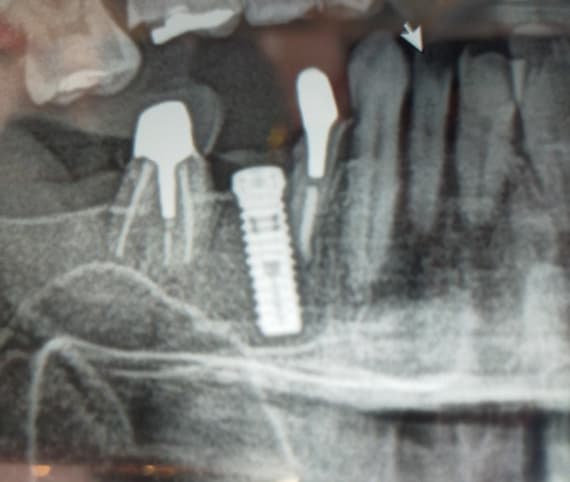
A 67-year old male presents with failed apicoectomy in mandibular incisors #24 and #25 as seen on the periapical radiograph (Fig 1). The patient is a nonsmoker and presents in good health.
Case Summary
Surgical Procedure
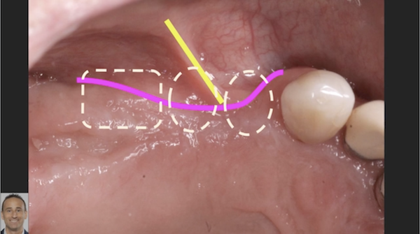
Mandibular incisors #24 and #25 were carefully extracted (Fig 2 and 3), and the sockets were missing a significant amount of bone on both the buccal and lingual walls. Treatment planning included grafting with a 0.5 cc of SynOss„ Putty and the use of a MatrixDerm® barrier membrane placed buccal, lingual and on the occlusal surface (Fig 4 and 5). Decortication was not needed, blood supply was sufficient. The membrane was used to help contain the graft and to prevent the migration of epithelial cells into the graft site. Radiographic appearance, SynOss„ Putty in extraction sockets can be seen in Figure 6.
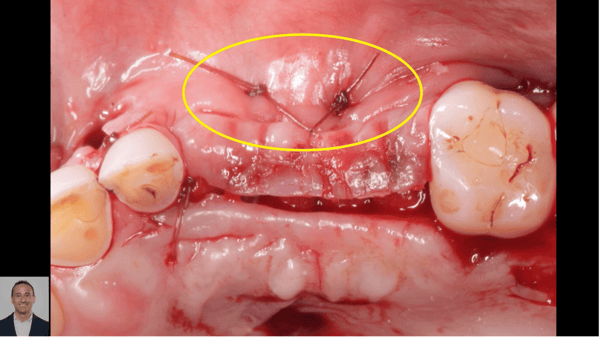
A continuous chromic mattress suture technique was used to close the surgical site. Primary closure was not achieved and the MatrixDerm® membrane was left slightly exposed. There was no attempt at primary closure in order to maintain attached gingiva and keratinized tissue (Fig 7).
Re-entry Follow-up Surgery
The two-month follow up tissue observation showed pink, healthy tissue with no inflammation. There were no post-operative complications (Fig 8). Upon re-entry 5 months after graft placement, adequate osseous fill was found and 6 mm of keratinized tissue was observed (Fig 9). Two, 3.0 mm diameter x 13 mm length implants were placed and primary stability was achieved. The implant placed in the #24 location had 3 mm thread exposure and implant #25 had 7 mm thread exposure (Fig 10). The threads were covered with a 0.5 cc SynOss„ Putty (Fig 11) and a MatrixDerm® Membrane was again placed buccal & lingual (Fig 12) and mattress suture used (Fig 13). Primary closure was not attempted.
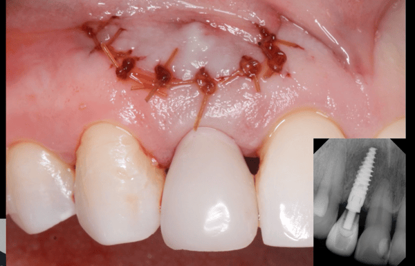
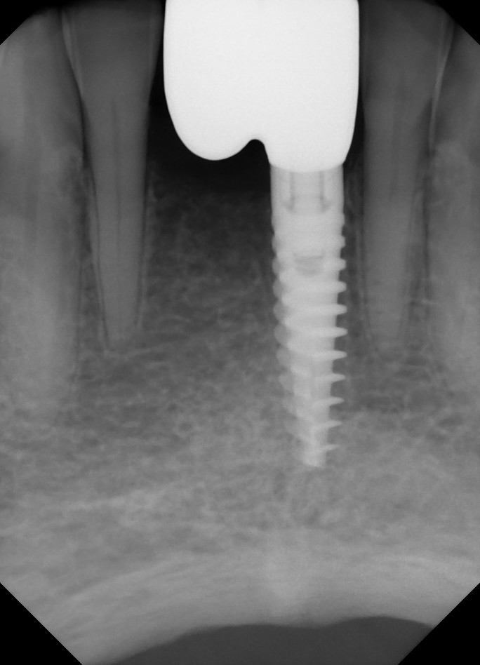
At the follow up visit the site exhibited excellent soft tissue response with 5mm keratinized tissue. Radiograph shows appearance of stable implants and graft (Fig 14). No inflammation around implants is observed (Fig 15 and 16). The temporary provisional restoration is seen in Figure 17.
Discussion
With defects in both the buccal and lingual walls, maximization of bone growth is imperative to building up the site for placement of dental implants. When inadequate alveolar bone height is present above the mandibular canal, dental implants are not an option. A barrier membrane must be used to contain the graft material and act as a barrier to epithelial down growth. In this case, SynOss„ Putty and MatrixDerm® membranes were used in both of the initial bone grafting procedures as well as in conjunction with implant placement at re-entry. This case demonstrates the versatility of the products™ handling characteristics to accommodate various clinical situations.
Conclusion
The conclusion is that vertical and horizontal bone growth was achieved with SynOss„ Putty with the aid of the MatrixDerm® barrier membrane by Collagen Matrix Dental. Dental implants were able to be placed and primary fixation was achieved in the grafted site.
Collagen Matrix Dental Products:
SynOss Putty Synthetic MineralCollagen Composite is a bone graft matrix with an additional characteristic that enables it to become moldable putty upon hydration. It is indicated for use in oral surgical applications involving bone repair such as augmentation or reconstructive treatment of the alveolar ridge and for the filling of periodontal defects in conjunction with products intended for Guided Tissue Regeneration and Guided Bone Regeneration, such as MatrixDerm® Membrane.
MatrixDerm porcine collagen membrane has been precisely enhanced to provide periodontal and oral surgeons with the ideal balance of properties to effectively address a host of clinical indications and surgical procedures. The programmed resorption time of MatrixDerm® supports bone or tissue regeneration for 6 to 9 months to allow remodeling of the defect site. The semipermeable membrane allows for nutrient exchange while providing a cell barrier to prevent epithelial down growth.


