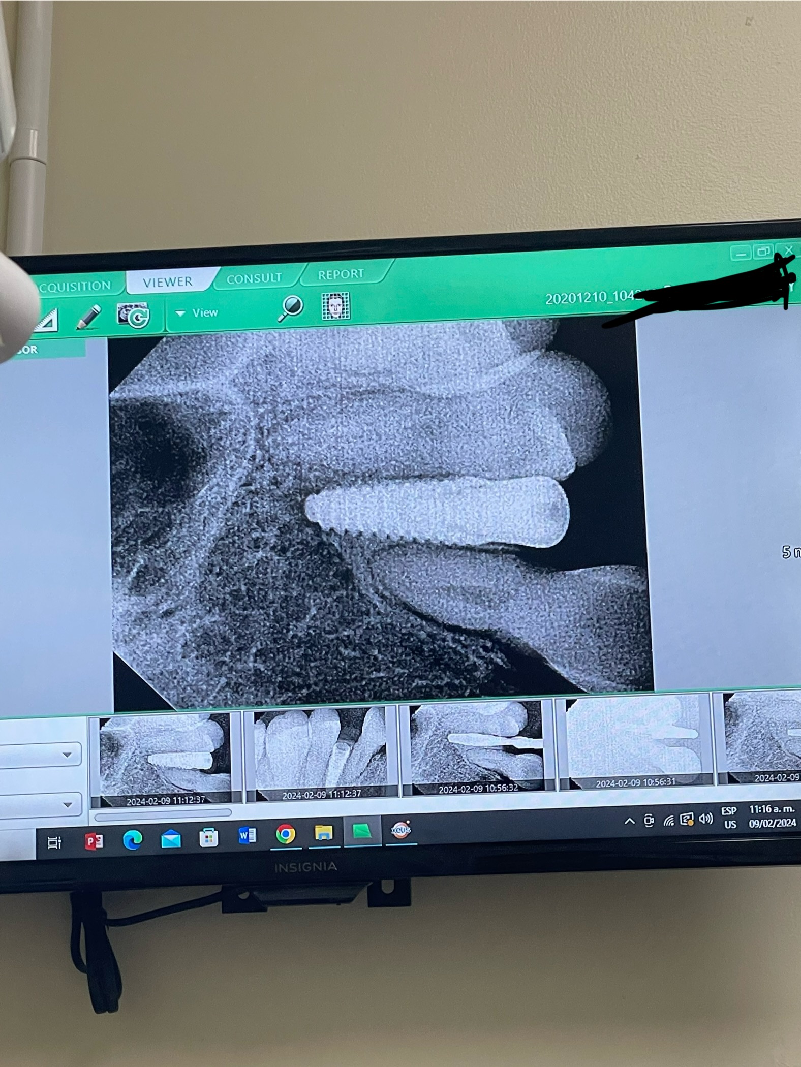
Graft failure with chronic apical abscess: Protocol?
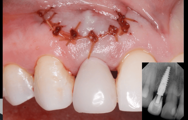
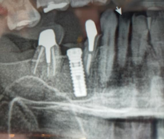
Tooth #30 had a chronic abscess and root canal failure. I extracted the tooth, debrided the socket, irrigated with saline and chlorhexidine and grafted with FDBA (Maxxeus) [freeze dried bone allograft]. A CollaPlug [absorbable collagen] was placed and sutured. This is how the area looks 3 weeks post-op. To me, the graft has poor density. Obviously, I will wait 3-4 months before even thinking about an implant. But I would like to know what is your protocol to clean and decontaminate a chronic apical abscess? I find these tend to damage the bone, and can affect implant success. What has been your experience?


17 Comments on Graft failure with chronic apical abscess: Protocol?
New comments are currently closed for this post.
Timothy Carter
7/11/2018
In my opinion that is normal. The graft is supposed to resorb and regenerate so a lack of density is to be expected during this phase
Neil Zachs
7/11/2018
Your graft looks awesome at the time of placement. Personally I would give it some time for bony turnover and see if the density gets better on its own. Worst case is in a few months if the fill is not getting more dense, you can open it up, debride and regraft. If you have good tissue coverage at that time ( Which you should) you can always use a resorbable membrane to exclude soft tissue invagination. I am curious to see what others say. I will say from my experience, the more long-standing, chronic infections always tend to heal slower and with less bony density
Neil Zachs
Periodontist, Scottsdale AZ
Don Callan
7/11/2018
suggest not to use chlorhexidine in any surgical site. Chlorhexidine kills fibroblast . Chlorhexidine has been shown to delay healing.
Doc
7/11/2018
As a periodontist I have been suggesting chlorhexidine as a post op rinse for 12 years now. I don't see any fibroblast inhibition. These in vitro studies don't hold up to clinical situations.
Francis G Serio
7/11/2018
A long time ago- 1988 I think- Dr. Robert Schallhorn broke Perio bone graft patients into three categories based on radiograph appearance of bone- fast, normal and slow healers- 6 months, 1 year, and 2 years until good radiographic evidence of bone regeneration could be seen. At three weeks, not much should be anticipated. Is the graft still in the socket or is the socket empty? Also hard to tell what is happening with non standard radiographs. Patience is the key. Biology takes the time it is going to take. Good luck.
Amit
7/11/2018
In a big socket with bone resorption already in play you need more of a scaffold since you'll have slow regeneration anyway. Using allografts will not give you the scaffold you are looking for since these resorb way too quickly and even the new bone formation will have a hard time maintaining itself. Try a xenograft mix for longer support or if you had a non RCT tooth extracted (which I can see you did not) you could have used it to produce Autologous Dentin Graft. That would have performed perfectly.
Robert J. Miller
7/11/2018
It appears that you left granulomatous tissue in the apical area that can be seen radiographically under the graft material. The pathogen load has now affected the surgical site and, upon re-entry, you will find nothing but a soft tissue complex. Open, debride completely, and regraft.
RJM
Erik
7/11/2018
I use clindamycin liquid form .8 cc as antibacterial vs peridex.
greg steiner
7/11/2018
I agree with Dr. Miller but there is no published histology on how cadaver bone grafts produce mineralization so what is happening at three weeks is just a guess. One thing I know for sure is that there is no histology that shows mineralized cadaver bone grafts ever resorb at all and that includes allografts.
LSDDDS
7/11/2018
Is that a retained 3rd molar root, open mesial margin on second molar?
Just saying...
Dr. Gerald Rudick
7/12/2018
When you remove a tooth that was sitting in an infected socket....what is the hurry that it has to be grafted immediately?........in my humble opinion, after tooth removal, antibiotic therapy and your choice if you want to use Chlorhexidine as a washing agent at the time of the surgery (causes no harm......wait 4 weeks before you re-enter the site and graft...... you will be going into a healthy site, the infection will be gone and most of the cells that are destructive to the bone will have been removed or resorbed......then go in, open a full thickness flap into the newly formed gingival tissues that completely close the opening, scrape the walls of the host bone and then apply your grafting material. The gingival tissues will close completely and your flap will be approximated ......now wait four months, and then go back and place an implant.
I agree with Robert Miller.... some granulomatous tissue may have been left... by waiting as I have suggested, you will have saved a lot of time and aggravation....good luck.
Doc
7/12/2018
I am in agreement 100%!!
Anytime I cannot fully degranulate a socket because of the volume of granulomatous tissue, or due to anatomical restrictions, I always follow this protocol and wait for 4 weeks. Exudate present - same thing, wait.
This protocol is much better that having an infected bone grafted, or an implant failure down the road.
Kaz Zymantas
7/15/2018
Just curious if there is any lit to back this up. I do the same but I only wait a week.
Matt Helm DDS
7/13/2018
Agree with Dr Rudick and Robert Miller 100%. In addition, I also think you're expecting results much too early, specially radiographically. You must remember that usually about 3 months -- and certainly no less than 6-8 weeks --must lapse before the bone reorganizes and there is sufficient calcium concentration in bone in order for it to appear radiographically. All you'll see prior to that is juvenile bone. That said, it would appear that the lesion is healing, at least radiographicaly. If, however, you find upon re-entry that it has not, and that it has also compromised the graft, you should completely debride again and close without grafting (making sure you have plenty of bleeding in the socket), and give the bone the necessary time to heal, as per Dr Rudick's suggested two-stage graft protocol. You might also want to consider using a laser to sterilize the affected bone. Good luck.
Dr. David Morales
7/17/2018
Thank you doctor, for sharing this case. After debriding soft tissue and feel I can achieve good results with a graft. I will use perioglas (novabone) combined with plasma from the patient. I have used just perioglas with great success. I have also used plasma by itself with great success. I usually wait 8 weeks before implant placement.
Debriding after extraction is the key for implant success.
Dr. David Morales
7/17/2018
I have been placing implants for twenty years. I have been using perioglas for 10 years with great success. I have used it without membranes and it functions very well.
Dr Bill Woods
7/18/2018
I would have made sure all pathology was removed from the site, including the tissue missed. You can feel the superior bone above the ISN with a curette or a smooth sided sinus curette. Visualize site. If there is any exudate I do not graft. I wait 8 weeks. Then full flap, graft usually with Corticocancellus bone. I do reconstitute graft with Clindamycin and have had good results in four months. Most patients can wait s few extra months for success. You can play with biology a little but can’t push it. It bites back. As the old saying goes, sometimes surgeons whistle while walking through the graveyard way too soon.


