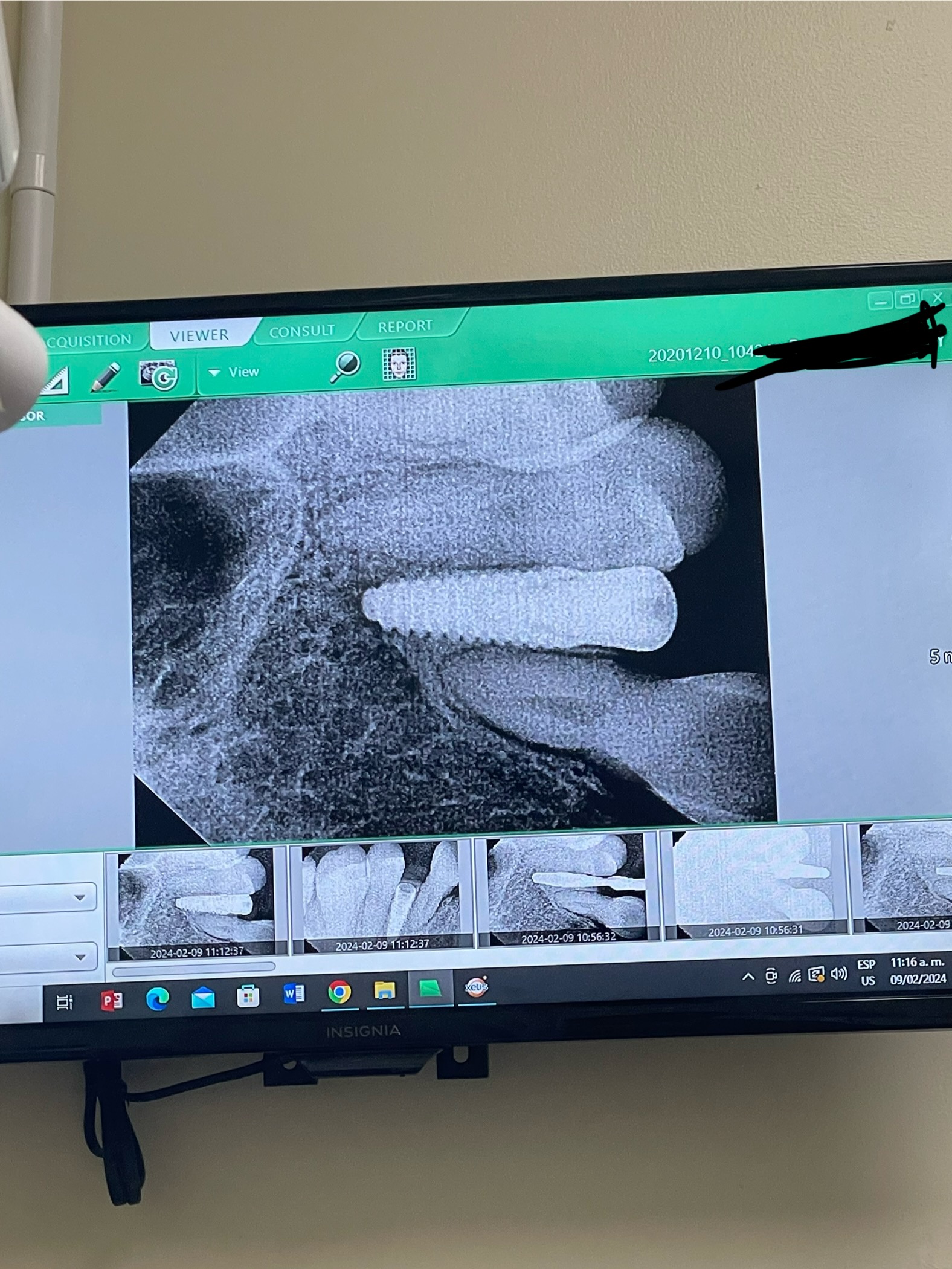
Implant in traumatic tooth case: options?

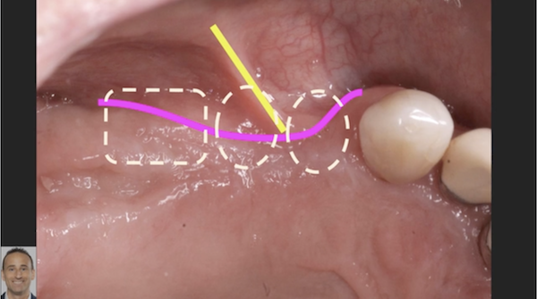
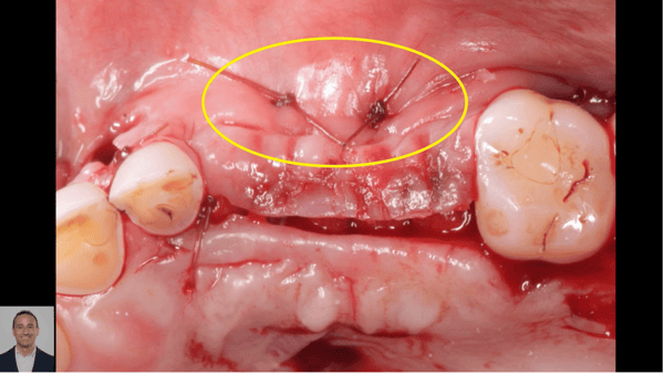
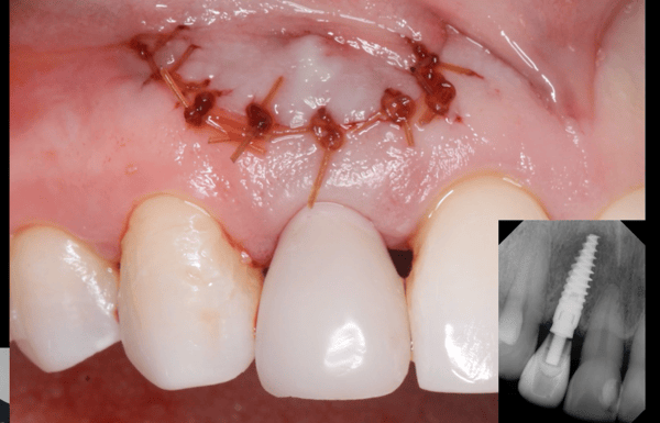
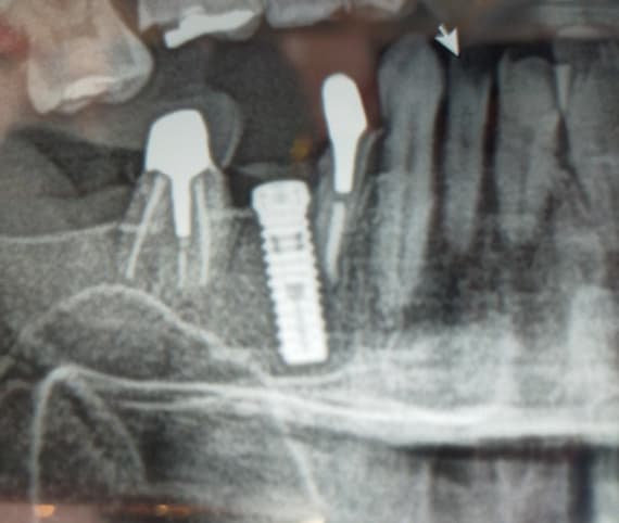
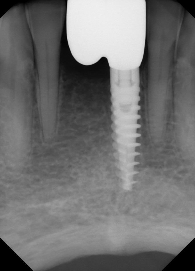
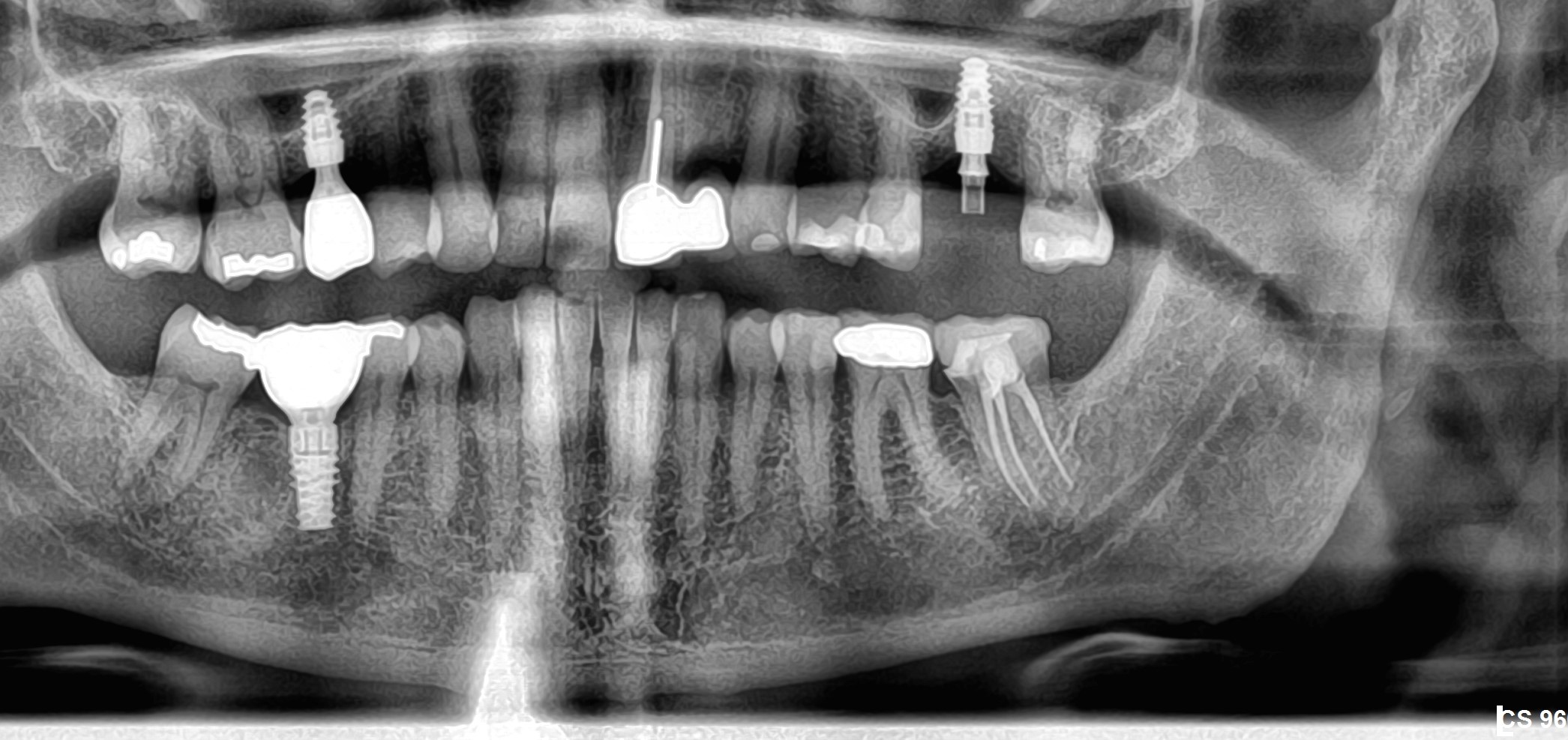
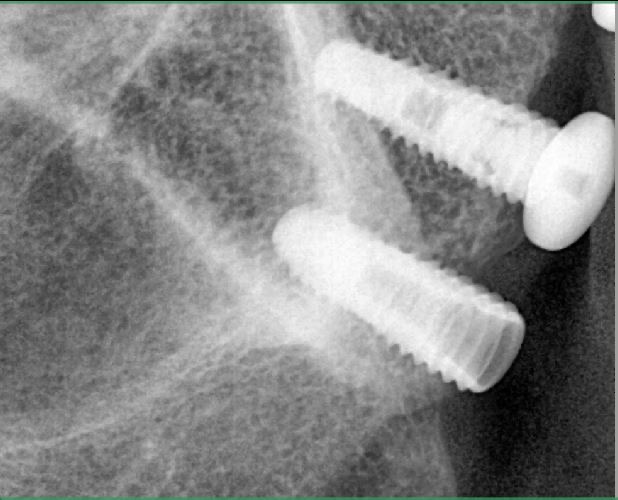
This case started with a traumatic tooth. RCT was failing with lesion and bone loss, and facial recession. The extraction was done, as well as, a ridge augmentation. I also tried to improve the recession by adding connective tissue graft to the area. The recession is improved partially. PRF was used with cort/canc bone mix. Since PRF was used, I went in at around 3 months after ridge augmentation. Fully flapped, placed implant. The newly formed bone was still kind of soft, implant had a cover screw and primary closure was obtained. I flapped case again trying to correct recession on #7, and noted the bone loss, around apical portion of implant. There is no mobility so far, but threads are not in the bone. I added bone around around it, but really nothing healed, including gingival graft. Again prf was used, and there is also fenestration on facial of root #7. I took a CT scan and it shows that angulation of implant is not perfect, but when originally it was placed, I was trying to adapt to crown angulation of #9 ( I didn’t want abutment to go too buccally) and with that augmented ridge the whole implant was visually in the bone, but it all resorbed after implant placement. There is still no bone around apical third of implant. Implant was 3.5X11. Patient is a slow healer. Not sure what are my options now? Take it out and graft and do over again?


19 Comments on Implant in traumatic tooth case: options?

New comments are currently closed for this post.
Leal
2/21/2018
By the looks of the CBCT this has completely failed. No possible way to fix it. Remove and start over with a PTFE titanium reinforced membrane and graft of your choice (preferably b-TCP). Wait for healing and place implant in bone.
Leal
2/21/2018
Sorry CT not CBCT. About the angulation, better to be of angle but in bone. A lot of implants now have dynamic screws that allow for a screw retained crown to be screwed in an implant that is up to 30 degrees off angle. The provisional crown access will be buccal but that is temporary.
N
2/22/2018
How would I present this option to the patient, I think she will have a heart attack if I have to do it all over. Any recommendations?
Matt Helm DDS
2/23/2018
Refer this to an OMS ASAP! FIRST, talk to your best friend oral surgeon and explain the failure. He will have to cover for you! Your implant is not even screwed into the body of the maxilla! See my other comment below. After you’ve secured your referral, explain to the patient that the infection has damaged a lot of bone and that this would now best be handled by an oral surgeon. Explain that you tried to help her, but the bone didn’t respond, it will take more grafting and more healing time because of the pre-existing large infection, etc. And remove that disaster implant asap, before it causes other complications! It would be a good idea for you to go to the OMS with her. It will show caring on your part. Very important now! This is a very touchy situation because it can easily turn into a lawsuit due to the grossly poor implant placement if it ever gets out. Talk to your OMS and show him the scans and put him up to speed, before you talk to the patient again! You have a disaster on your hands and you must handle it with kid gloves!
WJ Starck DDS
2/22/2018
Better a heart attack now than a stroke later followed by a letter from an attorney and the dental board. Don’t take it personally, it is the nature of dental implant that things don’t always go as planned, particularly when you have a compromised site to begin with. Calmly explain to the patient that this implant had failed, and it needs to be removed, the site grafted and redone. The patient won’t like it, and neither will you, but what you will learn over time is that the patient will be a lot *less* pissed than if you fool around with this for another 3-4 months, and it fails anyway. Diecussions like these will get easier for you over time.
roadkingdoc
2/22/2018
I am always cautious when discussing implant success with patients. My only garantee to them is i will do my best for them. When one fails my comment is your body doesn't like this implant and is rejecting it. I don't like it and you don't like it but we will have to do a little more work for success.
Julian O'Brien
2/22/2018
The presence of pain and/or looseness is the litmus test. Pus can sometimes arise from a loose cover screw or abutment so it is not a reliable indicator of fixture integration. Try placing a chair side temporary and await symptoms. If you attempt to remove a "firm" implant, you may find it laborious and destructive to the remaining bone. Plus you and the patient may have that lurking doubt that its removal was warranted. Should it be mobile, you could remove it, curettage the apical area and immediately place a wider implant. Failure is an integral part of success and your daily fare so your warning of that blame free outcome at the beginning is key to the patient's disappointment. A risky outcome explained at the beginning is informed consent, given later it is a horrid excuse! Compared to a throbbing infected tooth, a failed implant is small bickies.. A fibrous apical residual boneless zone is capable of harmonious stable integration but could also be re-enterd and grafted after a year of more when the biology and emotions have settled.
Matt Helm DDS
2/22/2018
Am I the only one here who notices that this implant is not even screwed into the body of the maxilla according to these scans?!? This implant looks completely outside the body of the maxilla, on the labial plate! N, do yourself a favor and refer this to an OS ASAP! And use a guide next time. I think you're practicing way beyond your abilities at this time. Get more training and you'll get there. But this case is a disaster! (no offense) That implant isn't even screwed into bone! And I won't even get into the whole discussion of you failing to diagnose and treat the large pre-op apical radiolucency of the extracted central incisor. That radiolucency is still present! Look carefully between the implant and the other central incisor! Send it out to an OS who you are on good terms with and hope he covers for you!
N
2/22/2018
Thank you everyone for all the responses. @Matt Helm - yes this is the case that I am not particularly proud of, but I have been placing and restoring implants for 12 years now and had done enough anterior cases. I am always taking additional implant trainings. This is really the first time I am seeing such a failure. I did debride the socket thoroughly - this is what I do routinely, and grafted the site with membrane and ridge augmentation with extra bone on the buccal and flap was released and gingival graft done. I have done this a few times and have scans that show that I was able to grow as much as 3- 5mm of buccal bone. In 4 month went to place implant, the grafting site wasn't thick enough, so I grafted buccal again this time with PRF. The only thing is that I went to place implant at 3 months of healing per patient rushing me, and I fully flapped case again, so when the implant was placed it was fully buried into the bone, but the grated site was still soft and even I added more grafting material, it all resorbed, this is why it appears now that the bone is missing. But there are many cases shown to properly osteof=grate not only in soft graft but having completely buccal exposed and fresh graft. I could have used the guide, but of course trying to save patient's money and since I am flapping I have more experience free hand than actually with guides. As for my consent, there is a clause that there is no guarantee and that implant may fail, but especially this patient, the whole process is taking about a year, and she hates wearing temp, she is in public sometimes. I made her a snap -on smile temporary, which is the best for her case but she still doesn't like it. She will not wear a flipper and there is no lingual space (tight occlusion) for ribbond. Any suggestions?
Matt Helm DDS
2/23/2018
One more thing. The recession on the #7 which you mention in your initial narrative is indicative of traumatic occlusion and possibly bruxism as well. A hard lower night-guard is definitely indicated! Learn to treat the whole patient, not just an implant. And if you think I'm being tough on you, just think how tough a jury would be when the plaintiff's attorney projects this CT on the screen and says "ladies and gentleman just look at how this Dr screwed this implant almost into the patient's upper lip". Think that's impossible? Think again! Or how tough any dental board would be upon seeing these same CT's, and how deaf they would be to all your bone-grafting explanations. Send this to the OMS and handle it with kid gloves! Your career is worth more than a moment of misplaced ego!
Chris Smith
2/23/2018
Hi, as for the temporary measure - maybe just bond with resin to the adjacent teeth proximally an acrylic or resin pontic. This way you avoid the palatal with restricted occlusal clearance. The implant - it ended up in the wrong position . Possibilities - osteotomy in wrong plane buccopalatally , inadequate reduction of the palatal crestal bone ( higher density bone than facially) which could deflect the implant buccally on insertion. You need 1.8mm+ to the facial for bone to stay. So for me, the implant is out. Most likely the second attempt with a little help from digital planning and fully guided implantation..... Costs always have to be considered , agreed .
Dr Kamil KS
2/23/2018
Hi, Implants failure can happen under the hand of any dental surgeon. One study at Eastman Dental institute showed failure rate was more under the hand of experienced & specialists. Perhaps as they become very confident, they may proceed too quickly or even may take short cuts which will end up with failure. Anyway, this case is a failure. What ever you do, it is best to prepare the patient & your self to go through new treatments & new implant placement. Good luck
Peter Hunt
2/23/2018
Perhaps we should appreciate that the standard of care is changing. There was a time when we all worked with periapical and panoramic radiographs to guide us. Now it's becoming standard to incorporate CBCT images into the evaluation. A natural evolution of that is to perform procedures such as this with a Guided Approach. Modern technology for this is dramatically reducing the time to prepare and the costs of a Surgical Guide. It makes implant placement safer, simpler and less stressful.
roadkingdoc
2/23/2018
" I was trying to adapt to crown angulation of number 9" Preparing an osteotomy using anterior crown angulation as a guide will get you an apical facial perforation almost every time. I have to remind myself of this each time i prep for an anterior implant. Doing implants without a CBCT image is very stressful and a potential legal problem. I sleep better with the CBCT.
VickyDDS
2/23/2018
Which labs do you use to design and fabricate your guide? I use Ankylos (Dentsply ) implants and not everybody able to to fully guided with that. The Dentsply guide is super expensive. I found one lab, and even after extensive planning with them, when I placed implants through the guide, the most distal ones seem to be to close to adjacent teeth. So thinking maybe there is better lab.
Adibo
2/24/2018
Completely agree with Matt Helm DDS. The implant is misplaced. The only question of mine, No matter of your experience, previous successes and the patient's glamourous job, the case has failed and needs a new competent person to look at it. At the end of the day you need to think for the well-being of the patient as well as your own. We have to accept that most of the complications are operator related and nobody is exemption.
FES, DMD
2/26/2018
You may, and I say may, have had success if you would have waited at least 3 more months before placing the implant. I'm assuming the patient is at least 40 years of age, if not older, although no age was given. In that age group, the blood supply to your graft was more peripherally dependent (periosteum) than centrally dependent. By raising a second flap, at the most crucial time of new bone formation, the resultant loss of the graft, was very predictable. Never let a patient dictate timing of treatment, especially when it contradicts the very basics of bone grafting physiology and healing. One other noticeable technical error, was the presence of an intact labial cortex on the CT. Just onlaying (any type of graft material) without creating perforating channels or decortification, is predictable for significant loss of graft volume or complete loss of the graft. Going to have to start over with this one.....
N
2/26/2018
@FES, DMD: yes the patient is over 40, and this is what I thought as well. I was hoping PRF would expedite the healing but it did not, definitely my mistake for letting patient rushing me. But again there are lot of reports that with PRF, the healingof bone at 3 months and implant placement. As for the decortication, I did do it with round bur. Should have drilled in deeper into the cortex?
Bruce
3/6/2018
Ok, you tried something super hard and it failed. It happens to everyone. The patient is not the easiest to deal with. I agree that it should be passed on to a friendly OMFS or periodontist, but why not try something simple instead like a traditional old school bridge.

Featured Products

OsteoGen Bone Grafting Plug
Combines bone graft with a collagen plug to yield the easiest and most affordable way to clinically deliver bone graft for socket preservation.
CevOss Bovine Bone Graft
Make the switch to a better xenograft! High volume of interconnected pores promotes new bone. Substantially equivalent to BioOss and NuOss.