Implant Case with Large Periapical Radiolucent Lesion and Infection: Thoughts?

Dr. Z asks:

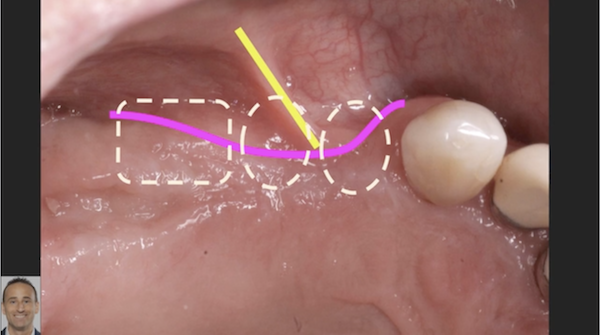
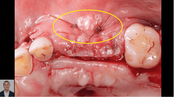
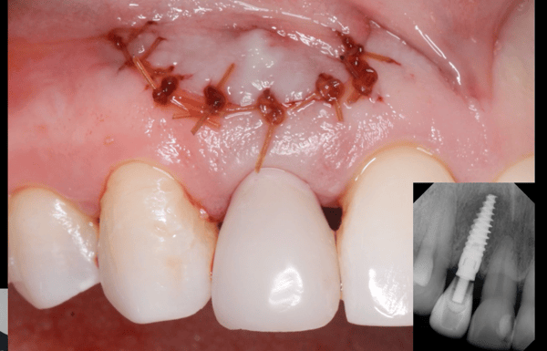
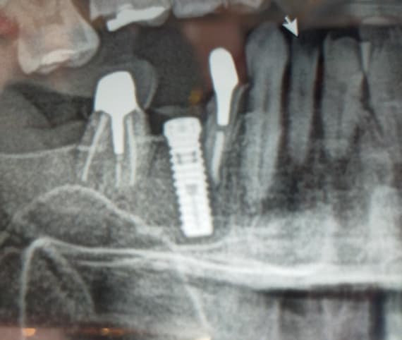
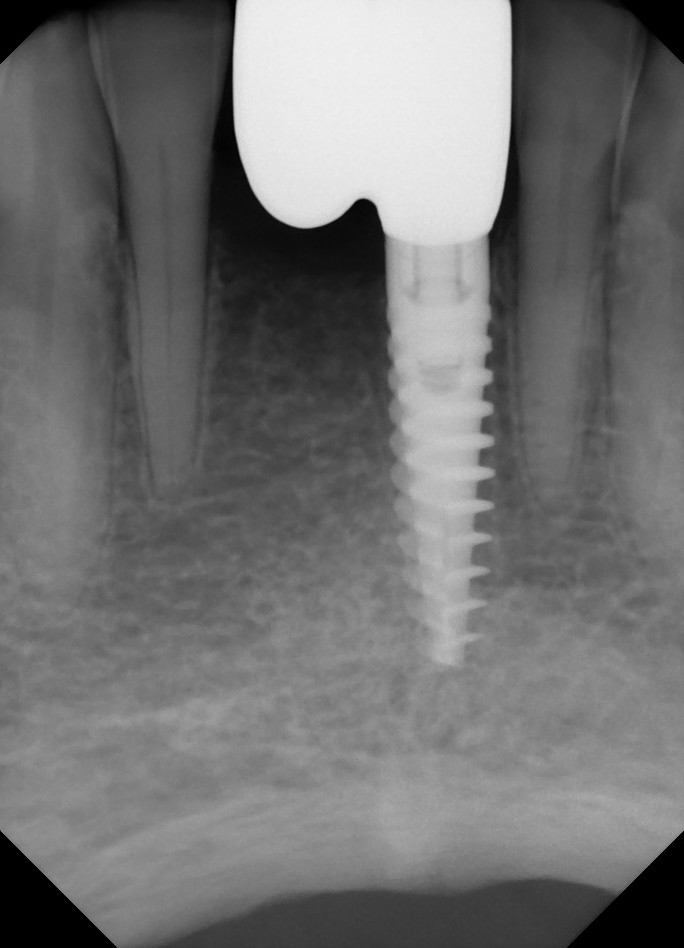
Please refer to case photos below.

I’ve just assessed a lady (referred to me) who had her uL2 extracted 2 years ago. There was a large periapical radiolucent lesion, and a couple of months later (4 months after the xla) went under GA to the hospital to have the area re-explored and irrigated due to numerous infections. (she is very nervous).

She wants an implant, and still complains of on/off infections in the area. I have assessed her for treatment this time under IV sedation.

I was going to access the area fully debride it, and graft it at the same stage….any thoughts if i should graft now or later after 3-4 months?….plus I have recently heard of more and more practitioners using bone substitutes such as bio oss instead of block grafts…(she has lost alot of buccal bone )..any one have any thoughts on whether I could get away with bioss and a membrane? She will need sedation, so i want to try to have minimum visits if possible.

Thanks.

Preop 2007

Post op 2010

19 Comments on Implant Case with Large Periapical Radiolucent Lesion and Infection: Thoughts?

New comments are currently closed for this post.
sb oms
9/6/2010
When you see a radiolucency like that, you are dealing with a through and through defect. In other words, there is no buccal or palatal plate. This is a complex situation. It looks like the adjacent central has major recurrent carries and poor marginal integrity.(first x-ray) I would consider removing this tooth, using it as the implant site, and making the lateral incisor a cantilever. If the lateral site is your choice, remember the following: 1. You have little to no bone here. This is a very challenging site that is prone to problems on an easy patient. You stated several times that this patient is challenging due to anxiety. There have been multiple infections here, so your soft tissue will most likely be compromised too. Poor soft tissue means poor blood supply for healing. This may not be the patient for you. 2. Does bio-oss have any role here?? Absolutely not. While an allograft may be of some assistance to you, i would be carefull with what i put into this site. 3. As i said above, your patient might be best off with an implant in the central with a lateral incisor canti-lever. I would assess the prognosis of the central before going ahead with anything.
TOBooth BDS Hons Msc
9/7/2010
Hi, i think its a difficult case. I dont think and implant in thelateral site is beyond us, but i woudl extract teh central first and make a flipper off teh soft tissue Ideally you would allow the site to heal 6-7 weeks re-enter, and you probably do have some alatal dehsicence in the lateral site. bio-oss biogide as its effectively a hollow 3 walled eefcet that will respond well, then after 6 months re-enter and place 1 or2 implant ideally in central at leat a 4mm diameter so if you do need to cantilever you can -non axial loading not ideal. Hopefully a 3-3.5mm can be placed in the lateral site it may need to be placed deep. Allo area to heal and reassess if need a sub - epithelial connective tissue graft ; if you hace 3mm plus attached around teh emergence profile reflect soem tissue from the palatal. Hope that helps if unsure refer on.
peter Fairbairn
9/7/2010
Hi Dr. Z as you know I suggested referral as it is a difficult case which could go wrong at ant time . But what would I do , when dealing with long term cysts a very vigorous curretage is advocated , back to bleeding bone so in this case definately sedation. Having done and seen many similar cases my route would be the use of Easygraft (or Crystal with HA) here and would currette and pack the site with it at the same visit.It is as you know a BTcp product ( available in the UK) with a poly-lactide coating which is held together with a medical bio-linker thus the graft is stable until the poly-lactide breaks down (3 weeks to a month). The bio-linker is bacterio-scidal ( for a very short time) and the graft is Bacterio-static so less of an issue isn this situation. Then at 4 months I would assess and place the implant with possibly another graft placement. But as I said a difficult case and best referred to a MFOS. Regards Peter
Mr. X
9/7/2010
let´s try it again. TOBooth BDS Hons Msc: You are ABSOLUTELY wrong. NEVER use BIO-OSS in that case. sb oms mentioned: "There have been multiple infection here." GENERELLY PLEASE INFORMATE YOUR PATIENTS ABOUT BONE SUBSTITUTES AND BIO-OSS. BIO-OSS HAS NOTHING DO TO WITH REAL LIVING BONE WHICH SHOULD BE YOUR AIM. BIO-OSS DOES NEVER RESORB. It is only a filler. There is no turnover in living bone. Well some bone grows around BIO-OSS and enables osseointegration for dental implants. With BIO-OSS you get only volume stability. BUT NOTHING MORE. Look at the scientific literature and the damaged patients. The user of BIO-OSS must be clear that every patient could sue you for damages at court. Use synthetical or allograft materials or allogenic bone or blood. PLEASE DO IT BETTER IN FUTURE! Department of Oral Maxillofacial Surgery, Ludwig Maximilians University, Munich, Germany. Abstract BIO-OSS is an allergen-free bone substitute material of bovine origin, used to fill bone defects or to reconstruct ridge configurations. Seventy one patients (39 female, 32 male) received 126 BIO-OSS implantations. Some health parameters or habits were documented to eliminate possible risk factors of influence. The diameter of jaw defects filled with BIO-OSS was measured. There was a significant influence of the defect size on the healing result. In X-ray controls, BIO-OSS served to identify the surrounding native bone. The density of the BIO-OSS areas was higher than in control sites. These radiological results were supported by bone biopsies. Histologically, the permanency of the BIO-OSS was still recognizable after 6 years and longer. The ingrowth of newly formed bone in the BIO-OSS scaffold explained the increased density of the implanted regions. There were no clinical signs of BIO-OSS resorption. Therefore, we can assume that form corrections achieved by BIO-OSS insertions will last. PMID: 10186966 [PubMed - indexed for MEDLINE] etcetera # paul carie May 31st, 2009 I can’t believe you guys. I had bio oss used on me 5 years ago. Never resorbed, still having chunks that have migrated everywhere taken out, gross sinus problems because of migration into the sinus. Dysguesia also. Why would any of you use this product? My dentist has photos of chunks he has been taking out. The company should be sued as well as the people who use this garbage. # david salzman September 13th, 2009 I have bio oss attaching and spreading everywhere. Some has been taken out, the implant was taken out. I now have a glob of this garbage attached above #16, some in the soft palate by 16. Salty bitter taste coming from there as well. Do any of you folks know of someone who can remove some of these particles. My ENT just removed several pieces from my upper lip. The bio oss was originally placed in #14, obviously did not stay there. Cannot believe anyone in good conscience would use this stuff. Please let me know if any of you know of someone in your profession who would take this on.
Sav
9/7/2010
Thanks guys...your input has been very valuable..i will definatley refer to a more experienced collegue. Thank You Savan
sb oms
9/7/2010
i know this is not the original question, but Bio-OSS is an excellent product when used correctly. I have used it in the sinus and for lateral ridge augmentations where slow - very slow- resorbtion and re-modelling is preferable. to those who have had bad experiences, i'm skeptical of the reasons and skill with which it was used. i direct you to Steve Wallace's work at NYU. They have the largest collection of sinus histology anywhere, using any materials. Bio-Oss has shown admirable results - equal to autogenous bone in terms of implant survival. To those who bash the product based on anecdotal evidence of a botched case, I am sorry. This is a good weapon to have in your arsenal.
TOBooth BDS Hons Msc
9/8/2010
Sorry chaps, i use this routinely, and its a good product. Dr X i think you maybe need to read your article! The aim of bio-oss or any augmenation in conjunction with a good volume of keratinized tissue is to prevent breakdown to threads. Undoubtedly it doesnt convert to bone completely but no product does, period! Provided there is no active infection, ie suppuration, this lesion if curretaged to bone like Peter said is a brilliant place to augment especially a cyst cavity right? as its not a conventional dehiscence, bio-oss cannot slide of the site. Wait 6 months and radiogrpah and implant. I do alot of cases like tis, Mr X quoting andom papers or should i say abstracts doesnt impress-mayeb you shoudl read it and critically appraise. Or alternatively look up 'critical sized defect' then you mayne you can understand more.
Dr cdic
9/8/2010
Difficult case, but gp can tackle such kind of cases if delta with proper care.
Dr cdic
9/8/2010
Difficult case , but gp can do if done with proper care and follow proper protocol
Richard Hughes, DDS, FAAI
9/9/2010
For some reason it appears as if alloy is in the site. I would not even place an implant is said site. I have had failures in sites where retrofills were once present.
Robert J. Miller
9/9/2010
The problem here is not the remnants of the retrograde filling, but rather the granulomatous mass that it is suspended in. Apical granulomas are high in inflammatory infiltrate and contain a phenotype of cells that are, in turn, self promoters of additional inflammation. If this tissue is not removed completely, we end up with a classic retrograde peri-implantitis that cannot be cleared with antibiotics. Over the past decade, I have found that the most effective instrument for granuloma removal is an ablative laser (Er,Cr;YSGG or Er;YAG). These lasers will selectively remove the soft tissue, have a high bacteriocidal kill, and will induce a different phenotype of healing cells to promote bone regeneration (photobiomodulation). The single most important factor that can be gleaned from this radiograph is the facial-incisal line angle. If this bone is intact, you can still acheive outstanding results with immediate implant placement. However, I would recommend a flapless approach to maintain the periosteal microvasculature of this isthmus of bone. Flapping here will almost guarantee loss of this line angle. We do these cases under a surgical microscope. Following debridement, we will tuck a small resorbable collagen membrane through the fenestration to extend 1 mm outside its border. We do not reflect the coronal border, however. The implant is placed and a fully resorbable alloplast or autograft is placed (i.e. bTCP or autogenous). We do NOT use a xenograft (Bio Oss). Ca+ ions released from these products have bacteriostatic properties. Bio-Oss does not resorb and therefore will not contribute in this manner. We have been able to acheive outstanding functional and aesthetic outcomes in these cases through the years with this protocol. RJM
Stanley Markman DDS
9/10/2010
My concern with the description is that of periodic infections. Did the patient actually develop these infections? Did you see any of them? Perhaps the patient is reporting periodic pain and thinks that they are infections. If in fact she is actally reporting pain than you may be dealing with peripheral neuropathic pain. At the orofacial pain clinic where I teach, we periodically see patients with post operative neuropathic pain.
Mr. X
9/10/2010
Scarano didn´t find histologically any resorption of Bio-Oss after 4 years. Unchanged particles of Bio-Oss were found. BIO-OSS DOES NEVER RESORB. Scarano et al. (2004) bei einem 50-jährigen Patienten, bei dem nach ca. vier Jahren ein Implantat (nach einer Implantatfraktur) entfernt werden musste, im histologischen Präparat unveränderte Bio-Oss- Partikel
Paul
9/12/2010
extract, debride, wait a few weeks for healing, then place mfdb (lifenet from salvin.com) with membrane. wait 4 months and eval. may have to graft twice with large defects.
Sav
9/12/2010
The patient does confirm periapical exudate..indicating active infection....Have refered case to more experienced surgeon, and has been initially decided to expose area, thoroughly debride and graft with autogenous bone (poss ring graft from chin)....however wil only know what next step to take once the area has been opened...the xla of the ul1 has not been ruled out...will keep you all posted on the findings nad progress..thanks again Sav
hwbrueggen
9/13/2010
Unfortunately ths is a common problem.Ilove reading the various responses regarding possible solutions.Every time I think I'm getting pretty good I recognize how much more indepth some drs knowledge is than mine. Personally I would certainly remove the central as well as the lateral. I would reflect a flap from the mesial of the extracted central to the distal of the adjacent cuspid.After curretting the defect thoroughly I would "freshen' the walls with a large round bur. I would place a biomend extend membrane from the lingual to the buccal extending it at least 2mm beyound the borders of the defect.Through the buccal I would suture the membrane to the palatal tissue with gut. In the past I would have filled the defect with 50-50 mfdba and bovine,with a 2mm sandwich of non resorable HA on the buccal and lingual.Bio oss would be perfect for the sandwich layer as well.Now I would use Infuse or Osteocell and still sandwich.Then I would reposition the membrane coronally and bucally and secure it on the buccal with 2 tacks. If I could not achieve primary closure and I probably could not I would cover it coronally with Alloderm. After thoroughly debriding the central socket I would place an immediate implant. At four months I would take a c-scan and remove the two tacks without a flap and if there was adequate bone I would place an implant in the lateral position. If I did not have a c-scan in office I would be comfortable with "bone sounding".If there was not adequate bone, and there certainly might not be, I would cantilever off of the central if good occlusion was achievable in centric and excursive movements.If I wasn't comfortable with the cantilver, I couldn't place an implant in the lateral position, and ths patient was excessively phobic I would consider a three unit bridge from the central implant to the natural cuspid. Implant to natural dentition would be my last option but if the cuspid shows no mobility, and I suspect it doesn't,I would consider it before resorting to a second bone graft. I know there are many other approachs which would work, some possibly better than this, but this is how I routinely handle this situation. Prior to Infuse and Osteocell I would expect about 7-8out of 10 to have enough bone. With the new products I expect all of them to be successful; we'll see. Except for the swelling I've really enjoyed Infuse. Though I've had many years of experience I am a GP and at some point I had to do my first case like this. If you are comfortable doing the procedures I described you should be just fine.I hope this helps you next time.
gpkaralis dds
9/15/2010
So the second Rad is flipped on the 2010 post op and it looks like the Crown on 9 was replaced and looks ok. What am I missing? I think with bone sounding right off, you'll be able to tell that there's no buccal plate, then lay the flap, should be a real fun dissection, end of the day maybe, so if you need more time you have it. Currete, bone file, good bleeding, assess volume requirements, appropriate graft, barrier, tension free primary closure. I mean she healed pretty good. I don't know maybe I'm not paranoid enough, would this have happened if the extraction was handled aggressively and a graft and barrier were placed initially? The height isn't bad, the width is going to be the challege insical to the radiolucency, you're probably not going to get adequate width that high, that'll be where the esthetic challenge arises.
Mr. X
10/7/2010
Excuse me, I made a fault with one word in my article from September 7th, 2010. "Please use synthetical or allograft materials or autogenous bone or blood". The result will be living bone. Did anybody know something about chronic sinusitis maxillaris with Bio-Oss? I do not mean any bacterial infections. Had anybody experiences with immune defences of patients caused by this bovine material? Could you imagine that Bio-Oss causes scars in the soft tissue? Has Bio-Oss different lot variations with problems for the patients? Did anybody take biopsies of that cases which shows the reaction of Bio-Oss? How much cow is inside of that product?
Mr. X
10/16/2010
Unfortunately nobody has answered yet. Then I would like to ask something else. It exists different hydroxylapatites for example synthetics or bovines (from cows). Which of both will resorb in a humane bone and will change it to a living bone?

Featured Products

OsteoGen Bone Grafting Plug
Combines bone graft with a collagen plug to yield the easiest and most affordable way to clinically deliver bone graft for socket preservation.
CevOss Bovine Bone Graft
Make the switch to a better xenograft! High volume of interconnected pores promotes new bone. Substantially equivalent to BioOss and NuOss.