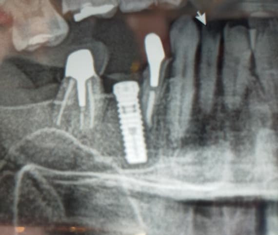
Incorrect angle of implant: has it failed?
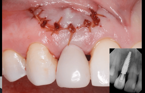
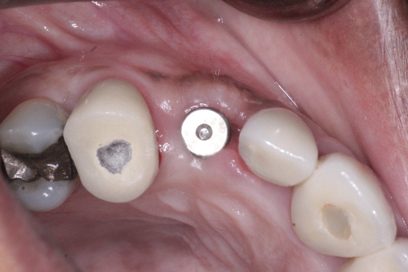
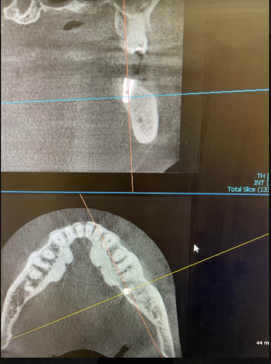
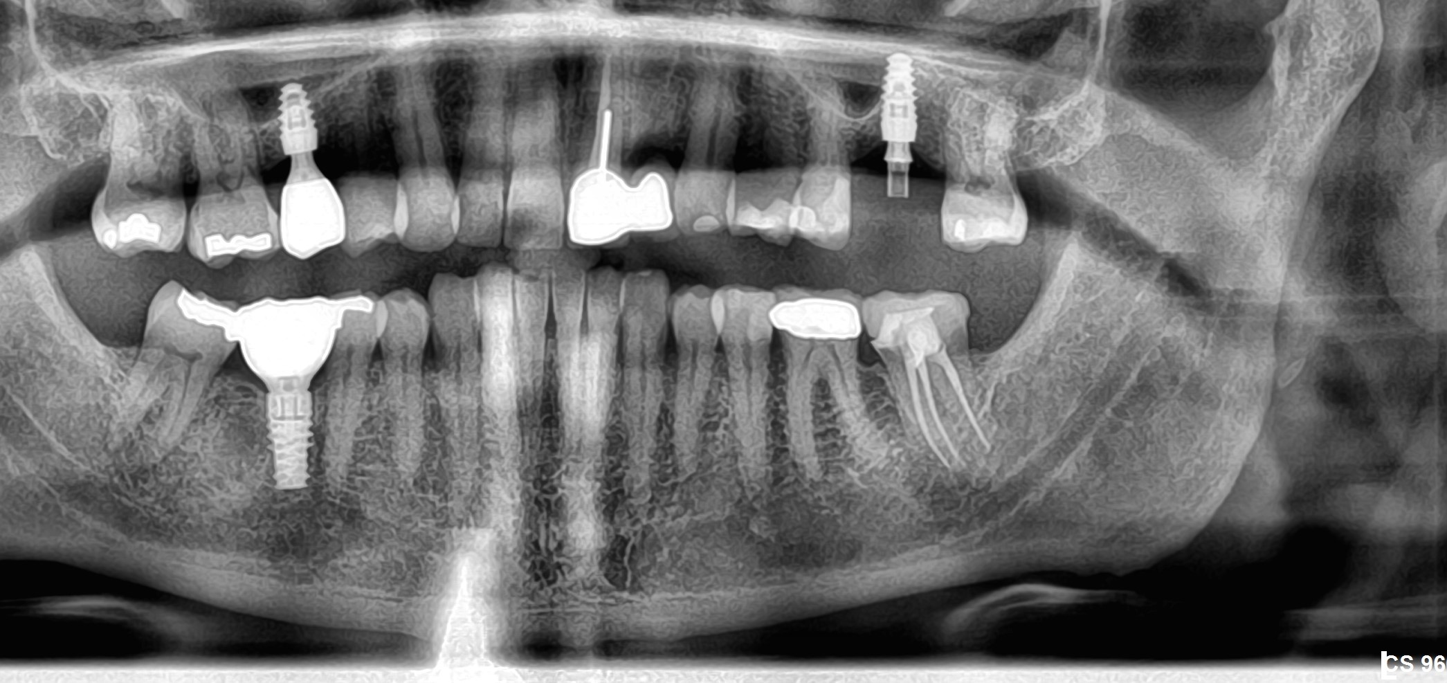
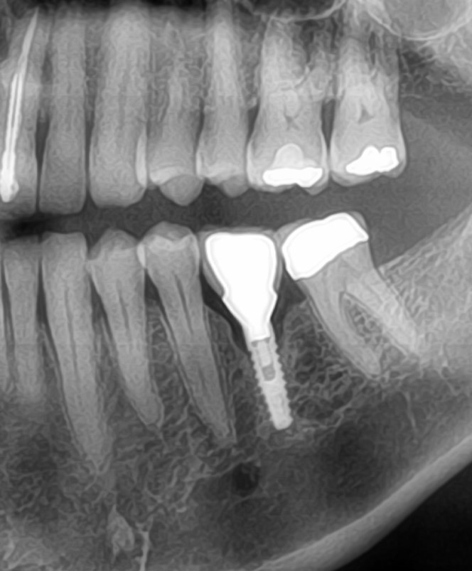
I extracted #9 and placed an implant and restored with a crown. This is the sequence: 1st x-ray -7/26/2017 #9 extr. 2nd,3rd, 4th x-rays-11/16/2017 osteotomy and implant placement. Implant angle is too mesial, yet platform was still within normal limits to restore. 5th x-ray-3/22/2018 redid crown to improve appearance. 6th x-ray-5/2/2018, checked aesthetics. 7th x-ray-11/21/2018, symptomatic. I checked the occlusion but it was WNL. I checked for for swelling but was WNL. It was sensitive to percussion from lateral. Has this implant failed? Is Nasopalatine an issue?


18 Comments on Incorrect angle of implant: has it failed?
New comments are currently closed for this post.
Peter Hunt
1/8/2019
You have a nice series of radiographs here but it appears as though the implant is starting to fail. It may be that there is lack of a labial plate. It would be useful to have a CBCT cross-section view to see the relationship to the labial bone.
It may be necessary to remove the implant, to regenerate the region and then re-treat. With a situation like this I would prefer to place the implant in a surgically guided mode.
Dok
1/8/2019
Does this case look stable clinically ? Is it infected, hurting, pocketed or mobile ? If so, easier to deal with now before there is more bone resorption. Have a good local periodontists do the removal/grafting/replacement implant and you just re-do the prosthetics. All free to the patient if it were my practice.
Gregori M Kurtzman DDS
1/8/2019
Looks like the mesial of the implant is in the incisive canal. If the implant is stable (no mobility) I would suggest flap the area, clean out whats in the incisive canal and pack osseous graft in it close it and let it heal. also if there is any labial dehiscence I would graft that too. To access the incisive area its a palatal flap
DrH
1/9/2019
Invasion in to the incisive canal is the most concern I have at this time, leading to sensitivity. I would prefer to look at this issue first. Thank you.
Don Rothenberg
1/8/2019
Looks like #10 has an apical lesion.
I would have an endo consult.
Is #10 sensitive to percussion or cold?
DrH
1/9/2019
Upon her complaint, tests were completed on all anterior teeth. #9 was the only issue and w/ sensitivity.
Bernard Tahon
1/8/2019
In my opinion, the poor design of the standard abutment (shoulder is not high enough) and convex deign of the temp crown is the issue. There is no room for soft tissue, and the biological with is not respected, so the bone remodels.
Tim Hart
1/8/2019
I agree with Dr. Tahon regarding the existing stock abutment/insert. A custom abutment would be a more effective approach to manage a more gradual and tissue-kind emergence profile. As long as the margins on your custom abutment are sub-G to an equivalent amount as a prepared natural tooth (i.e. 0 .7 to 1.0 mm apical to the free gingival margin) you really do not have to worry about cementation. If you are still concerned about cement below the margin, you could put a venting hole in the lingual of your new crown. Additional benefit is that you would be able to re-angulate the actual "prep" portion of the abutment to be vertical and centered between 8 and 10. That being said, I agree with the others that the implant is probably failing, but you could consider a custom abutment for your retreatment (or a 3-unit bridge, depending on how the soft tissue looks).
Dr H
1/9/2019
Tibase was utilized in this case. The abutment cemented to Tibase and polished prior to being screwed into implant. Crown cementation was at the margin. No gingivitis at time of exam. Sensitivity seems to be more apically.
Joseph Kim, DDS, JD
1/8/2019
The implant may or may not be ailing, but the lateral percussion sensitivity is not well described. How are you tapping this crown "laterally"? From the facial, lingual, or mesio- or disto-incisal corners? What is the intensity of the pain? An obvious improvement to minimize discomfort would be to allow more soft tissue space between the mesial bone and the overcontoured crown, and even consider thinning out the shoulder/ledge of the ti-base as well.
DrH
1/9/2019
Lateral (facial and lingually only). Slight sensitivity, yet with the addition of what I see on Xray, this concerns me.
Dr J
1/9/2019
IMO the bone loss that we're seeing isn't due to disease, but due to remodeling. You don't have room for biological width there. Therefore, bone resorption will happen to provide that space.
DrH
1/9/2019
Will that explain the sensitivity, while I do not observe gingivitis in the area? Tissue is pink and looks healthy
Andy
1/9/2019
I agree with placement into incisive canal
DrH
1/9/2019
I am starting to think along the lines with you.
Wadhwani c
1/9/2019
Radiographs are a poor way of evaluating implant health- See Malloy Ijomi 2018, and also Terry Walton's paper. Luterbacher's work with Lang suggested (perio2000)BOP 2/3 times over consecutive recalls has 100% positive predictive value for peri-implant disease (mucositis or implantitis ) the AAP has adopted this as a criteria for evaluating health. Issue is that few can predictably use the correct pressure when using a perioprobe - too much and BOP will occur as a result of trauma- too little and the sensitivity of the test goes down. Read: Cha article just out in IJOMI.
The rads shown are poorly standardized. There may or may not be an issue not possible to determine.
DrH
1/9/2019
Thank you
D-r Yaromirov
1/23/2019
Take down the crown and try 20Ncm torque on the implant. If it hurts you got a problem.
If it don't hurts try to change that abutment with custom one.
A CBCT would be a good idea.
p.s. did you check the contact with ?pponent tooth precisely? (sorry about that last one but I ahve to ask)


