Increasing the zone of keratinized and attached tissue: What is the best surgical intervention for this?
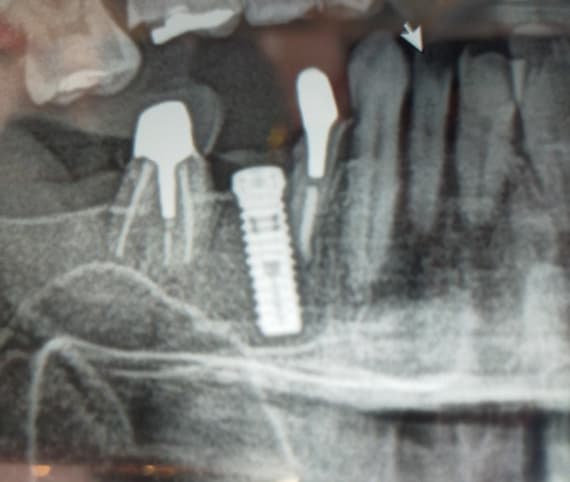
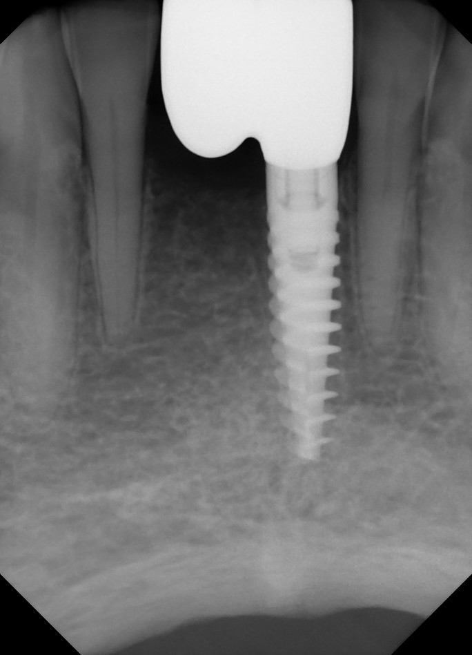
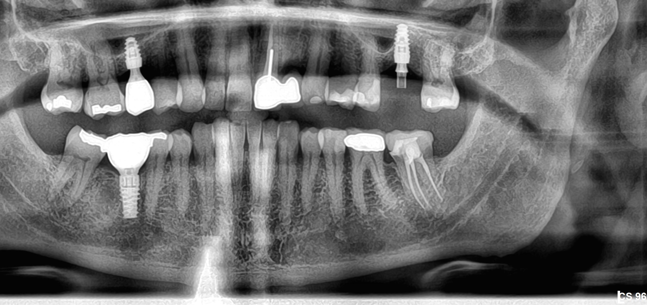
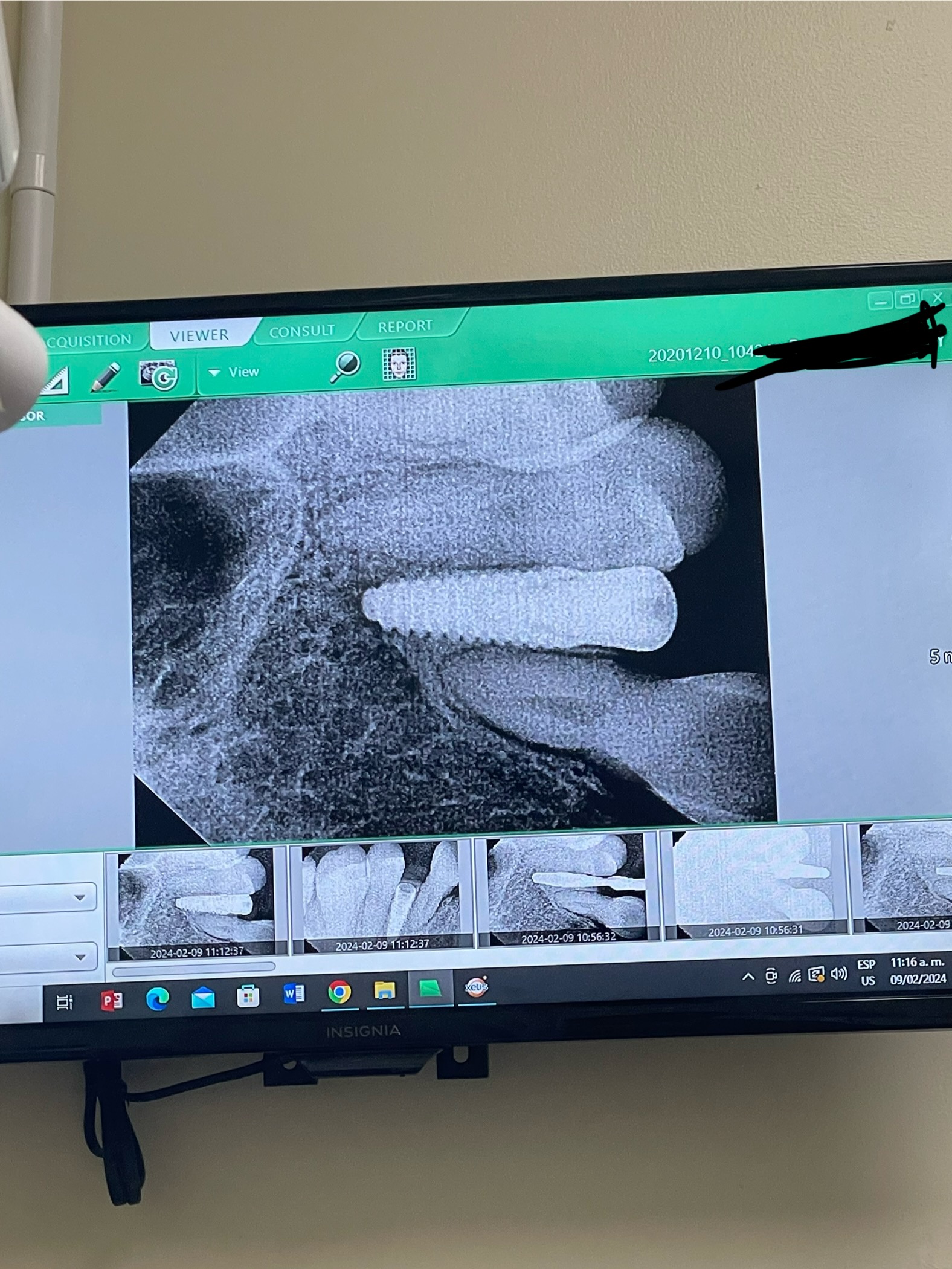
I have a patient with marginal bone loss and thin, nonkeratinized and mobile tissue around an implant. I would like to improve the health of this implant by increasing the zone of keratinized and attached tissue. What is the best surgical intervention for this? Would the best treatment be a connective tissue graft? What kind of flap design should I use? Would a free gingival graft be the best technique? For future cases is it better to increase the zone of keratinized tissue before or after the implant is installed?
13 Comments on Increasing the zone of keratinized and attached tissue: What is the best surgical intervention for this?
New comments are currently closed for this post.
Tr-Dt
4/15/2013
Very Good issue.how can we have attached mukoza? Can we gain it with CTG? Whats the indication for ctg
peter Fairbairn
4/15/2013
These are difficult and can lead to big disappointments so best way of treating is maybe refer to someone who has dealt with them . I rarely say that but that is the best option .
Best to sort the keratenized tissue out at the exposure phase and you will need to have bony coverage of the Implant for a good result.
Peter
Hazem Torki
4/15/2013
A partial thickness flap with a connective tissue graft would be a successfull procedure if you have enough tissue to cover the graft completely. Sometimes a full thickness flap can be done and the graft is sutured to the flap, but this is more difficult due to the decreased blood supply which will be from decortication of the buccal bone. But its an easier procedure.
Hazem Torki
4/15/2013
To correct the tissue problem before or after implant placement is decided by whether you only have tissue defects or bone and tissue defects. Some schools prefer to solve the hard tissue problem first, others prefer to solve the soft tissue problem first. If you only have a soft tissue defect, correcting the problem before placement is sometimes better.
CRS
4/15/2013
Is the marginal bone loss an early peri-implantitis? Without an xray or photo could be treated with a flap, bone graft and ct graft. Is this in the esthetic zone, then I would use a closed tunneling technique. Is there pocketing? I guess I need a diagnosis prior to suggesting treatment. If it is simply needed keratinized tissue then a CT graft is indicated . I like to evaluate this need prior to placing the implant crown, sometime I like to do this at implant placement or at the bone graft stage.. Hope this helps!
juan rumeu
4/16/2013
When raising a question like this it is nice and very helpful to show the picture and the xRay of the implant. It is very difficult to help you out on this matter without this two things.
Dr shyam mahajan Aurangab
4/16/2013
Very important ,problem faced in few cases. I had not realized it till I noticed my own few cases showing inflammatory tissue around implant .Most of the time lack of keratanised tissue was main reason. Can anybody suggest books or internet links for soft tissue management.
Tom Wierzbicki
4/16/2013
As CRS mentioned, it is difficult to treat if there is no diagnosis. A photo, radiograph, probing depths, and baseline readings are required to help with the diagnosis.
If the issue is just keratinized tissue, a connective tissue graft or free gingival graft are required. However, the clinical scenario will dictate if a CTG or FGG is a better option.
If in doubt, refer to a periodontist.
CRS
4/16/2013
In my experience, a connective tissue graft is more predictable if you need thickness vs a free gingival graft if there is nothing to build off of. Without a photo it is impossible to advise. You can also do a pedicled graft depending on the clinical situation is there a frenum involved? Work with a colleague you trust, good luck.
David Morales Schwarz
4/17/2013
The best and easiest approach to deal with such a case is a palatal free gingival graft, FGG to obtain a band of adhered, thick, keratinized tissue around the neck of the implant.
The intervention in most of the cases can be done after delivering the provisional or final prosthesis, Only in very extreme cases, in which the vestbulum has completely disappered, I would recommend doing the procedure prior implant installation.
1.-A linear incision 2mm. Apical of the recession will give access to raise a split thickness graft in which care must be given to detach all muscular fibers from the underlying periosteum.
2.- Tissues should be apically repositioned.
3.- Harvesting of an FGG from the palate, of an adequate size and thickness.
4.- Fixation of the graft.
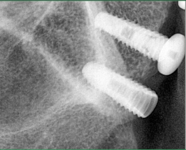
The most difficult part of the procedure is suturing both the host and donor tissues. to make this easier I have developed Ti selftapping microscrews which are recently commercialized by a spanish implant company (Bioner).
John Kong, DDS
4/19/2013
You can do a free gingival graft, CT graft or simply perform a split thickness flap and apically position it. Ideally, you can increase Keratinized Gingiva before implant placement or at uncovery.
Kaz Zymantas
4/21/2013
The literature shows that there is no difference in success if there is attached gingiva or just mucosa around the implant. The big factor appears to be mobility of tissue. Nevertheless, I attempt to get a band of att ging around the implant most of the time. I use a technique similiar to David Schwartz suggestion. I place what is known as a poncho graft where a small opening is made in the graft to totally enclose the implant at the crest. A big problem is how to keep the tissue approximated to the bone. I only do these procedures when I have a long healing abutment that can help to resist the displacement of the pac. After the surgery I place Perio pac mixed with Schlein's powder. The pac is compressed around the abutments and onto the graft for 3 weeks. The pac has tannin which helps reduce bleeding locally. Within a day the pac is very hard and a good barrier to prevent anything from dislodging the tissue. In most cases the end result is immobile keratinized tissue surrounding the implant.
David Morales Schwarz
4/21/2013
Inmmobile mucosa helps in mantaining a stable soft tissue seal around a dental implant, nevertheless if the band of adhered tissue has an adecuate width, is thick enough and is composed of resistant kerathinized ephithelium the clinical stability of the restoration will be much better.
Even though there are reports such as:
- Interventions for replacing missing teeth: management of soft tissues for dental implants. Cochrane Database Syst Rev. 2007 Jul 18;(3).
which are not conclusive about the role of attached kerthinized tissues around dental implants .
There are many recent studies recognizing the role of adhered kerathinized tissue in perimplant health:
- Five-year evaluation of the influence of keratinized mucosa on peri-implant soft-tissue health and stability around implants supporting full-arch mandibular fixed prostheses. Clin Oral Implants Res. 2009 Oct;20(10):1170-7.
- Width of keratinized gingiva and the health status of the supporting tissues around dental implants. Int J Oral Maxillofac Implants. 2008 Mar-Apr;23(2):323-6.
. Influence of soft tissue thickness on crestal bone changes around implants: 1-year prospective clinical trial. Int J Or Maxf Impl. 2009 24(4):712-9.


