Managing aesthetic implant case: advice?

I am having little trouble with one of my cases, and I would be grateful for some advice.

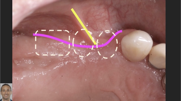
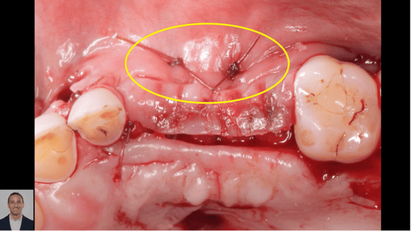
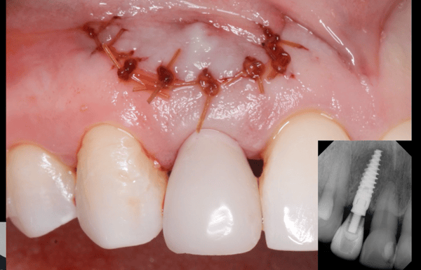
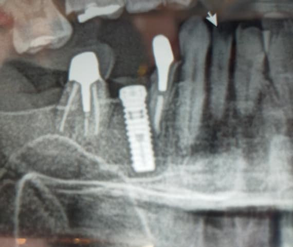
Tooth #10 [maxillary left lateral incisor; 22] was extracted last December. I performed socket preservation using bio-oss collagen and x2 collagen plug. During the exo appointment, I attempted to remove the tooth atraumatically using only periotomes. However, the tooth was ankylosed and it was very difficult to remove. At 4 months post exo, I installed a 3.3 NP x 10mm Straumann BL implant on the 22 site. There was a small defect distally exposing 2 threads, so Bio-oss/bio guide was placed on the distal defect as well as on the buccal site to improve the contour.

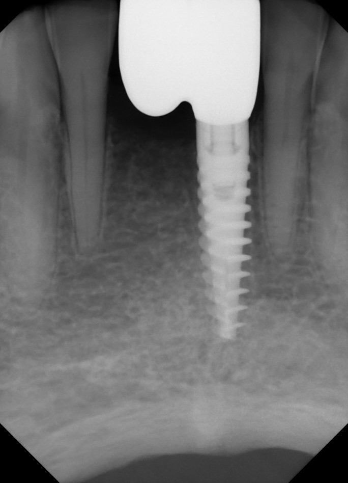
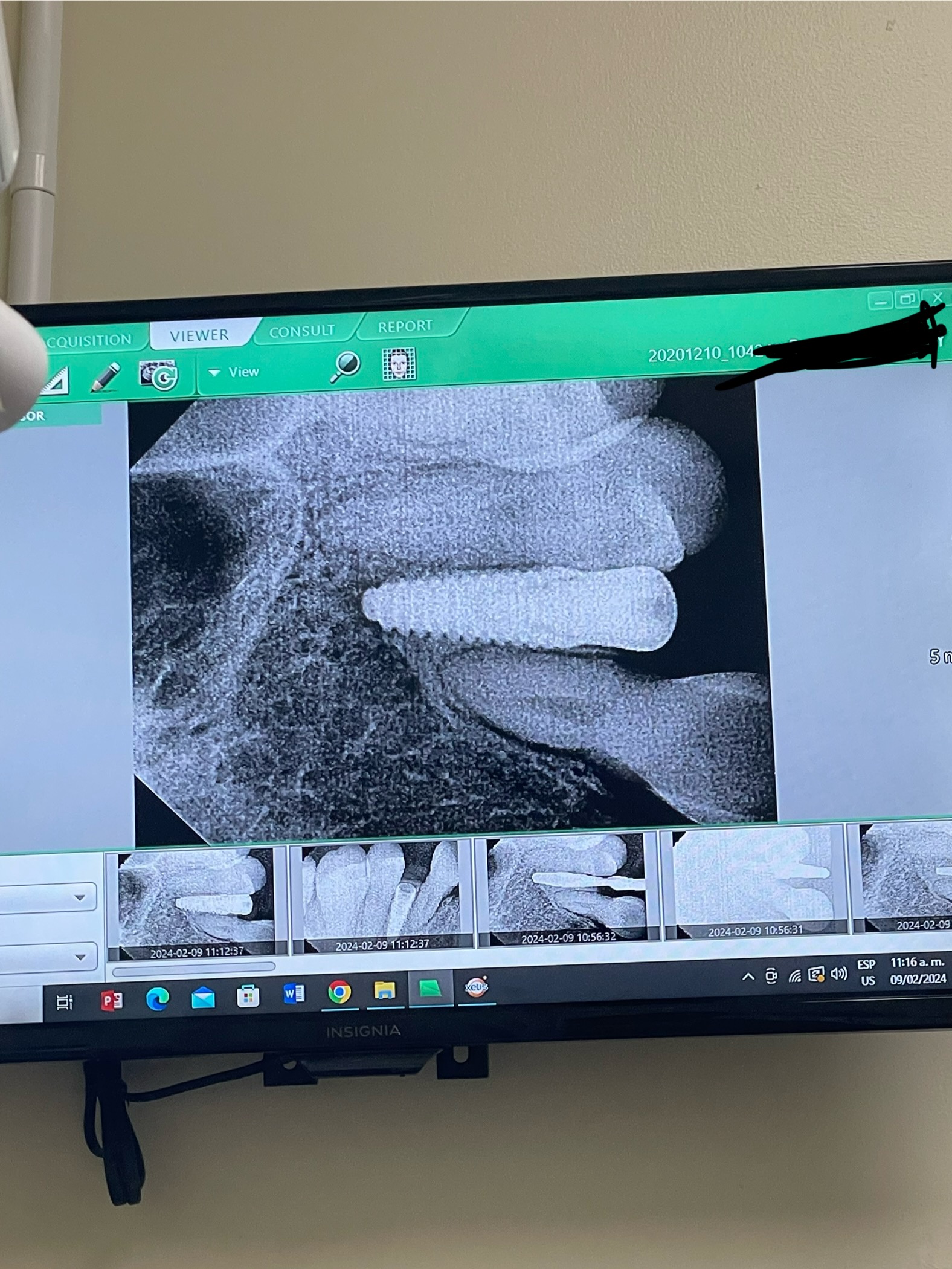
4 months post treatment, an x-ray was taken and I noticed some defects where Bio-oss was placed.

I have a couple of questions:

Q1) What would you expect to see on the distal during uncovering, and what would you do rectify? If you see the defect, would you graft again and attempt to do a primary closure again?

Q2) About the soft tissue deficiency, would you consider a connective tissue graft (CTG) during the second stage to improve the volume, eg Buccal contour?

Q3) Any other suggestions to improve the aesthetics?

Thanks in advance for your comments!!


![]a](https://osseonews.nyc3.cdn.digitaloceanspaces.com/wp-content/uploads/2015/06/x-ray-b4-exo-e1435502615500.jpg)a
![]y](https://osseonews.nyc3.cdn.digitaloceanspaces.com/wp-content/uploads/2015/06/b4-x-ray.jpg)y
![]x-ray-immediately-after](https://osseonews.nyc3.cdn.digitaloceanspaces.com/wp-content/uploads/2015/06/x-ray-immediately-after.jpg)


![]sa-4_2](https://osseonews.nyc3.cdn.digitaloceanspaces.com/wp-content/uploads/2015/06/sa-4_2.jpg)


![]Frontal View After](https://osseonews.nyc3.cdn.digitaloceanspaces.com/wp-content/uploads/2015/06/frontal-view-after.jpg)Frontal View After

10 Comments on Managing aesthetic implant case: advice?

New comments are currently closed for this post.
peterFairbairn
6/29/2015
Yes I always prefer graft material to fully bio-absorbso all you are left with is TRUE host bone for a number of reasons . The bone can turn over naturally un-impeded by foreign material , all you need is a radiograph to assess the case long term ( as no radio-opaque material remaining and the soft tissue attachment is improved . There is also a reduced host response to a biocompatible material for improved healing response. It appears that the material distal to the Implant is HA in granulous tissue which can lead to the soft tissue appearance . Best solution is to clean and re-graft as soft tissue surgery may not offer best long term outcome that host bone will . Peter
drt
6/30/2015
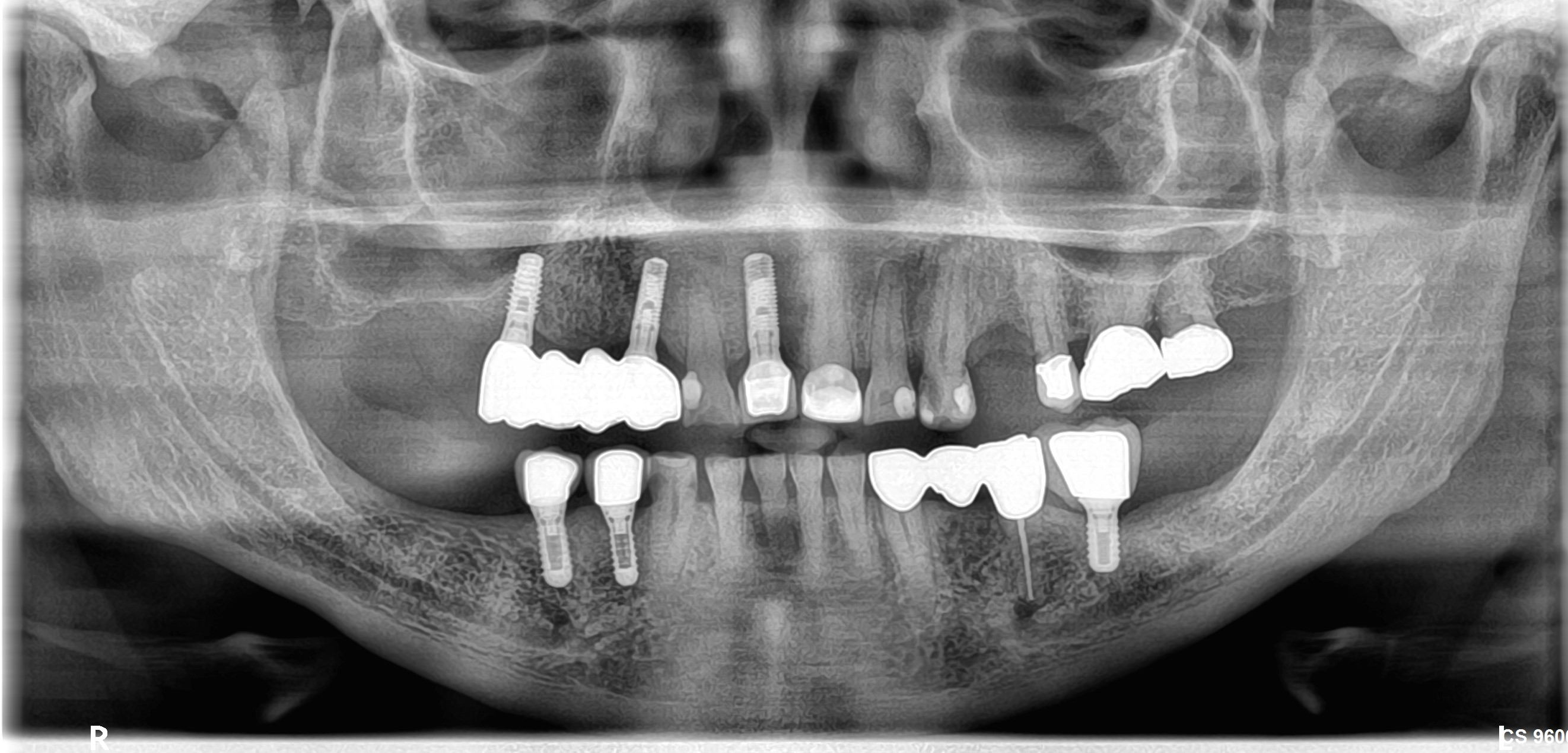
What specifically are you concerned about esthetically? There is generalized staining, apparent visible calculus (UR6, #3) and decay (UR2, #7). SEVERE loss of VDO likely due to porcelain wear but also bruxism (note the exostoses). A 3.3x10 can get beat up pretty badly by a bruxer. I'm not trying to be mean, but we really need to consider the whole mouth not just the hole in the mouth.
CRS
6/30/2015
Looks like the bony support for the papilla was lost possibly during the extraction, but soft tissue regeneration can be improved during the temporization period. What is the brown stuff over all the teeth is it an enamel defect or calculus. Why were the teeth veneered? The implant looks okay and I would have used human allograft at extraction with bio OSS onlay at exposure if needed. But more concerning are the veneers with the recession. The implant should be fine, probably the most esthetic thing there. There is a good connective tissue base. I would replace the veneers with proper contour at the gingival base, see what grows back, then do connective tissue grafts where needed. Plan comprehensively on this one. There seems to be some significant attrition how did that happen?
Dr. Gerald Rudick
7/1/2015
From the xrays and photos, I think you have done an excellent job. On the mesial /occlusal aspect of the implant, there seems to be some nonmineralized graft material.... that just may become mineralized with time. The soft tissue looks good, and there is a hint of papillas, so you will probably not have black triangles when you install a crown....manipulate your temporary crown to develop nice gingival contour before taking the final impression Pay particular attention to the occlusion, and keep the contacts very light, as this patient appears to be a bruxer. If you will access Implant News & Views,Vol 14 No. 5 Sept/October 2012,I published an article entitled " TanGer Technique for Atraumatic Root Removal in the Esthetic Zone" which describes a very simple, inexpensive method to remove anterior roots without using periotomes or other instruments that will damage the very delicate labial bundle bone...so you will avoid these problems in the future. Good luck and keep up the good work. Gerry Rudick Montreal, Canada
will
7/1/2015
Thanks for your comments. brown color is just staining from savacol the CHX mouth rinse. the patient used to have alchol/drug problems and she has been cleaned for over 3-4years. lot of damage were done during that period. pt was informed about the her dentition but her main chief complain was to have her tooth in the front. what exposure techinique may be prefer for this case? i know there are many eg semi lunar, reverse peninsula, I insion and more. i would normally do the palatal crestal incisionfollow by palacci incision to move some bulk of gum to the mesial. also, i am planning to place temp crown for about 6 month prior to place the permant crown. then if the soft tissue bulk is not sufficient then consider CTG. what do you think about this approach?- also, how long do you guys wait to place temp crown after exposure? i was going to take impression 2wks post exposure to fabricate acrylic/ resin based temporary. i really appreciate your comments Thanks Will
John Manuel, DDS
7/1/2015
The Upper Left Central appears too narrow and the Lateral Incisor #10 Space appears disproportionally wide, so the esthetic problems need a wax up and review. This appears to be a Class III Skeletal, Div 2 case with the lower cuspids forward enough to overload the upper laterals which is probably the cause of the Upper Lateral Incisor damage, and the Division 2, Palatally inclined Central Incisors flex, chip and break from the Anterior interferences. Some form of protection is needed for any restoration in these areas.
PeterFairbairn
7/2/2015
Hi Will , yes that is a great protocol about the restoration thickening the gingiva up when fitting a healing cap by moving tissue from the palatal area as you have a very forgiving gingival type in this case . As you know my initial answer was to your questions the whole case depends on how much the patient has to spend essentially . Your concern was the appearance ( possible blueing ) and radiographical appearance of the distal grafted site as pointed out by Dr Ruddick , hence my initial response . But it work out fine just an observation that there is a good chance that the site is soft tissue with embedded HA which is fine although as I stated I prefer host bone . Just different ideals and protocols all work so maybe merely preference . All the best Peter
Hank D. Michael, DMD
7/2/2015
Couple of great comments already made. Very unstable occlusion and a lot of necessary dental reconstruction appears to be needed. Yes a CTG or papilla lift would be beneficial... always better to have too much tissue to start with. Once you open the site, try to excavate any non-integrated bone particles and re-graft void with allograft if possible. Depending on the boney defect that is observed, it may even be better to remove the dental implant and bone graft to build ridge up prior to doing a second dental implant where you know there are no boney issues at time of implant placement. You may also gain some height of soft tissue and possibly get a bit larger implant. It can be difficult to get bone to grow on the existing dental implant. It would be great if the patient could commit to some reconstruction. You may be able help mask any papilla defects with your other restorations - even things out.
Barrow Marks
7/7/2015
Thank all of you for your insightful comments.I have been placing implants and restoring them for a number of years now, and implantology has revolutionized our profession. However, Good treatment planning is always the foundation of any dental restoration. This implant was placed beautifully andall of the complex tasks of an implantologist have been accomplished here. However, I would like all my colleagues to give some thought here as to why that lateral incisor could not have been restored with a post and a crown. Another alternative is to crown the central incisors and the canine if you found the lateral incisor was not restorable. The canine has extensive decay and cosmetically this case would be vastly improved with crowns on the centrals bridged to the canine. By maintaining the lateral incisor, the issue of a black triangle as a result of loss of papilla would be circumvented.
Gregori Kurtzman, DDS, MA
7/7/2015
I would have used a 4.0mm diam implant instead of the 3.3 as there is plenty of width to accommodate it and easier to get a natural emergence from a 4.0 then a 3.3 in that space

Featured Products

OsteoGen Bone Grafting Plug
Combines bone graft with a collagen plug to yield the easiest and most affordable way to clinically deliver bone graft for socket preservation.
CevOss Bovine Bone Graft
Make the switch to a better xenograft! High volume of interconnected pores promotes new bone. Substantially equivalent to BioOss and NuOss.