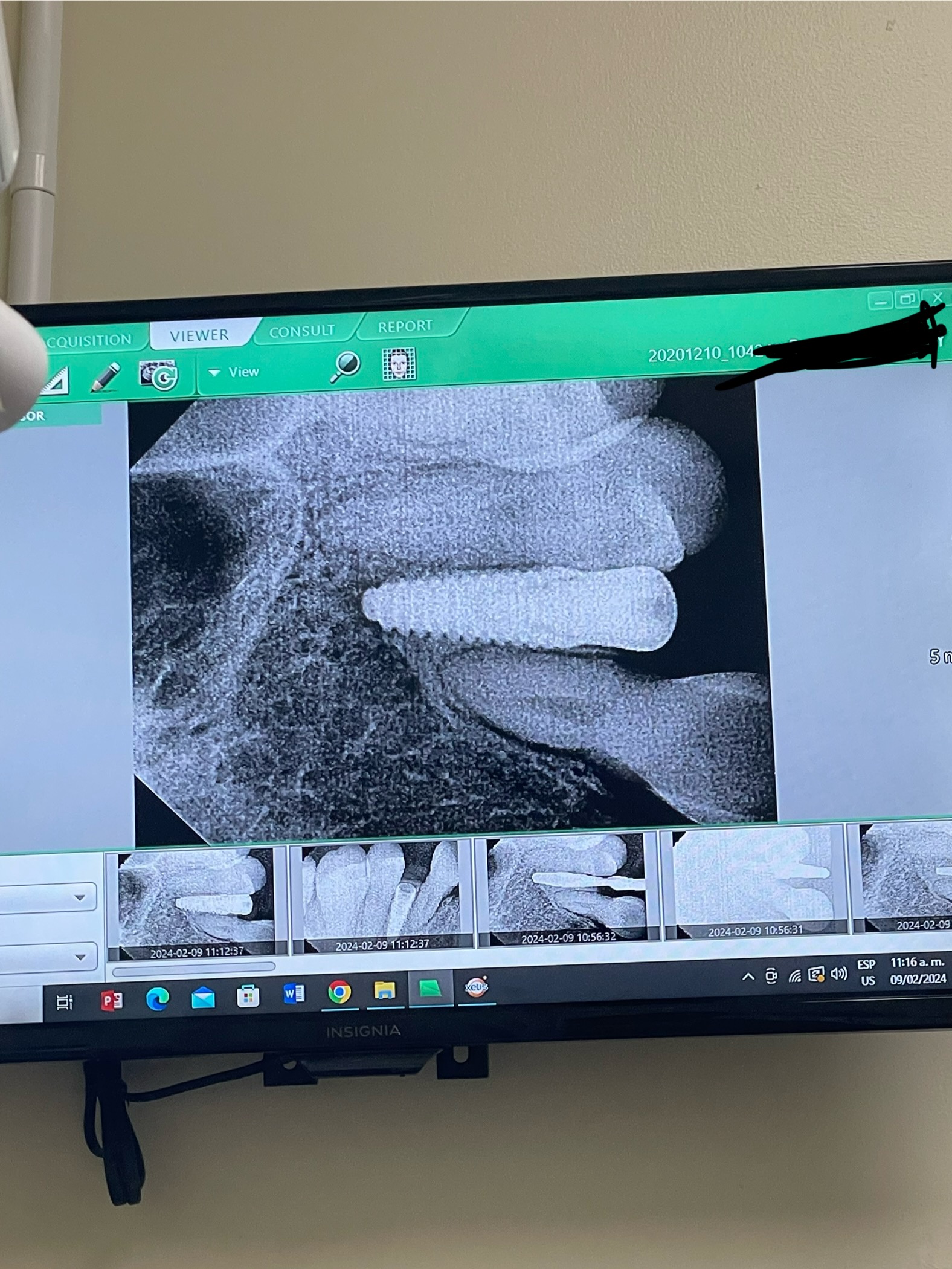
Nasal Inflamation after dental implant on #13: any ideas?

I have a 44 year old patient with no medical complications.  I extracted #13 [maxillary left second premolar; 25] and  installed an implant. The radiographic presentation was unremarkable.  I prescribed an antibiotic for one week.  The patient returned with the complaint of nasal congestion. Prescribed Augmentin and referred the patient to an EENT specialist [Eye, Ear, Nose, and Throat] who prescribed Flonase nasla spray.  He did not find any signs of infection or inflammation in the maxillary sinus.  The patient is still complaining of nasal congestion.  Any ideas what is going on and what I should do?


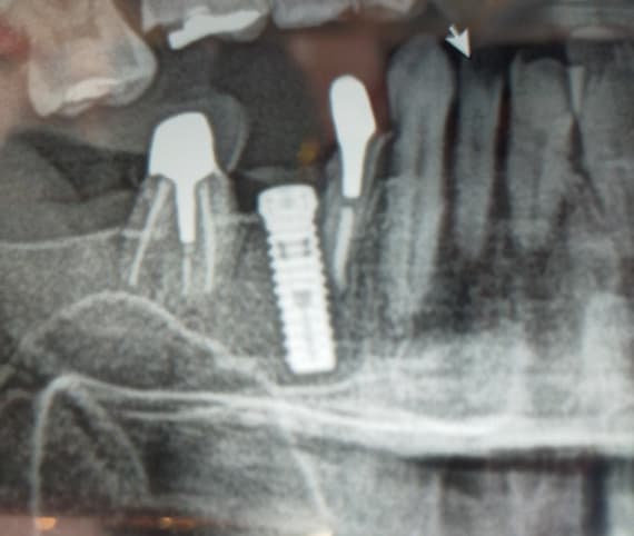
![]](https://osseonews.nyc3.cdn.digitaloceanspaces.com/wp-content/uploads/2012/10/old5-e1350249263857.png)Pre-op
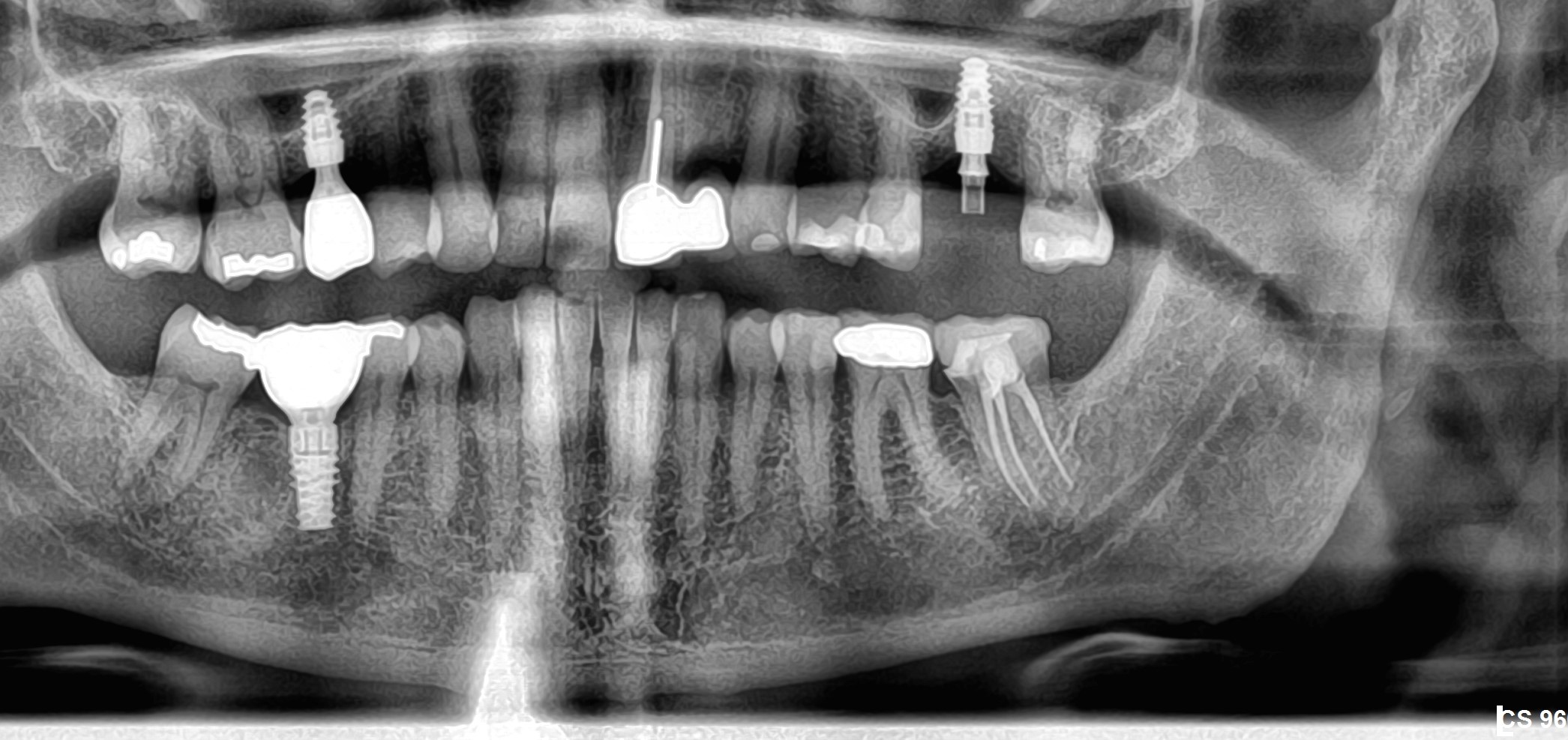
![]](https://osseonews.nyc3.cdn.digitaloceanspaces.com/wp-content/uploads/2012/10/new3-e1350249212172.png)One week after post-op

21 Comments on Nasal Inflamation after dental implant on #13: any ideas?

New comments are currently closed for this post.
CRS
10/14/2012
Nothing the nasal congestion is not related to the implant.
Posted doctor
10/14/2012
Patient had any sinus or nasal problems before this procedure not even cold. The M.D put on oral steroids for 3 days because he see severe inflamation inside the nose but no infection.
Peter Fairbairn
10/15/2012
The pre-op looks very congested ( the other side to your enlargement ) so I would think that this was an unrelated issues or if tooth related it would be the molar (26 ) that is the issue. The implant looks far away enough to be no issue. Regards Peter
Richard Hughes, DDS, FAAI
10/15/2012
This most likely is due to allergies and is coincidental. I suggest that the patient follow up with the ENT. It does not appear related to the implant procedure.
Alaa. M
10/16/2012
I think you have to make sectional CT for the area of the implant to exclude any factors related to implant such as connection with floor of the sinus which not very helpful with this OPG.
Ian
10/16/2012
The likelihood is the bone in that entire area is infected. If you have access to MRI you would probably see this.
Alan Sherrill DMD
10/16/2012
Was implant placement performed post extraction? Did you curette the socket and was there a granuloma present? Apically it appears the root was close to sinus but all things considered the implant is not the issue. Seasonal Allergies,chronic sinusitis or acute sinus infection are more likely.
Jfab
10/16/2012
The problem is not on the implant you placed,if you see on the other side the sinus looks congested. The problem on this congested side seems to be tooth #4 or #2 I suggest vitallity test on the right side on this teeth. Rx penicillin vk 500 and clindamycin 300 for 28 days Let the EENT take care of the sinus problem,you have to help doing the test and find wich tooth is causing the problem
Juan Rumeu
10/17/2012
I wonder what happened with the two silver points on the root canal treatment on the first upper left molar. the two silver points have dissapear. that means that you did a new root canal treatment on this upper left first molar. this is probably could be the cause why the patient has some nasal problems. The implant is not even touching the sinus so it is very improbable that could be the cause.
Keith VanBenthuysen
10/17/2012
I agree with Juan. I believe the problem is with either #14 or 15 not the implant.
CRS
10/17/2012
The sinus membrane is thickened above 14, 15. You could remove the crowns check for recurrent decay or determine if there is a fracture or a root canal is needed. My protocol is to have my endodontic colleague evaluate and treat under a microscope, fractures can also be picked up on cone beam but the RTC has to be performed under a microscope anyway. If there are only nasal and not sinus symptoms it is not the teeth, that is my point. It is reasonable to evaluate the molar crowns I have seen fractures requiring extractions or retreatment. This could be an early sinusitis developing due to pathology in the molars. You are the best judge as to the molar crowns I don't have the history. Be very careful the best treatment for the molars is under a microscope since there could be a fracture and you don't want to have an untreated canal (look at the curve) and you don't want to add to the patient's problem.Good diagnosis is key. I recently saw a similar problem the generalist tried to just crown a fractured upper molar with sinus symptoms, the patient required sinus surgery and I eventually had to extract, graft the defect, and I just placed the implant. A good initial diagnosis under a microscope would have saved the patient a lot of time and trouble. Good luck.
CRS
10/17/2012
I ' m sorry did you perform the root canal under a microscope? I failed to read Juan's comment there could be a untreated canal or fracture or 15 could be involved. Now it makes sense, 13 is not the problem.
Posted Doctor
10/17/2012
Patient had tooth #14 re RCT two months before implant, one root was calcified and no issues in last two months. Patient has seen new ENT, prescribed Ciprofloxacin 5oomg for 6 weeks.
CRS
10/17/2012
Under microscope?
Jfab
10/17/2012
On the pre-op the left maxillary sinus looks congested,on one week post-op many things happened Right maxillary sinus congested and the lef side clean. Silver points from 14 gone. Tooth 16 gone You better be clear on what your doing, multiple treatment done . The problem is not the implant is the retreat or the Xb on 16 . Next time be clear and give more information on what you have performed you did to many things and mislead the dx
CRS
10/18/2012
Excellent point if the molars are the problem then the patient will be on Cipro a long time. The teeth need to be ruled out. Best way I know of is by an endodontist you trust with a microscope. I suspect an untreated canal or a root fracture. Was the retreatment performed under a microscope?
Baker k. Vinci
10/22/2012
This is exactly why this should have been sent to an omfs. It took someone with a dental background to even come close here. An up to date omfs would have scanned the patient and ruled out all dental causes. We treat sino/nasal disease. If I were to guess, the patient was sent to the wrong specialty out of selfish pride and or ego. Is there any chance that the pilot drill was placed too zealously and affected the o/m complex? I waited as long as I could to respond, but as far as you knew, this was a dental problem; when are we going to start realizing that ent doctors are for the most part, "clueless" when it comes to this stuff. I got a patient today, that had an upper right third molar( not displaced into the sinus) removed via functional endoscopic sinus surgery. That's right, through the nose. Do you know why; because there was a lesion around it and he told the patient it would be "malpractice" to go through the lateral sinus wall. If anyone would like to challenge this coincidence, my email is on the site. I will send images and notes, with today's date on the information. Bvinci. Vinci oral and facial Surgery. Baton Rouge , La.
Baker k. Vinci
10/22/2012
The untreated canal shows up every time, with a high quality ct image. Especially in the axial plane. Bvinci
Baker k. Vinci
10/23/2012
Had new patient today who's dentist sent then to an ENT for pain at maxilla before placing an implant. One ct, too high and a referral to a neurologist later, guess what. She has an ABSCESED MOLAR, that had been treated twice, with a lucency > 10mm, barely expanding the sinus floor. I am not making this stuff up. Several have contacted me from this site, I don't have time to make this stuff up. These all originate at the poorly trained dentist office, that is afraid to let anyone know, they don't know it all. It doesn't upset me by the way. Keep um coming!!!! Bvinci
CRS
10/23/2012
It is really reassuring to know that my training as an oms can be uniquely helpful, I have seen some pretty clueless ENTs and mds, but I see that as n opportunity to teach them. The hard part is that my own dds colleagues don't refer out to me when they need it. We are supposed to be the place where the buck stops. My comments on the microscope are for during the actual performance of the root canal, yes a cone beam picks them up but it isI more practical during the procedure to use a microscope,. I learned his from a very talented enodontist.I've seen a lot of "neck abscesses"that we're really of dental origin! Good comments Bvinci.
leroy McKane
10/24/2012
are they as painful as they look?

Featured Products

OsteoGen Bone Grafting Plug
Combines bone graft with a collagen plug to yield the easiest and most affordable way to clinically deliver bone graft for socket preservation.
CevOss Bovine Bone Graft
Make the switch to a better xenograft! High volume of interconnected pores promotes new bone. Substantially equivalent to BioOss and NuOss.