Osteopetrosis: best way to proceed?
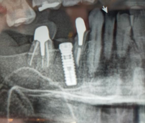
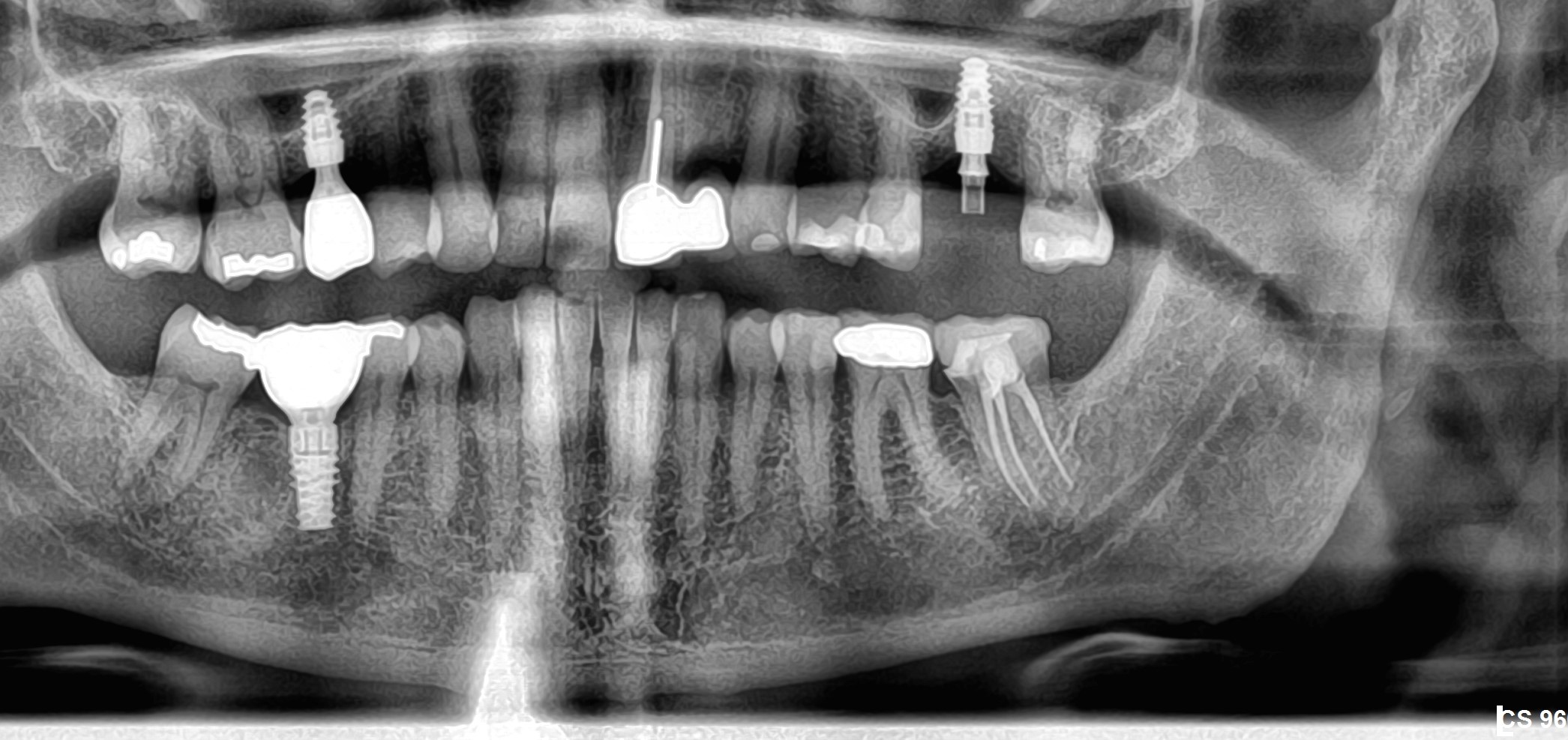
Two years prior I installed 2 implants in the maxilla of a patient with diffuse osteopetrosis [Marble Bone Disease; Albers-Schonberg Disease]. At the time of implant installation I noted that the bone at the implant osteotomy site was not normal. I took a biopsy and the histologic diagnosis was made. Now the patient has developed an apparent osteomyelitis 2 years later on the buccal aspect of the implants. There is an apparent infection. I do not have any experience in treating this kind of complication in a patient with osteopetrosis. How do you recommend that I proceed?
17 Comments on Osteopetrosis: best way to proceed?
New comments are currently closed for this post.
CRS
1/6/2014
Qualifying question was the patient informed of the diagnosis just after implant placement and the possible sequela with extractions or implant placement? And did you do a literature review? I feel that there could be an ethical question here of treating your own surgical complication once the diagnosis was made. The complication could be difficult to resolve I would have gotten the biopsy result back prior to placing the implants or removed them once it was obtained. If the patient knew the possible sequela after diagnosis you should be okay but now do you want to refer or manage this? The osteo in these cases can be difficult to resolve, possibly requiring HBO. This is somewhat similar to a bisphosphonate protocol due to the osteoclasts. For me it is an issue of rolling the dice and keeping the implants in this contraindications to implant placement. Has any medical or endocrine work up been addressed with this patient? Good luck, thanks for reading.
Dime Sapundziev
1/8/2014
What has been done is done. You shold manage the situation now.
If there is only exposed bone without inflamation the treatment shold be just to keep the area cleen as much as possible. Chlorchexidine and jodine solutions for rinsing 3-4 times daly.
If there is a sequesra formed should be removed with the implant and bone covered with thick layer of tissue, probably buccal fat pad.
HBO is not an issue here because there is no vascular component of these kind of dessease. The osteoclasts wold not start to function better if you increase the concentration of oxigen.
Your eforts should not be focused on healing the necrotic bone becaue that is not possible. You shold keep the condition asimptomatic by improvement of the local condition, good hygiene, remove all sharp edges and undercuts so the area can be easily accesable for maintainence of good hygiene.
Good luck.
CRS
1/8/2014
Dear Dr I think you may not understand how HBO is used for the infection which is developing you may want to check your facts. My comments are based on once a diagnosis is made, it is a choice to remove the implants since bone pathology is a contraindication to implant placement, thus patient was not a candidate. Just because an implant can be placed doesn't mean it should. Ironically these implants may eventually be lost anyway. This condition has variable presentation I'm not even sure who manages this but it is a significant disease and the patient needs to be worked up. The poster was prudent to biopsy and I applaud that but it was a poor decision to place the implants in light of the diagnosis. Peter's comments while often very helpful are in management if a different disease process which is caused by drugs not genetics. It is in the patients best interests to refer to an appropriate physician, and eventually remove the implants went clinically appropriate. This doctor made a risky decision now the sequela need to be treated for the patient's health not for the sake of the dental treatment I feel that this is part of the responsibility of being the treating surgeon. Now you are asking an inexperienced doctor to perform complicated management on a known sequela of this disease. I think better advice is to refer this case for appropriate management and be honest with the poster and patient. If the dentist did not discuss the pathology and sequela why bother to biopsy?
Peter Fairbairn
1/8/2014
Well done in your observation of the bone Quality ( Marble Bone ) which led you to take a core biopsy , which in turn led to the diagnosis of Osteopetrosis a rare disease .
Now you have another issue which is possibly an infection and hopefully not a full Osteomyelitis from your description .
I have had a similar issue on a patient on Fosamax which we have resolved by raising a site specific flap and cleaning the area , including Prophy jet cleaning of the Implant surface .
We then grafted with a BTcp and CaSo4 mix into which Tetracycline was added and the site was re-sutured after the mix had "set" .
Three years later and there has been no further issues and the bone has regenerated as all the graft material will have by now been bio-absorbed .
Mixing ABs into CaSo4 is often used in intra,bony infections by our Orthopedic friends with great results ( Stimulan , CaSo4 Pellets infused with ABS in the US )
Good Luck
Peter
peter Fairbairn
1/8/2014
Hi Dima it was great to meet you in Rome last year , we may be helped by a picture of the "infection " to get a clearer picture if possible .
Peter
CRS
1/8/2014
Peter I am going to go out of a limb on this comment, note that I will obtain Dr Sapundziev's article from our hospital database, so here goes. At first glance in Marble Bone disease the bone becomes denser and loses intra medullary blood supply due to the lack of osteoclast activity vs in Bisphos induced osteo bone is not remodeled and poor quality bone not removed. When there is an infectious process present, soft tissue and periosteal blood supply can be improved. Bisphos although a long half life, the drug can be removed, possibly the body recovering vs a genetic process which is uneffected and can worsen over time. There is a variability in expression in Marble Bone which was initially diagnosed upon bone biopsy. So my point is I think the patient should be referred for medical management by Md's and dental management by an OMS. We can't unscramble an egg, however the patient needs appropriate care now by both medical and OMS. I feel we need to be careful comparing two different disease processes since the etiologies are different even though the clinical picture or treatment may be similar. What I learned from this is to look up Dime's research on Bisphos and ORN what I hope the poster learned is that is is okay to remove implants in Marble bone once a diagnosis is made and prior to osteointegration and perhaps this problem could have been avoided. We can't even be sure that an osteomyelitis is developing or that the Marble Bone diagnosis was even discussed. So I am taking my own advice and checking out Dime's research and I am learning that there us a big world out there to learn from our international colleagues. I so far found only one article on dental implants and Marble bone, I just think that the risk was too significant to place implants, I tend to be conservative when weighing dental health with systemic issues. Happy 2014!
greg steiner
1/8/2014
Here is an article that may be of help.
J Oral Maxillofac Surg. 2010 Jan;68(1):167-75. doi: 10.1016/j.joms.2005.07.022.
Intractable bimaxillary osteomyelitis in osteopetrosis: review of the literature and current therapy.
Because both bronj and osteopetrosis are diseases of the osteoclast albeit one is drug induced and one is genetic I would think the treatment protocols would be similar. You did the patient a great service by diagnosing this disease. No one has much experience treating osteomyelitis in osteopetrosis patients so I would think and oral surgeon who has had success treating bronj would be a good referral. Greg Steiner Steiner Laboratories
CRS
1/11/2014
Thanks Greg!
Peter Fairbairn
1/13/2014
Yes Thanks Greg will look that paper up .
Happy new Year
Peter
Carmine Rappani
1/15/2014
Dear CSR ,
Thank you very much for yours kind advices. I think you should try to understand that inside of a scientific community is much more useful to show our mistake or unsuccessful case that only the successful cases: these last could seem “cool†but it is certainly not every constructive.
Of course I made a mistake taking a planning decision based on a wrong OPT lecture. However I want to clarify that patient’s doctor and the same patients nighters known about this localized asymptomatic disease. During the surgical implant I noticed a particularly bone aspect and I decided to do a bone biopsy . For this reason, the histological diagnosis was done only in a second time, when no failure or problems were recorded.
I want to emphasize that my error does not meant that I am not able to retreat the patient as it looks from your heavy words. This should not be part of a medical blog but ,may be, times are changing… Blog exists to improve our knowledges and to exchange our experiences. Therefore if you want to continue to argue my case, I ask you to moderate the words and your criticism.
The purpose for which I wrote these lines is to share my experience with other colleagues, to compare the case with who had a similar problem . I consider ethical to retreat by myself, the patient, moreover because I think I am able to do it despite your hasty judgements.
Dear Dear Dr. Fairbairn,Dr. Sapundziev,Dr Steiner,
Thanks for yours useful advices. 3 weeks after implants placement I received the bone biopsy results, and I informed the patients about he diseases and about of all the risks and possibles complications. The patient refused to removed the implants despite all the possible risks.
It was 24 months ago. 18 months ago we did the final prosthesys.
This singular case was discussed from a DDS, LMD during a Thesis in Oral Surgery in the Dental University of my city 12 months ago. The literature was verified we concluded that actuallly there are any scientific protocol which clinician can use to face this similar cases. The association of surgical curettage with hyperbaric therapy or laser stimulation looks very interesting, but I would like to have some advise by who had a similar case.
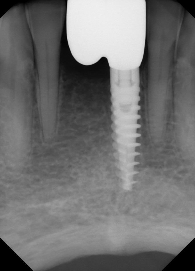
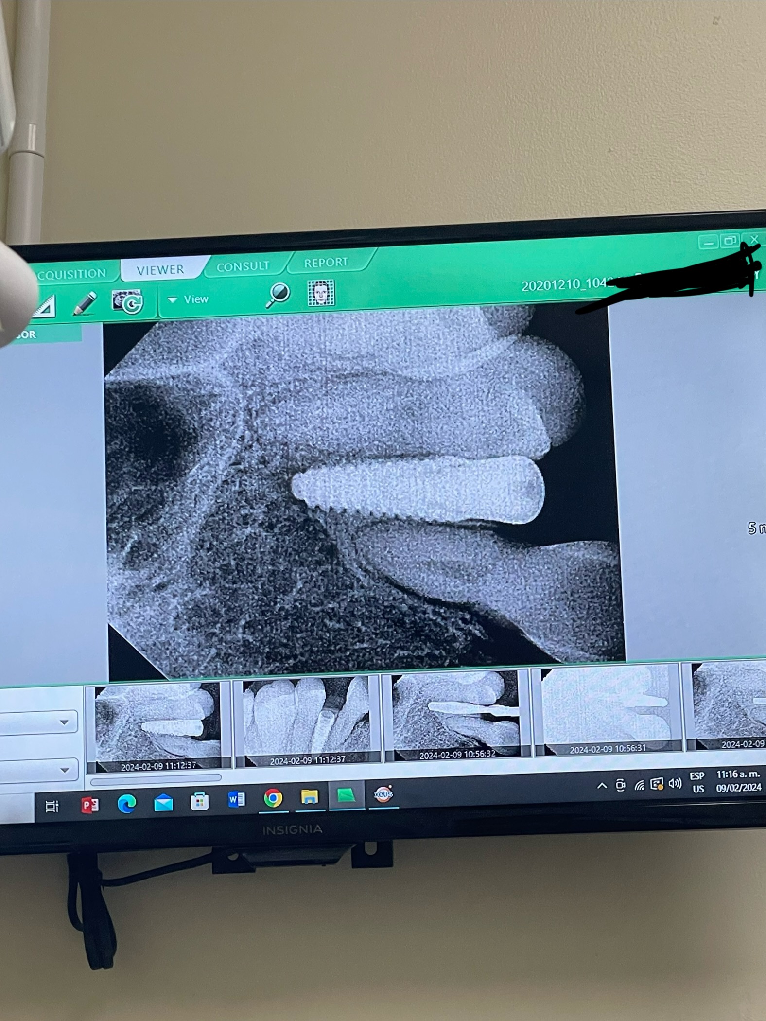
3 weeks ago, during the classical recall, we noticed a fistula in the apical zone, between the implants. A guttaperca cone was placed inside this fistula and an apical X-ray was done.
The path of the cone is anything but close to the implants. The mesial implant is non probed it it presented bone resorption involving the firsth two turns of the mesial implant. On the other hand the distal implant is in perfect condition (clinical and radiographic). . Prosthetics crowns are stable and peri-implant health is sound.
The radio transparency area correspond to the biopsy site .I am going to ask to the patient a CBCT, to evaluate the precise location and size of the lesion.
In conclusion I will follow your advice. I will open a large flap away from the lesion in order to remove all infected tissue. I will close a flap hermetically. Moreover I intend to stimulate the surgical site with photo dynamic therapy and , if it will be necessary I will propose him .hyperbaric therapy. I will be really grateful to any colleague who wants to bring his experience in the treatment of this case.
I will post all the pictures after the treatment.
Best Regards
Carmine “CL†Rapani
CRS
1/18/2014
Dear Carmine, thanks for listing the rest of the information that would have been helpful at the get go, and your defense of your actions, not sure why the defense was necessary. My comments are honest and there is no obligation to follow them, my opinion my tone. Sometimes I feel honest imput is more beneficial and advising someone who states they have little experience with this to refer I feel is prudent. I just would have removed the implant in light of the diagnosis once I knew there could be significant complications which quite frankly I would not want to have to manage. I usually don't allow a patient to make the call on whether or not to remove an implant which could have a significant future complication even if the error was mine in placement. unfortunately dentists do need to understand bone pathology if they are placing implants I fear that knowing the biopsy results and keeping the implant in may have caused this predicament. I hope you are not going down a slippery slope here, so good luck and please post your outcome for our scientific community so we can all learn. I hope my honest comments are taken in the manner intended.
Peter Fairbairn
1/16/2014
That would be nice , as we are all always learning especially in rare situations such as this .
Peter
peter Fairbairn
1/17/2014
Read so old Sottosnati research on CaSO4 lastnight and there was reference to Dressman in 1892 using the material successfully in the treatment of Osteomyelitis ....
Peter
Dime Sapundziev
1/20/2014
Dear Carmine Rappani,
Very hot discussion you brought out here with this interesting case.
I do not think that this forums are made for spreading out our frustrations but to exchange our experience and learn from each other. Some of us have the opportunity to work in bigger institutions and have a chance to deal with more complicated cases, some perform minor procedures but the common thing is that we deal with patients and here is the place to share our experience, successes, mistakes and failures.
Analysing your case I was thinking and I would share my toughts with you no intend to judge. It is clear that you recognized rear disease and took specimen for biopsy. If I were you I would stop at these point and wait for the results. But as I said in my statement what is done is done, can not turn back time.
Now when the implants are in function and patient presents with osteomyelitis these condition needs to be treated maybe you shuold consider to remove the implants.
I can share my case with you, what I have done in a patien who recived dental implants two years prior to administration of Zometa i.v. She presented with a sequestrum arond the implant with acute signs pus discharge, neurological disturbances ( numbness of the lower lop) and pain. I removed everything, did local drbridement. She recived antibiotics Amoxicillinum with clavulanic acid no HBO and condition resolved. Two years since the operation the lesion is heald.
As far as HBO is concerned dear colleague CRS I recommend you to have a closer look at the Oral and Intravenous Bisphosphonate-induced osteonecrosis of the Jaws second edition by Robert E. Marx. Personally I have treated more than 100 patiens with developed BIONJ an in none of them I used HBO. All of them are symptom free.
Hello Peter it was a great pleasure meeting you in Rome and I hope to see you soon on some upcoming event.
CRS
1/24/2014
Dear Dime I have the book will review. For the HBO I am picking up on the original post stating pus and infection and the fact that in marble bone there is little blood supply, however I don't know if it would be helpful with this disease. I also understand that BIONJ is not an infectious process but can have an overlying infectious component. It is sometimes hard to communicate online vs an in person discussion. I want to learn more from your experiences. Thanks for the very valuable information!
carmine
1/24/2014
Dear Colleagues,
It is very nice that we can exchange ours ideas and experiences. ☺
I find your experience very interesting and the only way to proceed here is probably the implants removal.
In my case , thus , the intraoral radiography has highlighted that the gutta-percha cone goes away from the two implants!
Only the mesial implant has a bone resorption in the mesial side. On the other hand the distal implant has not rx problems. Clinically I have no peri-implant disease. I am going to do an extended flap to expose the lesion and, if it will be necessary, I will remove the implants. I will keep in touch with you to discuss of the evolution of this case.
Gratefull, Carmine
peter Fairbairn
1/26/2014
Carmine do a little research into Stimulan ( US marketed CaSo4 pellets for carrying abs to infection sites) .
I have just started treatment on another similar case ( referred in ) again drug related cause but as with yours not right the site of the Implant ....
So interesting idea ...
Peter


