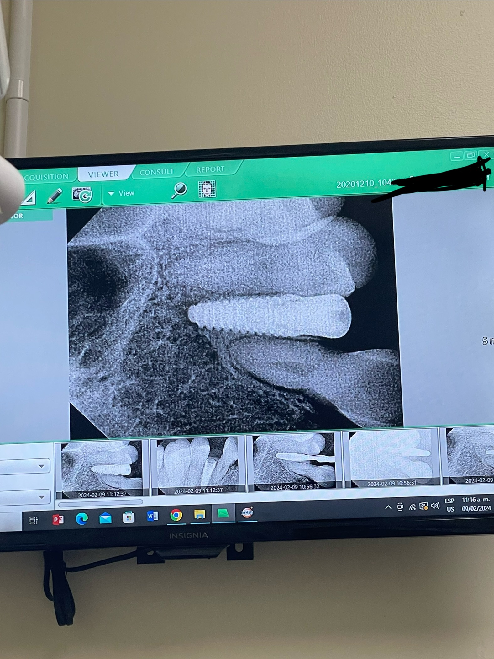
Resorbing graft over implant after 4 months: thoughts?
I placed an implant with buccal graft covered by non-PTFE membrane and buried it 4 months ago.
Graft thickness according to CBCT was 2.3mm.
I took another CBCT 4 months later and the graft has resorbed to 1.5mm and I can see the greyness of the fixture through the gingiva. I’m pretty sure when I expose the fixture for second stage it will expose some threads.
Should I re-graft the site with more particulate bone graft ? Will it osseointegrate against the current graft?
Thanks for your thoughts!

16 Comments on Resorbing graft over implant after 4 months: thoughts?
New comments are currently closed for this post.
nalmoc
2/24/2020
I'm afraid that the implant may not integrate well. Is there any reasons why you did not pick a longer implant? 100% of the buccal plate around that implant is grafted material while you have close to 10mm apical bone from inferior alveolar nerve canal.
For what you have now, at second stage if the implant has integrated, you should add more bone but also pay attention the quality of the soft tissue you have there. if you have thin biotype or all mucosa, you need KT graft as well for good long term prognosis for the implant
WJ Starck DDS
2/24/2020
A couple of problems with this case:
1) the implant is too wide for this ridge. You don’t say which tooth you were replacing, that’d be helpful
2) it will be nearly impossible to regenerate bone on the lingual of this implant. Think of the blood supply - the only place it will come from is the periosteum.
3) I would remove implant, perform a ridge split with grafting, then go back and place implant
Greg Kammeyer, DDS, MS, D
2/24/2020
Since we can only see one of the many slices on the buccal to gauge how much is native bone and how much is grafted bone, I wouldn't draw conclusions about predictability.
First I also wonder, why didn't you use a longer implant?
Second:Which membrane did you use? Collatape? Cross linked or Non-cross Linked collagen: Study them to learn about the resorption times.
Third: Did you fixate the membrane? The mentalis muscle will move the membrane and graft around, decreasing the quality and quantity of bone without fixation.
Fourth: Did you do an aggressive corticotomy? that area has low blood flow due to thick cortex and and with a thick cortex I would avoid a ridge spit there, unless you are trained, experienced and equipped to do block grafts.
Lastly, since the tissue is commonly thin there, there is likely a low level of vascularilty to the implant and a CTG is probably in order. We need both 2mm of bone and 2mm of soft tissue for long term stability.
Yes, certainly secondary grafting is in order. Scrape some autogenous bone from the area or use L-PRF to get some growth factors going.
No need in my opinion to remove the implant.
PerioProsth
2/24/2020
I do no tknow if want to redo it, but IF you can and if you want, I'd recommend to remove it and placed with a Longer and one size narrower and then do GBR.
IF you don't want to do it, you should flap, remove all the soft , mushy graft from the B, harvest allograft from the adjacent area and place it on the body of the implant and cover the rest with a long absorbing graft ( Cortical allograft or Xenograft) & a membrane before you load the implant. Consider a Biologic Agent during GBR, if you can.
i would prefer the first option, myself.
good luck.
Peter Fairbairn
2/24/2020
Ideally it would be perfect if all the graft resorbed and is replaced by host bone , I know the only bone I can regenerate is my own , so working with and understanding host healing is critical . The less we do the better the outcome the host is the miracle not us . Regards
Practical Caveman
2/24/2020
I much prefer to graft first and come back for the implant, (one miracle at a time). You can't count on getting all the bone you grafted with to become host bone. I just posted another case showing a tent-graft technique.
Dr Dale Gerke, BDS, BScDe
2/24/2020
The above points are relevant (a longer implant would be desirable – did you plan this with 3D before placing the implant and use a surgical guide?) - but perhaps consider this.
You have what you have. Depending on the age of the patient and circumstances, if it was me I would consider explaining to the patient that I was not happy with the outcome but if the patient was happy I would try to improve the situation at no cost to the patient. I mention that it has been shown that short implants do survive in the mandible – but always better to use the longest possible.
If the patient agreed to let me proceed then I would raise a buccal flap and check the graft integrity. If it was not real bone (ie it is residual soft graft material) I would scrape away whatever is not bone, then place 3 mm of Ethoss over the buccal aspect – with a few holes drilled into the mandible) and replace the flap. No membrane is required which makes the process quicker, cheaper and easier – but the bonus is - you do not use extra space with a membrane). I think you will find that considerable real bone will be laid down and without a major inconvenience to the patient. The point above about attached gingiva needs to be considered.
You would then need to test the integration of the implant at 3-4 months. If satisfactory then you can finish the restoration. If not you should follow the advice above.
I am presuming you have not done many implants. So my advice is to learn as much as you can with this case. Use 3D planning and a surgical guide and if you are not familiar with certain technics then seek help from someone who is (ie refer the patient but attend the surgery and learn).
Well done for asking about this case, it has prompted some interesting comments which you and others can consider in future.
Peter Fairbairn
2/24/2020
Yes Daot just keratinised soft tissue but attached keratinised tissue for long term stability . Regards le nice response as Dentists we sometimes get confused between radio opaque material and bone , which as I said can only be regenerated by the host not us Dentists . With this there is also an improvement in n
Peter Fairbairn
2/24/2020
Messed that up , computer went awry .....yes Dale , agree as Dentists we get confused with radio opaque material and host bone which can only be regenerated by the host not us . With host bone comes improved soft tissue , attached keratinised for long term stability . Regards
CLF
2/24/2020
Thanks for the responses everyone. I'm the dentist who placed this implant.
This is a fixture 4.5x10mm to replace a lower right first molar. A preop 3D scan was taken but no surgical guide was used. A collagen membrane was used without fixation.
With regards to using a longer implant, I felt a 10mm was sufficiently long. And with regards to going down a size I was worried if a 4mm would be too "narrow" for a molar.
I have engaged a local periodontist for his help to help me do a 2nd stage bone and soft tissue augmentation at no cost to the patient.
I'm just wondering what is the likelihood of regenerating good quality bone and soft tissue here? Will it just resorb over time again because there isn't good vascularity? I suspect the implant has integrated in the proximal and lingual areas but it has a dehiscence defect on the buccal.
Peter Fairbairn
2/25/2020
Hi CLF , fine , the right size implant to use here maybe a mm deeper would be ideal but no ideal world as the ideal would be a tooth . But looks OK and depends what you want as to host bone or foreign material with a little bone in between . Scans are not an ideal tool in assessment when using materials that do not fully resorb as they cannot detect bone just radio opaque stuff . Anyway you are on the right road getting some help as I am sure your patients interests are your primary concern. Good luck and yes the the host heals often despite what we do so hopefully will be fine . Regards
Peter Hunt
2/25/2020
The basics of generating bone in a situation like this are relatively simple. Open the region with a relatively wide flap, cortical bone in the region needs to be perforated with small round bur holes to generate blood flow in the region. A bone graft needs to be placed over the top of the implant and out over the adjacent bone. This should be over-built as it will always shrink down in healing. A membrane should then be placed over the top of the bone graft to contain it and thicken the soft tissue complex. The flap should be periosteally scraped or released so it can cover up and over the grafted region where it can be sutured.
You seem to have achieved what might be expected by this first procedure. Yes, there has been some shrinkage but that is to be expected. At this time if you are desiring more bulk then you can open flap and re-graft on top of what you already have in place. Serial augmentation like this can be combined with a second stage exposure and placement of a healing post.
In short, you are on the right track. Good luck with it from here. Best wishes.
CLF
2/25/2020
Thanks again for all the prompt responses here. Really good to learn just albeit unfortunately that our patients have to go through more procedures.
To Dr Peter Hunt: yes did all that but I suspect maybe my flap was not absolutely tension free and thus the graft might have slumped.
In any case since there have been some mentions that I can hope to regraft it I will go ahead with the help of a periodontist in my country, instead of removing the fixture (of course I will check for integration at 2nd stage). Thanks again for your opinions.
cnushet
2/25/2020
All above comments will help you to do better for the next case. Your implant selection was not perfect, you already realize that. The issue is now what you should do! I suppose you to go for second surgery and check the ISQ value. If you have 1 mm regenerated bone on the buccal side. You have also bone on mesial distal and lingual side. So ISQ value will guide you, this implant is stable or not. If good ISQ value go for prosthesis and think about KT graft. Incase of low ISQ value, I would prefer to place a new perfect size implant in perfect 3D position. Good luck.
mark simpson
2/26/2020
first of all I always place every implant one mm. below the crestal bone to start with. Secondly like those before you had a lot of room apically .
Zachary F
7/2/2020
At 4 months BTCP is well on the way to full resorption and immature bone difficult to see on radiograph. There are form stable resorbable TCP,s emerging that contain a trace radiopaque market allowing for improved monitoring in such cases.
Robert


