Ridge Split to Create More Buccolingual Space for Implants: Make a Surgical Guide?
Dr. M asks:
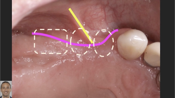
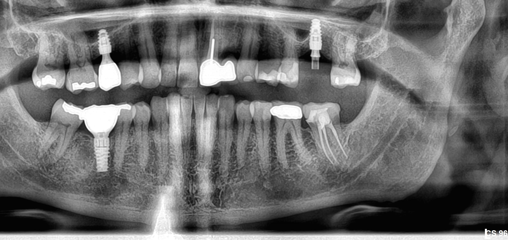
I have a patient with a thin alveolar ridge. I am planning on doing a ridge split to create more buccolingual space for placing the implants. I would like to use a surgical guide to do this. Is this the proper protocol? Do you have any recommendations on how I could make a surgical guide for the ridge split portion of the surgical installation of the implants?


23 Comments on Ridge Split to Create More Buccolingual Space for Implants: Make a Surgical Guide?
New comments are currently closed for this post.
matthew s
10/24/2011
You could preplan your future/desired shape and dimension of the jaws and then transfer it to a surgical guide/prosthesis (check your planning software). It could definitely help you in establishing the limits of the split and/or other grafting techniques.
Ian E
10/25/2011
Are you planning to place implants in the maxilla and mandible?
I have used a modified template in the Mx with ridge splitting. I have the lab wax out the buccal mucosa to ideal width and they fabricate a template to that width.
I also have tubes in the template for angulation and I place the implants into the ridge through the tubes.
Alejandro Berg
10/25/2011
just a friendly recommendation, dont do a split ridge specially in the 102 cut, not enough bone and you can get in to an unsolvable problem later.
cheers
Bruce GKnecht
10/25/2011
Well, this split is going to be a fracture and I hope you have bone scrws if the buccal plate fall off. There is a lot of D1 bone and this is very hard to expand.You may want to use a block from another donor site or lay some infuse on it. Yes it is always good to have a surgical guide. If for nothing else it gives you perspective to where you are in space. Good Luck. Make sure that the patient knows that there are very high risks and take photos for documentation.
dr. bob
10/25/2011
I would not ridge split here, agree with Bruce, block graft is the best choice.
gary omfs
10/26/2011
Cut off the top 5- 7 mm of the ridge. Turn it upside down. Fix it to the new crest with some screws. You can put in some temporary narrow implants if patient wants. Final implants after three months, together with removal of the other hardware. Ridge split works OK in the upper, but lots of bone resorption. Bad control in implant direction. Lots of plate fractures. Not my preferred technique, anymore.
Carter
10/26/2011
I believe that in the mandible you might consider making the classic toronto bridge with 4 or 5 implants lowering the bone until sufficient minimum thickness.
best regards
CD
10/26/2011
How about grafting first???? Is the objective 2 mini implants for mandibular overdenture? Do no harm and keep in the spirit of beneficence and look at the other options seriously. It is possible to create a surgical stent/guide while doing ridge splits. Asking the question about "proper protocol" seems irrelevant to your objectives for providing the patient excellent care. This of course depends on you skills, experience, and the particular case.
sergio
10/26/2011
Good chance to fracture if you go for ridge split.
I would avoid and instead, take down the sharp edge 2-3 mm to gain more b-l width.
CD, it takes more than 2 mini implants to hold down a lower denture. Lots of dentists complain about high failure rate of minis because they don't follow proper protocol.
dr.t
10/26/2011
I agree with Sergio on both points. 1. Unless the entire ridge is only a few mm thick (in which case a split is way too risky), just lower the crest until you get to wider bone. 2. Too much torque during function to only use two. Four minis are needed for a lower overdenture, minimum. Ideally I use 5-6.
ttmillerjr
10/27/2011
You cannot split this ridge, will not work. Have you had some education on ridge splitting? If you tried to split this ridge you most likely will wind up breaking off what height of bone your patient has. I know you want to do implant cases, but this is not a learning case.
Dr Samir Nayyar
10/28/2011
Hello
My humble advice to you is please don't play with the patient's remaining bone. Don't even try if you are not 100% confident to do the procedure. Just lower down the crest until you find minimal bone width & put 2 or 3 mini implants.
Best of Luck.......
sergio
10/28/2011
Again,
2 or 3 minis won't support lower full denture.
Why are there so many dentists that don't know the proper protocol and still spread the word!! That type of technique causes higher failure with angry disappointed patients.
Dr.S.Lin
10/29/2011
Ridge split can be done predictably , but you must be proficient at it.Use the 2 stage split procedure, well illustrate by Bicon implant site: http://www.bicon.com/cases/case-study/mandibular-ridge-split-two-stage-implant-placement-and-their-restoration-with-integrated-abutment-crown/
This procedure can eliminate Block graft in many cases. I agree with previous comments that lowering the knife edge crest will facillitate the procedure.
hope this helps. It can also be termed as Osteoperiosteal flap procedure,as Dr. Ole Jenssen has illustrated in his text book.
Richard Hughes, DDS, FAAI
10/29/2011
It is according toTatum, an expansion. Again real men do not place mini implants.
Richard Hughes, DDS, FAAI
10/29/2011
One may easily place blades, subperiosteal or a ramus frame, if they have the surgical and prosthetic skill!
Andrew HF Tsang
10/31/2011
Hi Dr. M,
Case looks good on the upper except as noted there is a recent extraction site.
For this upper, your guide could have been done with the patient wearing it during the CT scan- that would be a definative way to get a better visualization prior to the surgery. But it's not nescessary and more likely a hindrance to use a fixed guide. Make a well reflected flap to help orient your instruments. You can also place an impression coping on the first implant to act as a guide for your split.
A deep enough flap along the buccel will make you feel comfortable. Good luck hammering away
For the lower, I'd reduce the bone height than ridge augmentation. It's more predicatable and there is ample bone present. Guide is very helpful here to know your reduction for your final prosthesis and position your implants accurately.
Richard Hughes, DDS, FAAI
10/31/2011
This case can be done if on has the proper training, instrumentation, manual dexterity and is patient!
John Manuel, DDS
11/1/2011
I agree with Dr. Lin re: the two stage ridge split demonstrated on the Bicon site.
You cut at the crest, splitting the narrow attached tissue with maybe some slight Lingual overlap and cut the "trap door" or "window" thru the cortical plate. The tissue is then repositioned passively and 3 weeks are elapsed.
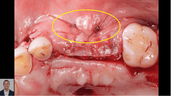
Second stage - You cut at the crest, trying to keep some attached tissue on each side of the flap. Do not lift the periosteum off the bone - it is needed for circulation. If you peel the periosteum off the bone, the trap door will be rejected. Anyway, you use large currette and/or splitting chisel to gently mobilize the trap door to the buccal. The implants are placed into the basal bone area 1/4 to 1/3 length and then the trap door is gently sutured up against the buccal of the implants. Collaplug strip is gently placed and sutures placed with care not to loosen the periosteum from the bone.
This gives you a huge ridge width with a nice wide band of attached tissue on top.
John
Dr. Jivan
11/2/2011
There are a couple ways to approach. I know for sure that some labs with a CT can fabricate a surgical guide that can guide ridge expansion (primarily using ridge expansion screws). In my experience ridge splitting can be very successful, but is very technique sensitive. In my hands I found the technique that works best is when it performed in 2 stages as described by John above.
Baker vinci
11/6/2011
Better yet, can you tx the many complications associated with the poor split. Success with grafting to increase width is predictable and would be the standard of care in my geographic locale. Nerve laterliztion would be next. Strongly encourage upgrading your cbct software, in that patient education is tough, with these images. How many ridge split cases like this one, have you done? Bv
Dr.H.Hamidifar
1/10/2012
There are several techniques and approach available for bone expansion and increasing bone width. The aim of this article is to review the current literature concerning bone expansion in narrow alveolar crest prior implantation. It is especially aimed to review the literature on various procedures and on their suitability in ridge-splitting as follow:
• Ridge-splitting with simultaneous implant placement utilizing chisels. This technique also mentioned as immediate lateral expansion.
• Ridge-splitting with simultaneous implant placement by means of tapered osteotome of increasing diameter. There is a modified approach for this technique introduced by santagata m. (MERE technique) 25, 26
• Delayed or staged lateral ridge expansion
• Nontraumatic bone expansion by means of increasing diameter bone spreader
• Ridge expansion osteotomy. (REO) 24
Dr.H.Hamidifar
1/10/2012
The narrower ridge in the anterior maxilla and premolars areas is often inadequate for placement of standard-diameter implants (4.0 mm). When ridge splitting is considered, the clinician should not compromise on the desired implant diameter simply to accomplish the implant insertion. The goal of surgery is to expend the ridge to allow placement of an appropriate size of implant for proper prosthetic contour and biomechanical support. Although small-diameter and narrow platform implants may be easier to insert when using ridge splitting techniques, their use should be limited to specific indications. Another possible indication is when the ridge width is adequate but the clinician desires to insert a wider diammater implant (>4.5 mm) for improved emergences profile or biomechanical support of the prosthesis.
The ridge-splitting techniques allows placement of implants in a narrow crestal ridge in a single procedure. Chiapaso and colleagues evaluated the success of different surgical techniques for
ridge reconstruction and success rates of implants placed in the augmented areas. The surgical success and the implant survival rates were as high as the guided bone regeneration and onlay graft procedure, with the advantage of a shorter treatment time. Sethi and Kaus reported the technique of maxillary expansion with simultaneous implant placement. They placed 449 implants in 150 patients in thin maxillary ridges of adequate height and comprising two separate cortical plates with intervening cancellous bone and observed them for a period of up to 93 months. A 97% implant survival rate after a 5-year observation period was found.
Splitting of the atrophic alveolar ridge to enable immediate implant placement is a complicated and demanding surgery, one that requires a long period of postoperative edentulism. However, the alternatives for patients with extremely thin ridges are often even less appealing. Many patients cannot wear complete dentures because of the loss of basal bone and unfavorable bone ridges. Widening of the ridge by more traditional grafting techniques usually requires unvasive surgery and hospitalization, with the attendant risks of donor site morbidity.
The bone quality also influences the ability to expand the ridge. Ridge splitting is more applicable to the maxilla than the mandible. The thinner cortical plates and softer medullary bone make the maxillay ridge easier to expand and simultaneous placement of implant. The lateral ridge expansion technique with simultaneous immediate implant placement is usually performed because it shortens the total treatment time.30,58 However,in the mandible, the risk of malfracture of the osteomized segment is greater because of the lower flexibility and a thicker cortical plates careful expansion of the buccal plate is essential when the lateral expansion technique is used because abnormal bone healing can result from undue trauma to the plate.. In such cases, Enislidis et al and Elian et al recommended a staged approach to avoid post-operative complications from malfracture of the buccal segment. Therefore, proper case selection and surgical technique is important when considering the use of ridge splitting techniques. In some cases, the narrow posterior mandible may be split and expanded successfully with simultaneous placement of implant especially when there are thin cortical plate and enough medullary bone. Favorable condition for the posterior mandible include a long edentulous span (missing molar and premolar), abundant bone height superior to the mandibular canal (>12 mm), and the presence of some cancellous bone between the dense outer cortical plates. If these conditions are not present, then the clinician may prefer onlay augmentation.This technique allows for delivery of an overdenture 6 to 8 months after the implant placement surgery. Nine months of healing is recommended for delivery of a fixed prosthesis to allow bone healing and implant osseointegration and reduce inplant failures.
The use of bone graft material and membrane in bone-splitting technique is controversial and completely depend on the situation and surgeon’s technique. Although some clinicians prefer to place particulate bone grafting materials around the implants and in the intercortical space. Scipioni et al25,26 have found that a bone graft is usually unnecessary. Wound healing in these cases is similar to the fracture repair of bone.61 The gap fills with a blood clot that organizes and is replaced with woven bone. This immature osseous tissue develops into load-bearing lamellar bone at the implant interface. Scipioni et al64 examined implants without primary bone contact in
a canine model. Narrow mandibular ridges were prepared with a partial-thickness flap and split with a chisel. The buccal plate was displaced and two 3.3-mm test implant were inserted. Following a 7-month healing period, the implants were examined clinically and histologically. The average ridge width increased from 2.4 to 6 mm. they found that the interproximal marginal bone healed to the level of the roughened titanium plasma-spray surface on the implants. The amount of direct bone contact to the implants along the mesial and distal surfaces was similar to the buccal and lingual surfacrs that approximated the adjacent cortices. Other studies have also examined the ability of healing bone to jump a gap and have revealed similar findings.
Several authors have suggested the use of a partial thickness flap to help immobilize the displaced buccal cortical plate. In contrast, Calvo Guirado et al believed that the periosteum is the best possible biologic membrane because it contains a rich supply of osteogenic cells.
The lateral ridge expansion technique is very effective for horizontal augmentations in severely atrophic posterior mandibular ridges. In the mandibular ridge which has low bone quality and a thin cortex, immediate lateral ridge expansion can be a useful procedure. Delayed lateral ridge expansion can be used more safely and predictably in patients with high bone quality and a thick cortex and narrower ridge in the mandible to avoid complete fracture of the buccal segments. In addition, delayed ridge expansion is recommended when the initial stability of the implants is poor.


